

Hip capsulolabral spacer placement for the treatment of severe capsulolabral adhesions after hip arthroscopy
- PMID: 24904779
- PMCID: PMC4044510
- DOI: 10.1016/j.eats.2014.01.003
Hip capsulolabral spacer placement for the treatment of severe capsulolabral adhesions after hip arthroscopy
Abstract
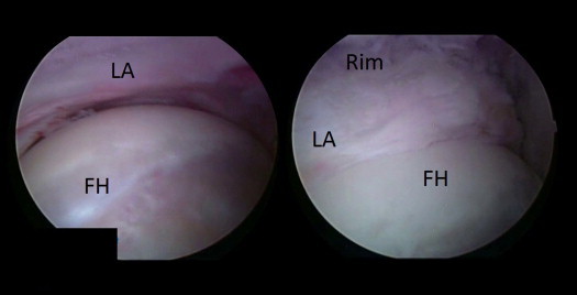
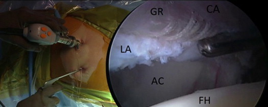
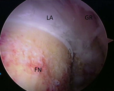
Recently, there has been a rapid increase in the number of hip arthroscopies performed. The increase in the number of primary surgeries has been associated with a similar increase in the number of revision procedures. The most frequent indications for revision hip arthroscopy are residual bony deformity (impingement), persistent labral pathology, and intra-articular adhesions. Our current understanding of capsulolabral adhesions is limited. Although adhesions between the capsule and labrum are common after hip arthroscopy, generally, they are mild and asymptomatic. However, in severe cases they may cause persistent synovitis and pain, and they may tether the labrum away from the femoral head, causing loss of the suction-seal effect. Such patients present with nonspecific symptoms such as persistent pain, giving way, catching, and pain in hip flexion. Magnetic resonance imaging can aid in the diagnosis by showing the absence of liquid in the capsulolabral recess, although the definitive diagnosis is based on dynamic arthroscopic evaluation. We present our approach to the lysis of capsulolabral adhesions with preservation of labral tissue and describe a technique that uses an iliotibial band allograft to prevent recurrence of such adhesions by maintaining space between the capsule and labrum.
Figures
References
-
- Lynch T.S., Terry M., Bedi A., Kelly B.T. Hip arthroscopic surgery: Patient evaluation, current indications, and outcomes. Am J Sports Med. 2013;41:1174–1189. - PubMed
-
- Philippon M.J., Schenker M.L., Briggs K.K., Kuppersmith D.A., Maxwell R.B., Stubbs A.J. Revision hip arthroscopy. Am J Sports Med. 2007;35:1918–1921. - PubMed
-
- Kelly B.T., Shapiro G.S., Digiovanni C.W., Buly R.L., Potter H.G., Hannafin J.A. Vascularity of the hip labrum: A cadaveric investigation. Arthroscopy. 2005;21:3–11. - PubMed
-
- Philippon M.J., Ejnisman L., Ellis H.B., Briggs K.K. Outcomes 2 to 5 years following hip arthroscopy for femoroacetabular impingement in the patient aged 11 to 16 years. Arthroscopy. 2012;28:1255–1261. - PubMed
LinkOut - more resources
Full Text Sources
Other Literature Sources
Molecular Biology Databases
