Review
. 2014 May 19:4:96.
doi: 10.3389/fonc.2014.00096. eCollection 2014.

The CK1 Family: Contribution to Cellular Stress Response and Its Role in Carcinogenesis

Affiliations
Review

The CK1 Family: Contribution to Cellular Stress Response and Its Role in Carcinogenesis

Uwe Knippschild et al. Front Oncol. .

Abstract

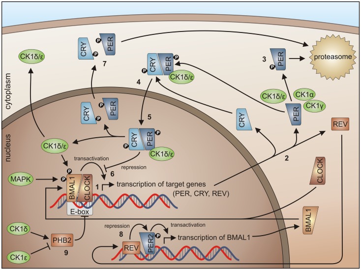
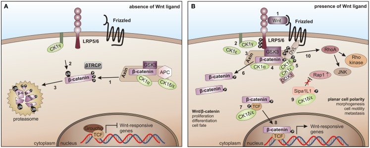
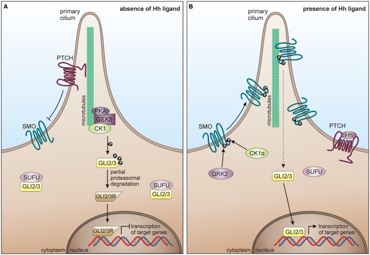
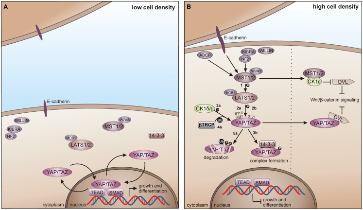
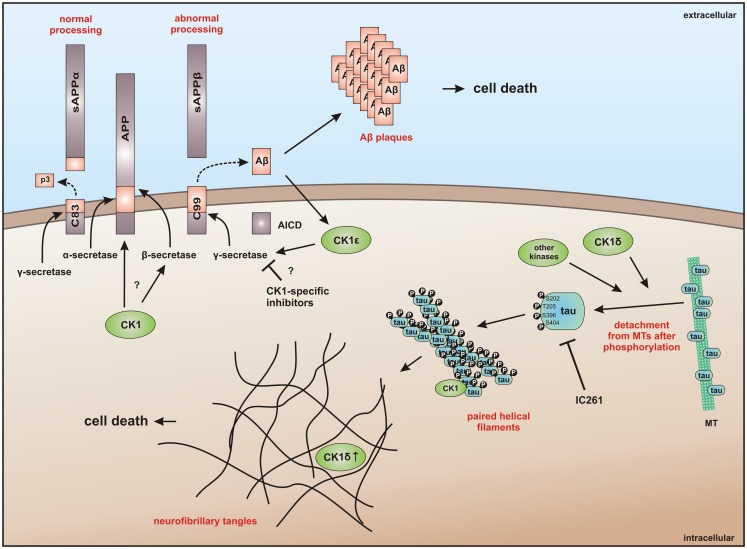
Members of the highly conserved and ubiquitously expressed pleiotropic CK1 family play major regulatory roles in many cellular processes including DNA-processing and repair, proliferation, cytoskeleton dynamics, vesicular trafficking, apoptosis, and cell differentiation. As a consequence of cellular stress conditions, interaction of CK1 with the mitotic spindle is manifold increased pointing to regulatory functions at the mitotic checkpoint. Furthermore, CK1 is able to alter the activity of key proteins in signal transduction and signal integration molecules. In line with this notion, CK1 is tightly connected to the regulation and degradation of β-catenin, p53, and MDM2. Considering the importance of CK1 for accurate cell division and regulation of tumor suppressor functions, it is not surprising that mutations and alterations in the expression and/or activity of CK1 isoforms are often detected in various tumor entities including cancer of the kidney, choriocarcinomas, breast carcinomas, oral cancer, adenocarcinomas of the pancreas, and ovarian cancer. Therefore, scientific effort has enormously increased (i) to understand the regulation of CK1 and its involvement in tumorigenesis- and tumor progression-related signal transduction pathways and (ii) to develop CK1-specific inhibitors for the use in personalized therapy concepts. In this review, we summarize the current knowledge regarding CK1 regulation, function, and interaction with cellular proteins playing central roles in cellular stress-responses and carcinogenesis.

Keywords: casein kinase 1; cellular stress; centrosome; disease; inhibitor; p53; signal transduction; tumorigenesis.


Figures

Figure 1
Figure 1
Structural presentation of CK1δ. (A) Phylogenetic relation between CK1 isoforms of Homo sapiens (CK1α, γ1–3, δ, and ε) and other members of the human CK1 family (TTBK1–2, VRK1–3). (B) Schematic alignment of human CK1 isoforms α, γ1–3, δ, and ε. Their molecular weight varies between 32 (CK1α) and 52.2 kDa (CK1γ3). In case transcription variants have been reported for one isoform, the molecular weight is given as range from the smallest to the largest variant. All CK1 isoforms are highly conserved within their kinase domains (light green box, 286 aa), but differ within their variable N- (4–40 aa) and C-terminal (39–122 aa) non-catalytic domains (dark green boxes) [according to Knippschild et al. (333)]. Ribbon (C) and surface (D) diagram of the molecular structure of CK1δ (PDB code 4HGT) modeled in complex with Mg2+-ATP at a resolution of 1.80 Å. The nomenclature is adapted from Xu et al. (24) and Longenecker et al. (25). Until today, crystal structures of human CK1 isoforms γ1 (PDB code 2CMW), γ2 (2C47), γ3 (2CHL, 2IZR, 2IZS, 2IZT, 2IZU, 4HGL, 4HGS, 4G16, 4G17), δ (4KB8, 4KBA, 4KBC, 4KBK, 4HNF, 3UYS, 3UYT, 3UZP), and ε (4HNI, 4HOK) are accessible as well. For reasons of clarity, we focused on CK1δ exemplarily, due to its superior relevance. The catalytic domain folds into two lobes primarily containing strands (N-terminal), respectively helices (C-terminal) forming a catalytic cleft between that represents the ATP binding pocket as well as a substrate binding site. KHD indicates the kinesin homology domain within L-9D. DD refers to a putative dimerization domain containing various amino acids of β1, β2, β5, L-5B, β7, and αB, whereas NLS displays a putative nuclear localization signal sequence at the junction between L-EF and αF. A tungstate molecule binding site identifies a specific phosphate moiety binding motif (W1). The active site contains a deep hydrophobic pocket (HPI) and a spacious hydrophobic region (HRII) (–28). All modeling and docking studies were performed using Schrödinger software (Maestro, version 9.3, Schrödinger, LLC, New York, NY, 2012; Glide, version 5.8, Schrödinger, LLC, New York, NY, 2012). The illustration of modeling results was generated by the PyMOL Molecular Graphics System (Version 1.5.0.4, LLC) (29).
Figure 2
Figure 2
Phosphorylation sites located in the C-terminal domain of CK1δ. (A) Alignment of the rat CK1δ C-terminal sequence with the human CK1δ transcription variants 1 and 2 C-terminal sequences (accession numbers L07578, NM001893, and NM139062, respectively) generated by using the program ClustalW (36, 37), showing conserved amino acids (gray) and obvious differences in the C-terminal domain beyond amino acid 399. (B) Domain structure of rat CK1δ (NLS: nuclear localization signal, KHD: kinesin homology domain). (C) Phosphorylation sites in the C-terminal regulatory domains of CK1δ rat and human transcription variants 1 and 2, that have so far been confirmed experimentally (–53). Kinases identified for phosphorylation of the C-terminal domain are shown for rat CK1δ (38, 39).
Figure 3
Figure 3
Centrosome-associated functions of CK1. For dynein-dependent transport along microtubules (MT), CK1ε phosphorylates the dynein intermediate chain (DIC) of dynein, likely IC138, thereby activating minus-end directed transport of membrane organelles along MT (62, 228). CK1δ and CK1ε are associated with the centrosome mediated through interaction with the scaffold protein AKAP450 (A-kinase anchor protein 450) (191, 193, 203). Both isoforms are related to Wnt-signaling and neurite outgrowth by phosphorylation of DVL (229, 230). In addition, CK1δ phosphorylates the end binding protein 1 (EB1), which is relevant for centrosome positioning during T-cell activation (74). Furthermore, a subpopulation of p53 in coordinated function with CK1 at the centrosome could ensure the integrity of the centrosome and thereby maintain genomic stability (–233).
Figure 4
Figure 4
CK1 isoforms in DNA damage-induced signal transduction. After induction of DNA damage (situation A) p53 and Chk1/2 are activated by ATR/ATM-mediated phosphorylation while the p53-regulatory component MDM2 is inhibited. The activation of Chk1 is supported by claspin whereas Chk1/claspin-binding is promoted by CK1γ1-mediated phosphorylation of claspin (162). The CK1 isoforms α, δ, and ε are able to activate p53 by site-specific phosphorylation (187, 234, 235, 237). Activated p53 in turn induces the expression of target genes like Bax (leading to apoptosis), p21 (leading to cell cycle arrest), and also CK1δ (autoregulatory feedback loop) (187). MDM2-mediated degradation of p53 can be activated via interaction with and phosphorylation by CK1α, but also through phosphorylation by CK1δ or ε leading to enhanced binding of MDM2 to p53. CK1δ-mediated phosphorylation of Ser-118 and Ser-121 however marks MDM2 for proteasomal degradation (, –241). In case Chk1/2 gets activated after DNA damage the phosphatase Cdc25, normally initiating cell cycle progression, is blocked by inhibitory phosphorylation and subsequent degradation. In the regulation of Cdc25 inhibition and degradation also CK1 isoforms α and ε are involved (141, 143). Signaling mediated by p53 can also be initiated by hypoxia (via CK1δ-regulated HIF-1α; situation B) (216, 243, 244) or DNA/RNA virus infection (via IFN and/or CK1α-related signal transduction; situation C) (236, 245). Depicted phosphorylation events refer to reported CK1-specific target sites.
Figure 5
Figure 5
CK1 in circadian rhythm regulation. By binding of the BMAL1/CLOCK heterodimer to the E-box, transcription of E-box-containing genes is initiated (1) (here shown for PER1-3, CRY1 and 2, and REV-ERBα), the transcripts are translated in the cytoplasm (2). Degradation of cytoplasmic PERs is triggered mainly by CK1δ and ε, but also by isoforms α and γ (3), while PER degradation is inhibited by binding of CRYs to PERs (4). Subsequently, complexes of CRY/PER and CK1δ/ε translocate to the nucleus (5). In the nucleus the CRY/PER complex represses the transcriptional activation of BMAL1/CLOCK target genes (6). CRYs and PERs finally shuttle back to the cytoplasm for proteasomal degradation (7). Repression of BMAL1 expression by REV-ERBα represents a second negative feedback loop (8). Together, these feedback loops are able to generate cyclic expression of BMAL1 and E-box-containing genes [for review see Knippschild et al. (219) and Cheong and Virshup (195)]. CK1δ and ε differentially effect expression of the period length modulator PHB2. Whereas CK1δ is able to promote PHB2 transcription, its expression is reduced by CK1ε (9) (8, 263).
Figure 6
Figure 6
CK1 in Wnt-signaling. (A) In the absence of the Wnt ligand, β-catenin is progressively phosphorylated by CK1α and GSK3 (1), recruited to β-TrCP for ubiquitination (2), and thereby primed for proteasome- dependent degradation (3). (B) After binding of Wnt to Frizzled and LRP5/6 (1), LRP5/6 is phosphorylated by CK1γ (positive regulation) and CK1ε (negative regulation) (2). It then recruits Axin and the β-catenin destruction complex to the membrane and inhibits GSK3 (3, 4). Wnt-activated CK1δ and ε phosphorylate Disheveled (DVL) and Axin (5), induce a conformational change in the β-catenin destruction complex and initiate the dissociation of various components (6). CK1ε cooperates with DDX3 in phosphorylating DVL (7). Also, TCF3 can be phosphorylated by CK1δ and ε thereby increasing its binding to β-catenin followed by the nuclear translocation of TCF3/β- catenin (8). The non-canonical Wnt pathway is positively regulated by CK1δ- and ε-dependent release of Rap1 from Sipa1L1 inhibition (9). The Rho/JNK signaling cascade is activated after phosphorylation of DVL (10) [adapted from Cheong and Virshup (195)].
Figure 7
Figure 7
CK1 in Hh signaling. (A) In absence of Hh ligand, PTCH localizes in the cilium and inhibits surface trafficking and cilia localization of SMO. GLI proteins are phosphorylated by PKA, GSK3β, and CK1, which lead to proteasome-dependent cleavage of GLI into a N-terminally truncated form, generating the repressor forms GLI2R and GLI3R. GLI2/3R translocate to the nucleus and inhibit translation of Hh target genes. Furthermore, SUFU prevents GLI from activating Hh target genes, by binding it in the cytoplasm and the nucleus. (B) In response to Hh, SMO is activated by GRK2 and CK1α-dependent phosphorylation and enters the primary cilium. Activated SMO orchestrates a signaling cascade, eventually resulting in the dissociation of the SUFU-GLI complex and the translocation of full-length GLI2/3 to the nucleus, where Hh target gene expression is induced.
Figure 8
Figure 8
CK1 in Hippo signaling in vertebrates. (A) In absence of growth suppressive signals YAP/TAZ promotes tissue growth and differentiation by regulating the activity of different transcription factors in the nucleus, including SMADs and TEADs. (B) Cell-density activated pathway regulation is controlled by multiple upstream branches by activating the core kinase cassette that represses YAP/TAZ driven gene transcription, either by degradation of TAP/TAZ or by forming physical complexes, preventing its nuclear access. Initially, MST1/2 is activated by various components and phosphorylates LATS1/2 (1), which in turn phosphorylates TAP/TAZ on Ser-311 or Ser-381 (2a). This phosphorylation primes YAP/TAZ for further phosphorylation by CK1δ/ε (3a) and consequent recruitment of and ubiquitination by β-TrCP (4a), priming YAP/TAZ for degradation (5a). However, LATS1/2 driven phosphorylation of TAP/TAZ on Ser-127 (2b) leads to the formation of 14-3-3-YAP/TAZ complexes, which accumulate in the cytoplasm preventing YAP/TAZ access to the nucleus (3b). Hippo pathway regulates Wnt/β-catenin signaling by inhibition of DVL, either by MST1/2-mediated prevention of CK1ε-dependent phosphorylation of DVL, or by direct inhibition of DVL by YAP/TAZ. ABCP: apicobasal cell polarity protein.
Figure 9
Figure 9
CK1 in neuro-degenerative diseases. It has generally been accepted that overexpression of CK1 plays an important role in neuro-degenerative diseases, especially in tauopathies, such as Alzheimer’s disease (AD). CK1δ is known to be up-regulated up to 30-fold on mRNA level in hippocampal regions of Alzheimer‘s disease (AD) brains (383). CK1δ plays a critical role in formation of neurofibrillary tangles through phosphorylation of tau at amino acids Ser-202/Thr-205 and Ser-396/Ser-404 (responsible for binding to tubulin) in human embryonic kidney 293 cells, thereby leading to a release of tau from MT and to destablilization of MT. Phosphorylation of these sites could be inhibited by the CK1-specific inhibitor IC261 (227). It is further known, that CK1 is associated to paired helical filaments in AD (384) and to tau-containing neurofibrillary tangles, in AD, Down syndrome, progressive supranuclear palsy, Parkinsonism–dementia complex and pallido-ponto-nigral degeneration (383, 385). The overexpression of constitutively active CK1ε, proposed to be involved in processing of amyloid precursor protein (APP) on γ-secretase level, results in an increase of amyloid-beta (Aβ) production, which is attenuated by use of CK1-specific inhibitors (386). In addition, Höttecke et al. (381) could show that the inhibition of γ-secretase by one of these inhibitors does not depend on CK1δ. An in silico analysis further revealed multiple CK1 consensus phosphorylation sites in the intracellular regions of APP, β-secretase, and γ-secretase subunits. Conversely, Aβ seems to influence CK1 activity (387). sAPPα/β: secreted amyloid precursor protein α/β; AICD: amyloid precursor protein intracellular domain.

References

    1. Bingham KD, Shaw DA. The metabolism of testosterone by human male scalp skin. J Endocrinol (1973) 57(1):111–21 10.1677/joe.0.0570111 - DOI - PubMed
    1. Agostinis P, Pinna LA, Meggio F, Marin O, Goris J, Vandenheede JR, et al. A synthetic peptide substrate specific for casein kinase I. FEBS Lett (1989) 259(1):75–8 10.1016/0014-5793(89)81498-X - DOI - PubMed
    1. Flotow H, Graves PR, Wang AQ, Fiol CJ, Roeske RW, Roach PJ. Phosphate groups as substrate determinants for casein kinase I action. J Biol Chem (1990) 265(24):14264–9 - PubMed
    1. Flotow H, Roach PJ. Role of acidic residues as substrate determinants for casein kinase I. J Biol Chem (1991) 266(6):3724–7 - PubMed
    1. Graves PR, Haas DW, Hagedorn CH, DePaoli-Roach AA, Roach PJ. Molecular cloning, expression, and characterization of a 49-kilodalton casein kinase I isoform from rat testis. J Biol Chem (1993) 268(9):6394–401 - PubMed