. 2014 Oct;26(10):2276-83.
doi: 10.1016/j.cellsig.2014.05.018. Epub 2014 Jun 3.

Epidermal growth factor inhibits transforming growth factor-β-induced fibrogenic differentiation marker expression through ERK activation

Affiliations

Epidermal growth factor inhibits transforming growth factor-β-induced fibrogenic differentiation marker expression through ERK activation

Xiaoying Liu et al. Cell Signal. 2014 Oct.

Abstract

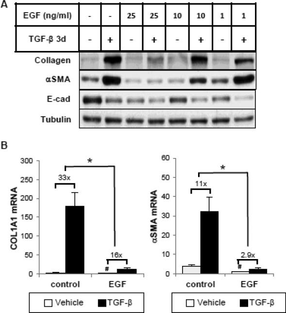
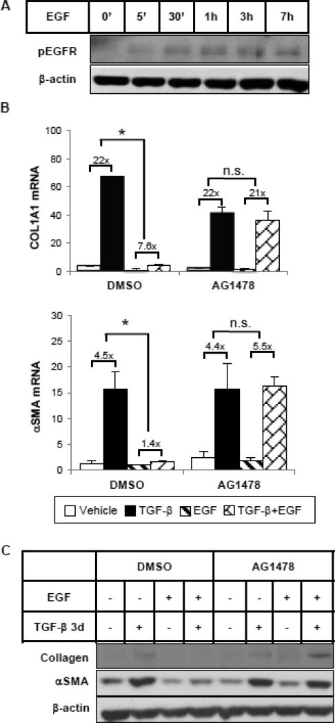
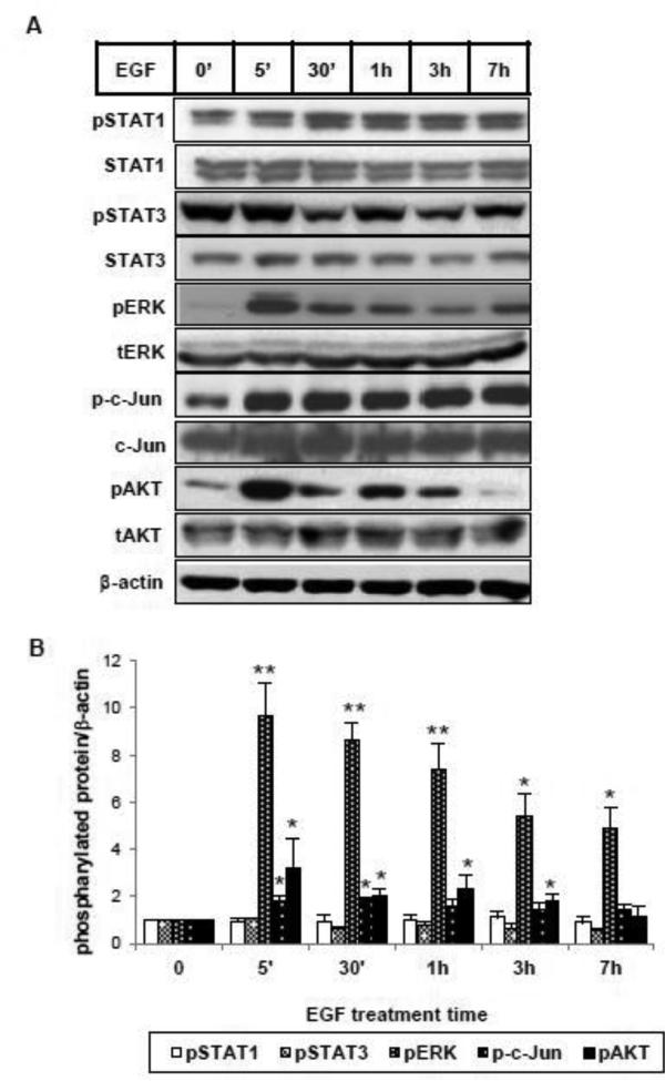
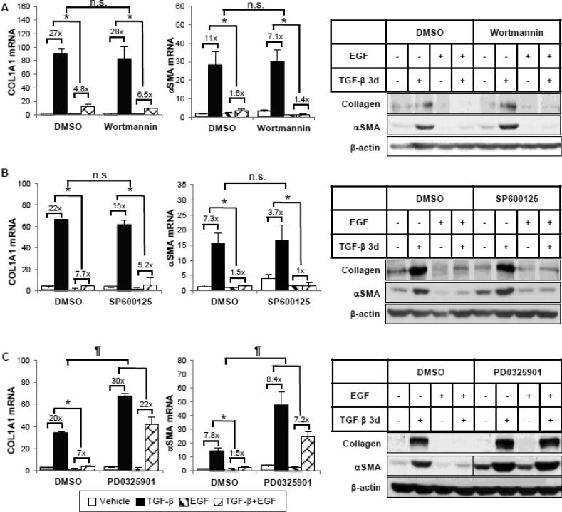
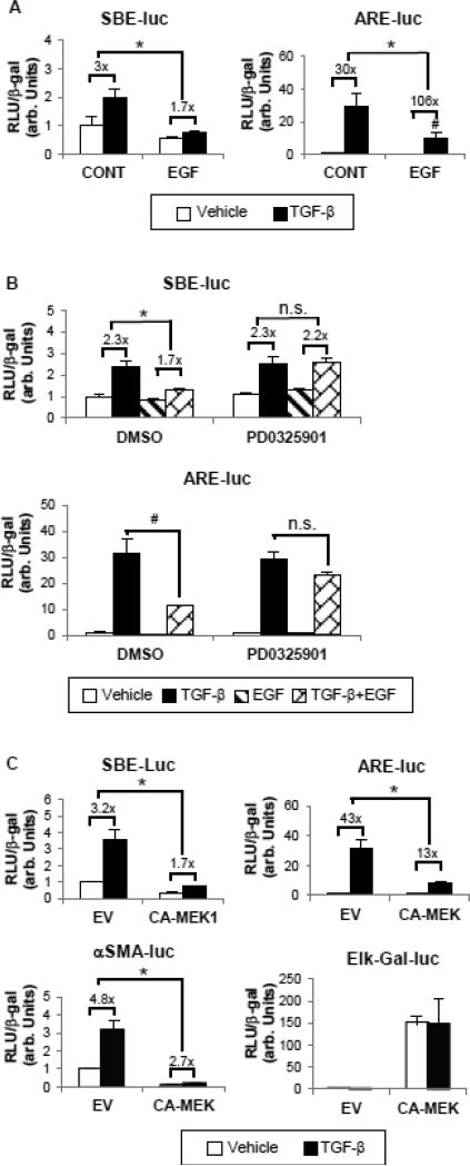
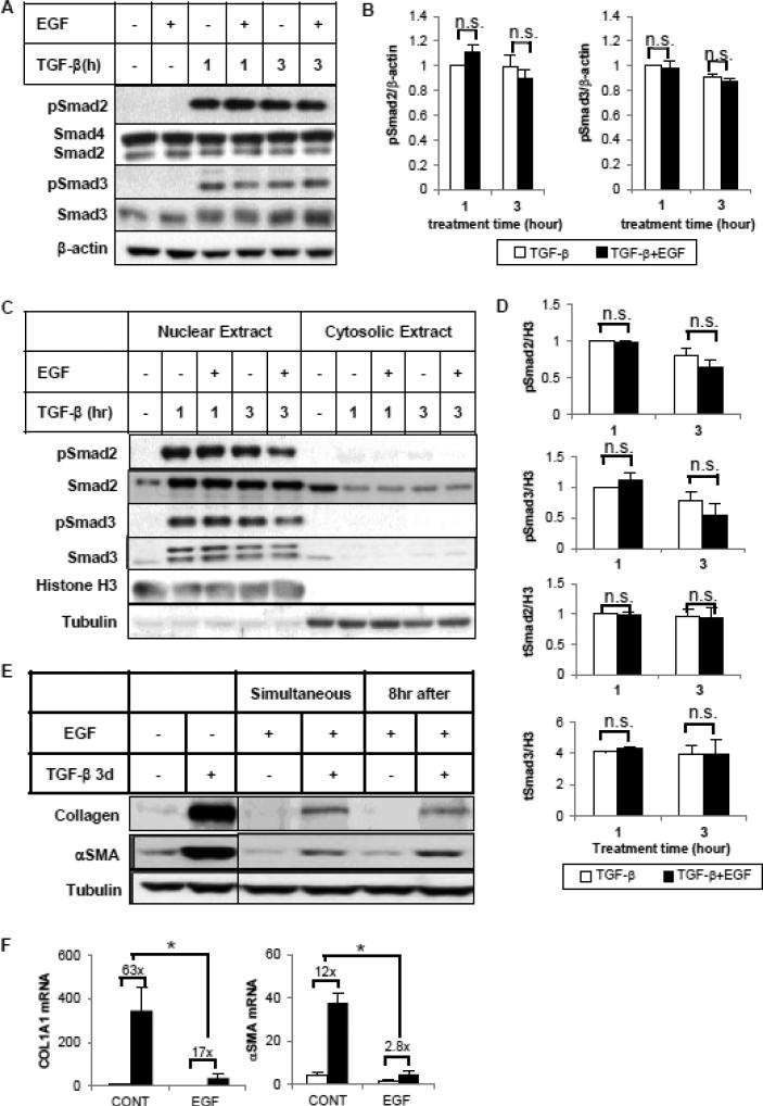
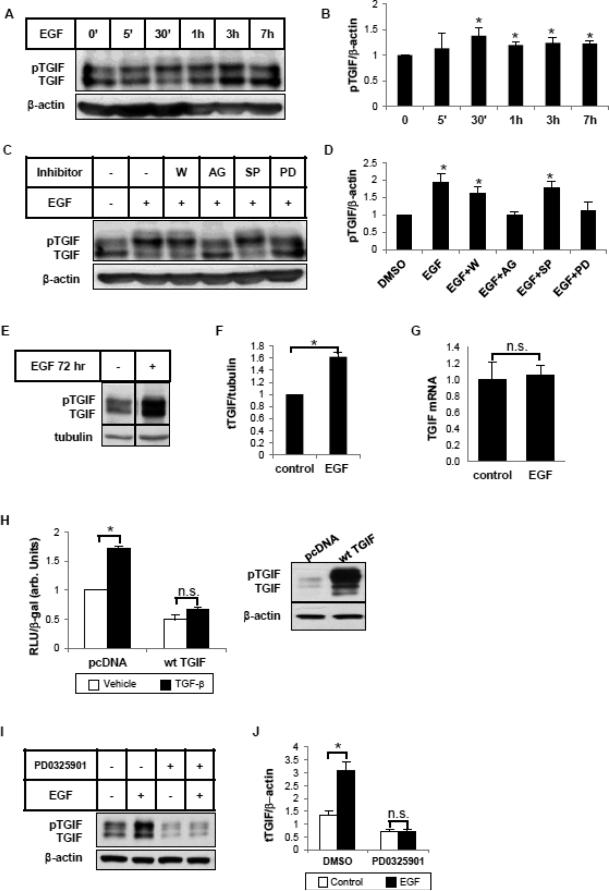
Transforming growth factor-β (TGF-β) signaling plays an important and complex role in renal fibrogenesis. The seemingly simple TGF-β/Smad cascade is intensively regulated at several levels, including crosstalk with other signaling pathways. Epidermal growth factor (EGF) is a potent mitogen for epithelial cells and is elevated in diseased kidneys. In this study, we examined its effect on TGF-β-induced fibrotic changes in human proximal tubular epithelial cells. Simultaneous treatment with EGF specifically inhibited basal and TGF-β-induced type-I collagen and α-smooth muscle actin (αSMA) expression at both mRNA and protein levels. These effects were prevented by inhibition of either the EGF receptor kinase or its downstream MEK kinase but not by blockade of either the JNK or PI3K pathway. Overexpression of a constitutively active MEK1 construct mimicked the inhibitory effect of EGF. Further, EGF suppressed Smad transcriptional activities, as shown by reduced activation of ARE-luc and SBE-luc. Both reductions were prevented by MEK inhibition. However, EGF did not block Smad2 or Smad3 phosphorylation by TGF-β, or Smad2/3 nuclear import. Finally EGF induced the phosphorylation and expression of TGIF, a known TGF-β/Smad repressor. Both the phosphorylation and the induction were blocked by a MEK inhibitor. Overexpression of TGIF abolished TGF-β-induced αSMA promoter activity. Together these results suggest that EGF inhibits two TGF-β-stimulated markers of EMT through EGF receptor tyrosine kinase and downstream ERK activation, but not through PI3K or JNK. The inhibition results from effector mechanisms downstream of Smads, and most likely involves the transcriptional repressor, TGIF.

Keywords: EGF; EMT; ERK; Fibrosis; Smad2/3; TGF-β.


Figures

Figure 1
Figure 1
Epidermal growth factor (EGF) inhibits basal and TGF-β-induced collagen I and α-smooth muscle actin (αSMA) mRNA and protein expression in human tubular epithelial cells (HKCs). (A) Subconfluent HKCs were serum starved for 24 hours before incubation with or without TGF-β (1 ng/ml) and/or EGF (1, 10 or 25 ng/ml) for 3 days. Whole cell lysates were immunoblotted with antibodies against E-cad, αSMA, type-I collagen and β-actin, respectively. β-actin was used as a loading control. (B) Cells were treated with TGF-β (1 ng/ml) and/or EGF (25 ng/ml) for 24 hours. Quantitative PCR was performed to assess the mRNA abundance of COL1A1 and αSMA. #p<0.05 comparing baseline activities (no TGF-β) with or without EGF, *p<0.05 fold induction by TGF-β comparing with and without EGF treatment (n=3).
Figure 2
Figure 2
EGF represses TGF-β-induced collagen and αSMA expression through EGFR. (A) HKCs were treated with EGF (25 ng/ml) for various times as indicated. Phospho-EGFR was examined by western blot. β-actin was used as a loading control. (B) Cells were pretreated with DMSO or EGFR tyrosine kinase inhibitor AG1478 (5 μM) for 1 hour, followed by incubation with TGF-β (1 ng/ml) and/or EGF (25 ng/ml) for 24 hours. COL1A1 and αSMA mRNA levels were examined by quantitative PCR. *p<0.05 fold induction by TGF-β comparing with and without EGF treatment (n=3). (C) Cells were treated as above but for 72 hours. COL1A1 and αSMA protein levels were examined by western blot. β-actin was used as a loading control.
Figure 3
Figure 3
EGF activates multiple signaling pathways in HKC. (A) Cells were incubated with EGF (25 ng/ml) for the indicated intervals. Total cell lysates were analyzed by western blot using specific antibodies against phosphorylated forms of STAT1, STAT3, ERK, c-Jun, AKT (pSTAT1, pSTAT3, pERK, p-c-Jun, pAKT) and their corresponding non-phosphorylated forms. β-actin was used as a loading control. (B) Densitometric analysis of pSTAT1, pSTAT3, pERK, p-c-Jun and pAKT was normalized to β-actin and expressed as fold change over vehicle alone. *p<0.05, **p<0.005 comparing with vehicle control (n=3).
Figure 4
Figure 4
EGF requires the ERK pathway, but not PI3K or JNK, for its inhibitory effects. HKCs were pretreated with DMSO or (A) PI3K inhibitor wortmannin (50 nM), (B) JNK inhibitor SP600125 (20 μM) or (C) MEK inhibitor PD0325901 (100 nM) for 1 hour, then incubated with TGF-β (1 ng/ml) and/or EGF (25 ng/ml) for 24 hours or 72 hours. COL1A1 and αSMA mRNA levels were examined by quantitative PCR after 24-hour treatment. COL1A1 and αSMA protein expression after 72-hour treatment was examined by western blot. β-actin was used as a loading control. The vertical line in the middle of the blot in (C) indicates the removal of irrelevant lanes within the same blot. *p<0.05 fold induction by TGF-β comparing with and without EGF treatment. n.s. or p<0.05 comparing percentage inhibition of TGF-β fold induction by EGF between DMSO and the inhibitor group (n=3).
Figure 5
Figure 5
EGF decreases Smad2 and Smad3 transcriptional activities via ERK activation. (A) Cells were transfected with either ARE-Luc plus FAST-1 or SBELuc reporter constructs to detect the transcriptional activity of Smad2 and Smad3, respectively. 24 hours after transfection, cells were exposed to 1 ng/ml TGF-β with or without EGF (25 ng/ml) for another 24 hours. *p<0.05 fold induction by TGF-β comparing with and without EGF treatment. #p<0.05 comparing absolute activities induced by TGF-β in the presence or absence of EGF treatment (n=3). (B) Transfected cells were pretreated with DMSO or PD0325901 (100 nM) for 1 hour before incubation with TGF-β and/or EGF for 24 hours. *p<0.05 fold induction by TGF-β comparing with and without EGF treatment. #p<0.05 comparing absolute activities induced by TGFβ in the presence or absence of EGF treatment (n=3). (C) A constitutively active MEK1 construct (CA-MEK1, 0.5 μg) or its empty vector (EV) was overexpressed in HKCs along with ARE/SBE-Luc or αSMA promoter reporter construct (SMA-Luc). Elk-Gal-Luc was used to determine the ERK functional activity. *p<0.05 fold induction by TGF-β compared with EV group. In all experiments, the luciferase activity assayed in triplicates was normalized to β-galactosidase (β-gal) to control transfection efficiency.
Figure 6
Figure 6
EGF does not block TGF-β-induced Smad2/3 C-terminal phosphorylation or nuclear translocation. Cells were treated with TGF-β (1 ng/ml) in the absence or presence of EGF (25 ng/ml) for 1 or 3 hours. Whole cell lysates (A, B) or subcellular fractionated extracts (C, D) were analyzed by western blot with antibodies against phospho-Smad2 (Ser465/467), total Smad2, Smad4, phospho-Smad3 (Ser423/425) or total Smad3. β-actin was used as loading control of whole cell lysate. Histone H3 and tubulin served as markers for nuclear and cytosol separation. (B, D) Densitometric analysis of phospho-/total Smad2/Smad3 was normalized to β-actin or Histone H3 and expressed as fold change over TGF-β alone. (E) EGF inhibits TGF-β-induced collagen I and αSMA expression when it is added 8 hour after TGF-β. HKCs were treated with TGFβ (1 ng/ml) for 3 days. EGF (25 ng/ml) was added simultaneously or 8 hours after TGF-β treatment. Whole cell lysates were examined for collagen I and αSMA expression by western blot. Tubulin was used as a loading control. The vertical line in the middle of the blot indicates the removal of irrelevant lanes within the same blot. (F) HKCs were treated as above for 24 hours. COL1A1 and αSMA mRNA levels were examined by quantitative PCR. *p<0.05 fold induction by TGF-β comparing with and without EGF treatment (n=3).
Figure 7
Figure 7
EGF induces the phosphorylation and expression of TGIF. (A) HKCs were treated with EGF (25 ng/ml) for indicated periods of time. Whole cell lysates were examined for TGIF using an antibody that recognizes total TGIF. β-actin was used as a loading control. (B) Densitometric analysis of pTGIF was normalized to β-actin and expressed as fold change over control. *p<0.05 compared with control (n=3). (C) Cells were pretreated with the inhibitors, wortmannin (W, 50 nM), AG1478 (AG, 5 μM), SP600125 (SP, 20 μM) and PD0325901 (PD, 100 nM) for 1 hour before 30 min treatment with EGF (25 ng/ml). Whole cell lysates were examined for TGIF by western blot. β-actin was used as a loading control. (D) Densitometric analysis of pTGIF was normalized to β-actin and expressed as fold change over DMSO control. *p<0.05 compared with DMSO control (n=3). (E) Cells were treated with EGF (25 ng/ml) for 3 days. Whole cell lysates were examined for total TGIF. Tubulin was used as a loading control. The vertical line in the middle of the blot indicates the removal of irrelevant lanes within the same blot. (F) Densitometric analysis of total TGIF was normalized to tubulin and expressed as fold change over control. *p<0.05 compared with control (n=3). (G) Cells were treated with TGF-β (1 ng/ml) and/or EGF (25 ng/ml) for 24 hours. Quantitative PCR was performed to assess the mRNA level of TGIF. (H) A wild type TGIF construct (wt TGIF) was overexpressed in HKCs along with αSMA promoter reporter construct (SMALuc). 3 hours after transfection, cells were exposed to 1 ng/ml TGF-β for another 24 hours. The luciferase activity assayed in triplicates was normalized to β-galactosidase (β-gal) to control transfection efficiency. * p<0.05 compared with vehicle control (n=3). Western blots confirmed the overexpression of TGIF. (I) HKCs were pretreated with DMSO or PD0325901 (100 nM) for 1 hour, then incubated with EGF (25 ng/ml) for 72 hours. TGIF protein expression was examined by western blot. β-actin was used as a loading control. (J) Densitometric analysis of total TGIF was normalized to β-actin and expressed as fold change over DMSO control. *p<0.05 compared with non-EGF-treated control (n=3).

References

    1. Burns WC, Thomas MC. The molecular mediators of type 2 epithelial to mesenchymal transition (EMT) and their role in renal pathophysiology. Expert Rev Mol Med. 2010;12:e17. - PubMed
    1. Poncelet AC, Schnaper HW. Sp1 and Smad proteins cooperate to mediate transforming growth factor-beta 1-induced alpha 2(I) collagen expression in human glomerular mesangial cells. The Journal of biological chemistry. 2001;276(10):6983–92. - PubMed
    1. Hu B, Wu Z, Phan SH. Smad3 mediates transforming growth factor-beta-induced alpha-smooth muscle actin expression. Am J Respir Cell Mol Biol. 2003;29(3 Pt 1):397–404. - PubMed
    1. Barrientos S, et al. Growth factors and cytokines in wound healing. Wound repair and regeneration : official publication of the Wound Healing Society [and] the European Tissue Repair Society. 2008;16(5):585–601. - PubMed
    1. Lu X, Kang Y. Epidermal growth factor signalling and bone metastasis. British journal of cancer. 2010;102(3):457–61. - PMC - PubMed

Publication types

MeSH terms