. 2014 Aug;33(8):550-65.
doi: 10.1089/dna.2013.2198. Epub 2014 Jun 6.

Genomic analysis and differential expression of HMG and S100A family in human arthritis: upregulated expression of chemokines, IL-8 and nitric oxide by HMGB1

Affiliations

Genomic analysis and differential expression of HMG and S100A family in human arthritis: upregulated expression of chemokines, IL-8 and nitric oxide by HMGB1

Ashok R Amin et al. DNA Cell Biol. 2014 Aug.

Abstract

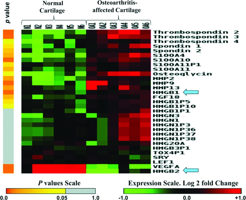
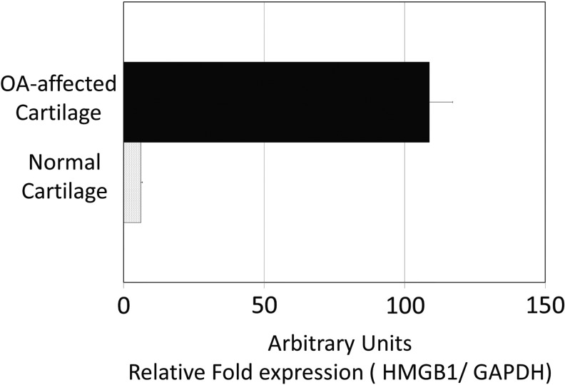
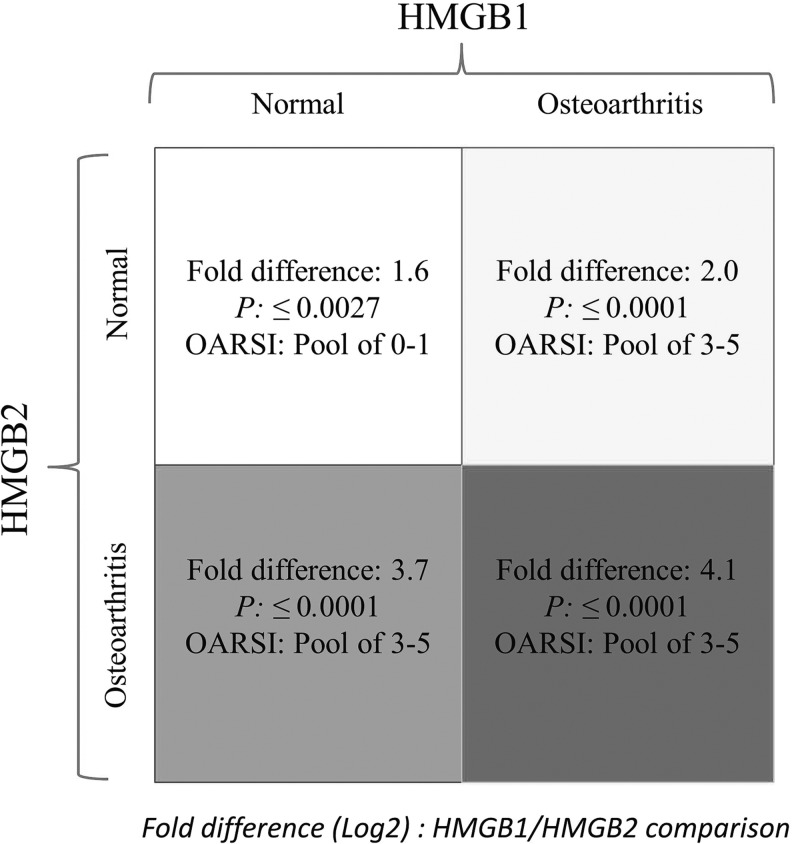
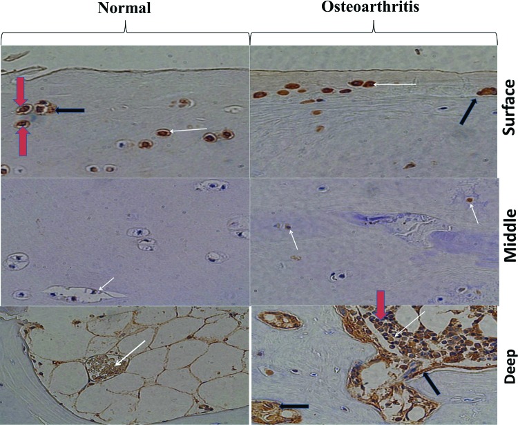
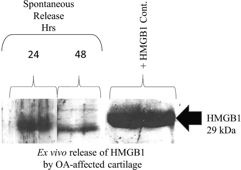
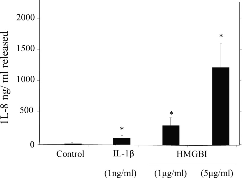
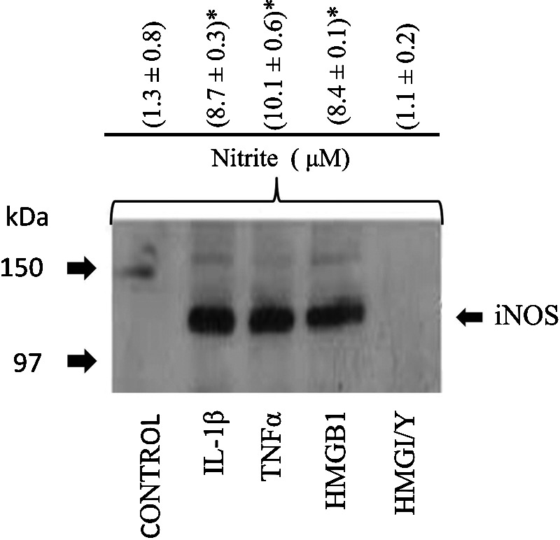
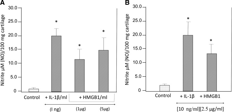
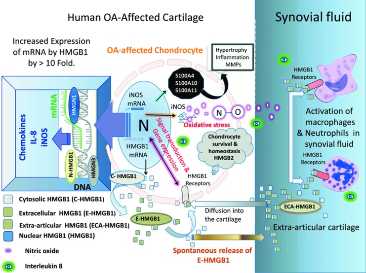
We applied global gene expression arrays, quantitative real-time PCR, immunostaining, and functional assays to untangle the role of High Mobility Groups proteins (HMGs) in human osteoarthritis (OA)-affected cartilage. Bioinformatics analysis showed increased mRNA expression of Damage-Associated Molecular Patterns (DAMPs): HMGA, HMGB, HMGN, SRY, LEF1, HMGB1, MMPs, and HMG/RAGE-interacting molecules (spondins and S100A4, S100A10, and S100A11) in human OA-affected cartilage as compared with normal cartilage. HMGB2 was down-regulated in human OA-affected cartilage. Immunohistological staining identified HMGB1 in chondrocytes in the superficial cartilage. Cells of the deep cartilage and subchondral bone showed increased expression of HMGB1 in OA-affected cartilage. HMGB1 was expressed in the nucleus, cytosol, and extracellular milieu of chondrocytes in cartilage. Furthermore, HMGB1 was spontaneously released from human OA-affected cartilage in ex vivo conditions. The effects of recombinant HMGB1 was tested on human cartilage and chondrocytes in vitro. HMGB1 stimulated mRNA of 2 NFκB gene enhancers (NFκB1 and NFκB2), 16 CC and CXC chemokines (IL-8, CCL2, CCL20, CCL3, CCL3L1, CCL3L3, CCL4, CCL4L1, CCL4L2, CCL5, CCL8, CXCL1, CXCL10, CXCL2, CXCL3, and CXCL6) by ≥10-fold. Furthermore, HMGB1 and IL-1β and/or tumor necrosis factor α (but not HMGI/Y) also significantly induced inducible nitric oxide synthase, NO, and interleukin (IL)-8 production in human cartilage and chondrocytes. The recombinant HMGB1 utilized in this study shows properties that are similar to disulfide-HMGB1. The differential, stage and/or tissue-specific expression of HMGB1, HMGB2, and S100A in cartilage was associated with regions of pathology and/or cartilage homeostasis in human OA-affected cartilage. Noteworthy similarities in the expression of mouse and human HMGB1 and HMGB2 were conserved in normal and arthritis-affected cartilage. The multifunctional forms of HMGB1 and S100A could perpetuate damage-induced cartilage inflammation in late-stage OA-affected joints similar to sterile inflammation. The paracrine effects of HMGB1 can induce chemokines and NO that are perceived to change cartilage homeostasis in human OA-affected cartilage.


Figures

<b>FIG. 1.</b>
FIG. 1.
Gene expression array of Damage-Associated Molecular Patterns (DAMPs) and High Mobility Groups proteins (HMGs) in normal and OA-affected cartilage. Heatmap shows up- and down-regulated transcripts from normal (N) and OA-affected (OA) cartilage. Gene expression arrays were performed from cohorts of six pools of normal and six pools of OA. Gene expression profiles are shown in rows. Color toward red indicates higher expression, and color toward green indicates lower expression as compared with median expression as shown on the scale. The statistical analysis is described on the left-hand side of the figure in the form of a heat map in yellow and gray, where n=6. The significant p-values (p≤0.05) are represented in yellow. The detailed characteristics of clinical samples are described in Supplementary Table S1. Details about the fold changes and statistical values for each gene are described in Supplementary Table S2. Color images available online at www.liebertpub.com/dna
<b>FIG. 2.</b>
FIG. 2.
qPCR of mRNA of HMGB1 in normal and OA-affected cartilage. Total RNA was isolated and quantitated from three normals and three OA-affected cartilage samples from different donors. The data are presented normalizing the values with GAPDH mRNA (housekeeping gene) and SD.
<b>FIG. 3.</b>
FIG. 3.
Bioinformatics and statistical analysis of HMGB1 and HMGB2 in normal and OA-affected cartilage. HMGB1 and HMGB2 share 80% homology at the amino-acid level (Sparvero et al., 2009). The gene expression of HMGB1 and HMGB2 was compared in both normal and OA-affected cartilage to examine their expression during the disease process. The figure describes the ORASI score of the cartilage in normal and OA-affected cartilage and the corresponding expression of HMGB1 and HMGB2 with their p-values.
<b>FIG. 4.</b>
FIG. 4.
Immunohistological staining of HMGB1 in normal and OA-affected cartilage. The figure shows the surface, middle, and deep region of the normal and OA-affected cartilage. Cartilage samples were stained (brown) with anti-HMGB1 antibodies+chromphore. The nuclear staining is represented in blue. Some cells show both blue and brown staining of nuclear HMGB1 as indicated by the red arrow. The white arrow shows the HMGB1-positive cells with cytoplasmic staining The black arrow shows HMGB1 staining in the extracellular milieu. Color images available online at www.liebertpub.com/dna
<b>FIG. 5.</b>
FIG. 5.
Spontaneous release of HMGB1 by OA-affected cartilage. Human OA-affected cartilage was incubated in ex vivo conditions. The spontaneous release of HMGB1 was determined at 24 and 48 h. An equal amount of supernatant was loaded onto the gel. One lane of the recombinant HMGB1 that was loaded onto the gel is shown on the right-hand side of the figure. Western blot of HMGB1 was performed as described in the “Materials and Methods” section. A 29 kDa signal of HMGB1 corresponding to the positive control in the Western blot analysis was observed as shown by an arrow.
<b>FIG. 6.</b>
FIG. 6.
Regulation of IL-8 by HMGB1 and IL-1β in human chondrocytes. Human OA-affected chondrocytes were seeded in triplicates for 24 h as described in the “Materials and Methods” section. One μg/mL of IL-1β or 1–5 μg/mL of HMGB1 was added to the cells. The production of IL-8 was estimated at the end of 72 h. Data are expressed as mean±SD (n=3). The p-values for IL-8 are compared with the nonstimulated cells, where *p≤0.05.
<b>FIG. 7.</b>
FIG. 7.
Effect of HMGB1, IL-1β, TNFα, and HMGI/Y on NO in human chondrocytes. Human OA-affected chondrocytes were grown for 24 h before they were stimulated with IL-1β (10 ng/mL), TNFα (1000 U/mL), HMGB1, or HMGI/Y (2.5 μg/mL) for an additional 24 h. Cell extracts were prepared, and an equal amount of total protein was analyzed for expression of iNOS by western blotting as previously reported. The 130 kDa iNOS is shown by an arrow. In a similar experiment, NO levels were estimated at 48 h in triplicate as shown in the upper panel. Significant levels of NO were calculated using Student's t-test. (*p≤0.01) where n=3, and the values were compared with the control cells.
<b>FIG. 8.</b>
FIG. 8.
Paracrine effect of HMGB1 on NO in human cartilage in vitro. Human OA-affected cartilage was incubated in triplicate in ex vivo conditions in two separate experiments (A, B) in the presence of IL-1β and HMGB1 for 48 hrs. The levels of NO were estimated in the form of nitrites, where n=3. The p (*)-values between control-unstimulated cells and IL-1β or HMGB1-treated cartilage was≤0.01.
<b>FIG. 9.</b>
FIG. 9.
The doppelganger life of HMGB1 in OA-affected cartilage: architectural factor, extracellular signaling, sterile inflammation, and cartilage homeostasis. The up-regulation of mRNA for HMGB1, HMGN1, and HMGN3 and a decrease in expression of HMGB2 in human OA-affected cartilage exhibited the interaction among HMG. HMGB1 is observed within the nucleus (N-HMGB1) of all nucleated cells. It was also observed in chondrocytes. There are typically ∼106 HMGB1 molecules per cell. The HMGB1 protein was observed in the cytosol (C-HMGB1) of chondrocytes and extracellular milieu (E-HMGB1), where it can adopt its role as a cytokine (Yang and Tracey, ; Yang et al., 2005). The extracellular HMGB1 (1) may bind to its receptor (red) and induce signal transduction (Wahamaa et al., ; Sparvero et al., 2009). HMGB1 exhibited superinduction of the CC and CXC family of chemokines, IL-8 and iNOS gene expression (by ≥1000%), as shown throughout this study. (2) may interact with matrix and or chemokines (CXCL12) and exhibit other functions (Diener et al., 2013); (3) be released outside the cartilage (ECA-HMGB1), where it can enter the synovial fluid in the joints (Kokkola et al., 2002). ECA-HMGB1 may (1) activate other inflammatory cells and/ or (2) might also penetrate back into the cartilage and activate the HMGB1 receptors on chondrocytes (Klune et al., 2008). Increased levels of HMGN1 and HMGN3 facilitate gene transcription (Bustin, 2010) and superinduction of chemokines, iNOS and IL-8, in OA-affected cartilage. NO and IL-8 can enter the synovial fluid. IL-8 can attract inflammatory cells. Most of the HMGB1-induced genes are involved in pro- or anti-inflammatory activity, chemotaxis and cell/matrix adhesion, matrix metabolism, and cartilage homeostasis as referenced in Table 1. Increased levels of nitric acid and oxidative stress have long been recognized to alter catabolism in OA-affected cartilage, degradation, and cartilage repair (Amin and Abramson, 1998; Abramson et al., 2001a). This oxidative environment in OA-affected cartilage may create an environment for post-translational modification of HMGB1 depending on its oxidation state within the cartilage (Janko et al., 2014) as described in some in vitro experimental settings. Oxidation of di-sulfides of HMGB1 has been reported to favor an inflammatory response, and terminal oxidation of HMGB1 may promote inflammation resolution (Yang et al., 2012; Diener et al., 2013; Janko et al., 2014). The decreasing level of HMGB2 is well documented in arthritis-affected cartilage and is shown throughout this study. HMGB2 plays a critical role in chondrocyte survival and cartilage homeostasis (Taniguchi et al., 2007; Taniguchi et al., 2009). The human OA-affected cartilage also showed increased expression of other DAMPs such as S100A4, S100A10, and S100A11. They have been reported to have catabolic and proinflammatory activity in chondrocytes (Yammani, 2012). Color images available online at www.liebertpub.com/dna

References

    1. Abramson S.B., Amin A.R., Clancy R.M., and Attur M. (2001a). The role of nitric oxide in tissue destruction. Best practice & research. Clin Rheumatol 15,831–845 - PubMed
    1. Abramson S.B., Attur M., Amin A.R., and Clancy R. (2001b). Nitric oxide and inflammatory mediators in the perpetuation of osteoarthritis. Curr Rheumatol Rep 3,535–541 - PubMed
    1. Adjaye J., Huntriss J., Herwig R., BenKahla A., Brink T.C., Wierling C., Hultschig C., Groth D., Yaspo M.L., Picton H.M., Gosden R.G., and Lehrach H. (2005). Primary differentiation in the human blastocyst: comparative molecular portraits of inner cell mass and trophectoderm cells. Stem Cells 23,1514–1525 - PubMed
    1. Alaaeddine N., Olee T., Hashimoto S., Creighton-Achermann L., and Lotz M. (2001). Production of the chemokine RANTES by articular chondrocytes and role in cartilage degradation. Arthritis Rheum 44,1633–1643 - PubMed
    1. Allen S.J., Crown S.E., and Handel T.M. (2007). Chemokine: receptor structure, interactions, and antagonism. Ann Rev Immunol 25,787–820 - PubMed

Publication types

MeSH terms