Review
. 2014 Jul 11:16:155-85.
doi: 10.1146/annurev-bioeng-071813-104553. Epub 2014 May 28.

Photoacoustic microscopy and computed tomography: from bench to bedside

Affiliations
Review

Photoacoustic microscopy and computed tomography: from bench to bedside

Lihong V Wang et al. Annu Rev Biomed Eng. .

Abstract

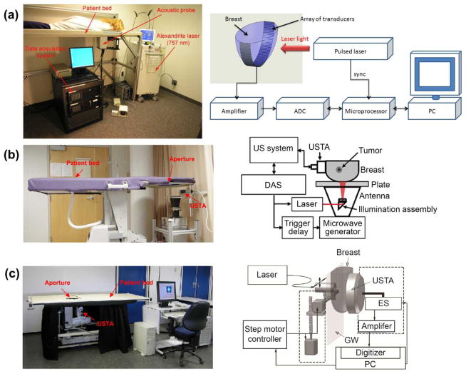
Photoacoustic imaging (PAI) of biological tissue has seen immense growth in the past decade, providing unprecedented spatial resolution and functional information at depths in the optical diffusive regime. PAI uniquely combines the advantages of optical excitation and those of acoustic detection. The hybrid imaging modality features high sensitivity to optical absorption and wide scalability of spatial resolution with the desired imaging depth. Here we first summarize the fundamental principles underpinning the technology, then highlight its practical implementation, and finally discuss recent advances toward clinical translation.

Keywords: biomedical imaging; cancer diagnosis; human imaging; label-free imaging; small-animal imaging.


Figures

Fig. 1
Fig. 1
Optical setups of OR-PAM working in a (a) back-reflection and (b) transmission mode.
Fig. 2
Fig. 2
Photoacoustic maximum amplitude projection (MAP) images of (a) vasculature structure in a mouse ear, (b) melanin in melanoma cells, and (c) cell nuclei. Reprint with permission from Refs (30; 34; 38).
Fig. 3
Fig. 3
Optical setups of AR-PAM under (a) bright-field and (b) dark-field illumination. Note the light diffusion inside the objects. MF: multimodal fiber. UT: ultrasonic transducer.
Fig. 4
Fig. 4
PACT images of athymic mice acquired noninvasively at various anatomical locations: (a) the brain, (b) the liver, (c) the kidneys, and (d) the bladder. BL, bladder; BM, backbone muscle; CV, cortical vessels; EY, eyes; GI, GI tract; KN, kidney; LV, liver; PV, portal vein; SC, spinal cord; SP, spleen; and VC, vena cava. Reprint with permission from Ref. (51).
Fig. 5
Fig. 5
Simultaneous, multi-wavelength PA and ultrasonic endoscopy. (a) The endoscope carries out circumferential sector scanning by rotating a scanning mirror, reflecting both ultrasonic waves and laser pulses. At each angular step of the mirror (~1.42°), both the first (wavelength λ1) and second (wavelength λ2) pulsed laser beams are independently fired through an optical fiber and the acoustic pulse is generated by an ultrasonic transducer. The ensuing PA and ultrasonic echo waves are detected by the same ultrasonic transducer. (b) Photograph of the PAE probe. (c) Definition of Cartesian and cylindrical coordinate systems. (d) A volumetric image comprising consecutive B-scan slices. (e) A representative cross-section of (d) along the x-y plane, which shows the 270° angular FOV of the endoscope. Reprint with permission from Ref. (54).
Fig. 6
Fig. 6
Optical setups of OD-PAT using (a) Mach–Zehnder interferometry and (b) phase contrast detection. BF, bandpass filter; BFD, balanced photodetector; BS, beam splitter.
Fig. 7
Fig. 7
Optical setups of OD-PAT, with (a) a high Q polymer microring resonator and (b) a Fabry-Perot polymer film as the pressure sensor. DM, dichroic mirror; EL: excitation laser; PB, probe beam; PD, photodiode; PF, polymer film; WG, wave guide.
Fig. 8
Fig. 8
Photoacoustic maximum amplitude projection (MAP) images of vasculature structures in (a) a mouse embryo and (b) colorectal tumor (LS174T) acquired by a FPI-based PACT system. Reprint with permission from Ref. (60; 61).
Fig. 9
Fig. 9
Structural and functional microvascular imaging by OR-PAM in a nude mouse ear in vivo. (a) Structural image acquired at 570 nm. (b) Vessel-by-vessel sO2 mapping based on dual-wavelength (570 nm and 578 nm) measurements. The calculated sO2 values are shown in the color bar. A1: a representative arteriole; V1: a representative venule. Reprint with permission from Ref. (80).
Fig. 10
Fig. 10
Single-cell temperature imaging with photo-thermal heating. The cell was loaded with metal particles and heated by a CW laser. (a) through (c) show the cell temperature images before, during, and after heating, respectively. The cell is pseudo-colored based on its PA-recovered temperatures. Reprint with permission from Ref. (81).
Fig. 11
Fig. 11
PA imaging of sO2 and blood flow in a mouse ear. Maximum amplitude projection (MAP) image of (a) a structure and (b) sO2. Scale bar, 250 μm. MAP image of blood flow (c) speed and (d) velocity with directions. (e) Trace of sO2 and (f) blood flow speeds along the main vascular trunk. (g) Velocity profile indicated by the dashed line in (d). Reprint with permission from Ref. (90).
Fig. 12
Fig. 12
PA flow cytometry. (a) Schematic. (b) Absorption spectra of whole blood (red) and platelet-rich plasma (blue). (c) Example of PA positive, negative, and combined contrasts from circulating clots of different compositions. (d) PA signal trace dynamics obtained with PA fluctuation flow cytometry in different vessels in normal and pathological conditions leading to RBC aggregation. Reprint with permission from Ref. (97).
Fig. 13
Fig. 13
Clinical PAT platforms for human breast imaging. (a) LOIS-64 PAT system (112). (b) the modified Philips iU22 system (118). (c) Twente PA mammoscope (119). ADC, analog to digital convertor; DAS, data acquisition system; ES, element selection; GW, glass window; MP, microprocessor; USTA, ultrasonic transducer array. Reprint with permission from Refs. (112; 118; 119).
Fig. 14
Fig. 14
PAT of a canine brain through a whole adult human skull. (a) PAT image of the human skull only. (b) PAT image of a canine brain acquired through the human skull. (c) Differential image of (b) and (a). (d) Image from (c) after high-pass filtering. (e) Photograph of the human skull from a top view. (f) Photograph of the canine brain cortex. Reprint with permission from Ref. (115).

References

    1. Bell AG. On the production and reproduction of sound by light. Am J Sci. 1880;20:305–24.
    1. Wang LV. Photoacoustic imaging and spectroscopy. xx. Boca Raton: CRC; 2009. p. 499.
    1. Gusev VE, Karabutov AA. Laser optoacoustics. xvii. New York: American Institute of Physics; 1993. p. 271.
    1. Maugh TH. Photoacoustic Spectroscopy - New Uses for an Old Technique. Science. 1975;188:38–9. - PubMed
    1. Kruger RA. Photoacoustic Ultrasound. Med Phys. 1994;21:127–31. - PubMed

Publication types

MeSH terms