. 2014 Jun 5;157(6):1292-1308.
doi: 10.1016/j.cell.2014.03.066.

Eosinophils and type 2 cytokine signaling in macrophages orchestrate development of functional beige fat

Affiliations

Eosinophils and type 2 cytokine signaling in macrophages orchestrate development of functional beige fat

Yifu Qiu et al. Cell. .

Abstract

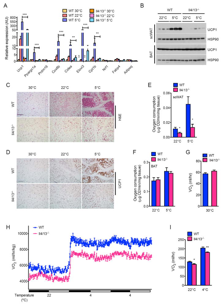
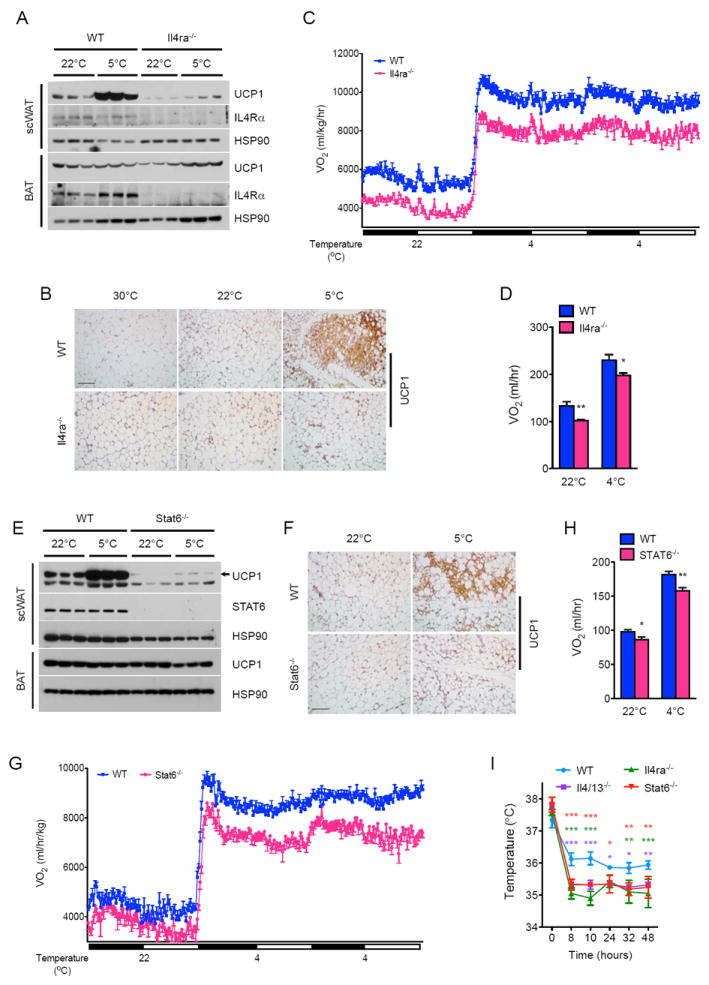
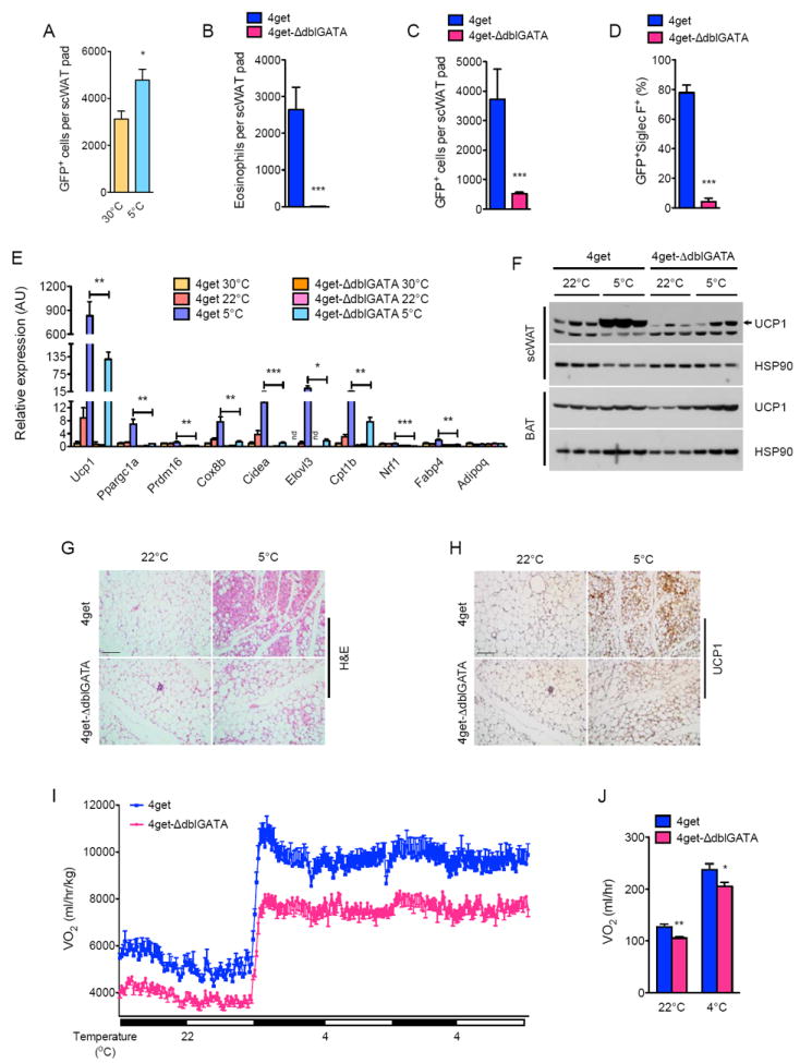
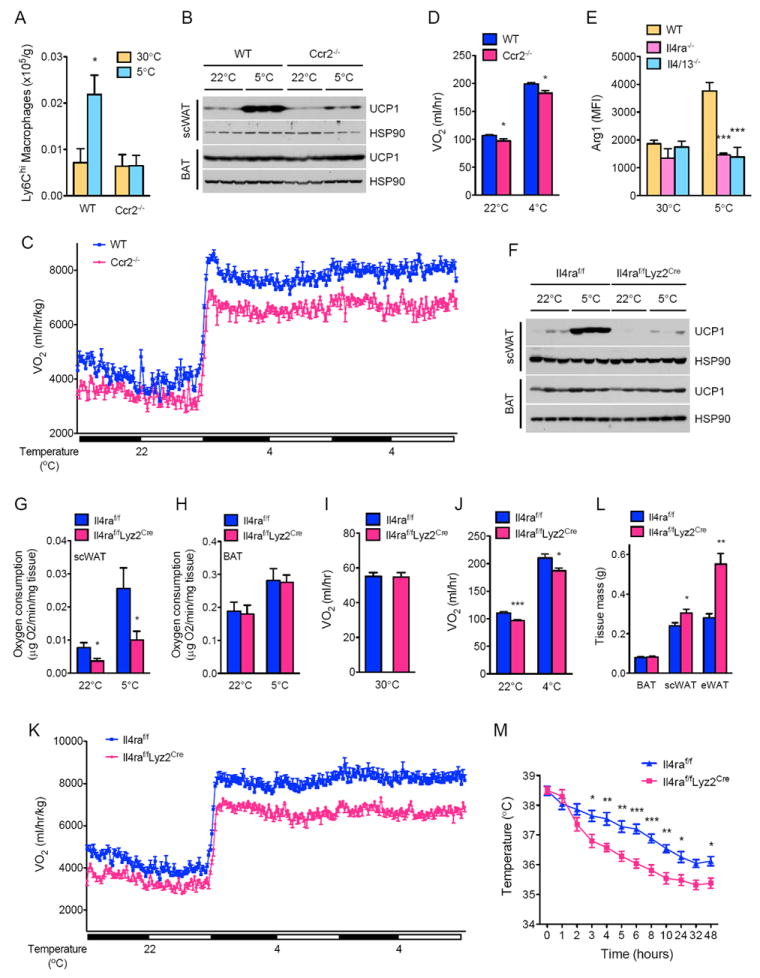
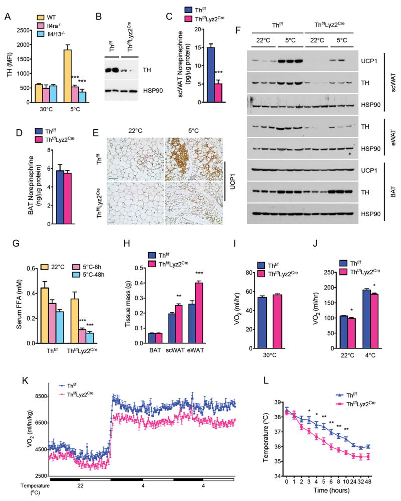
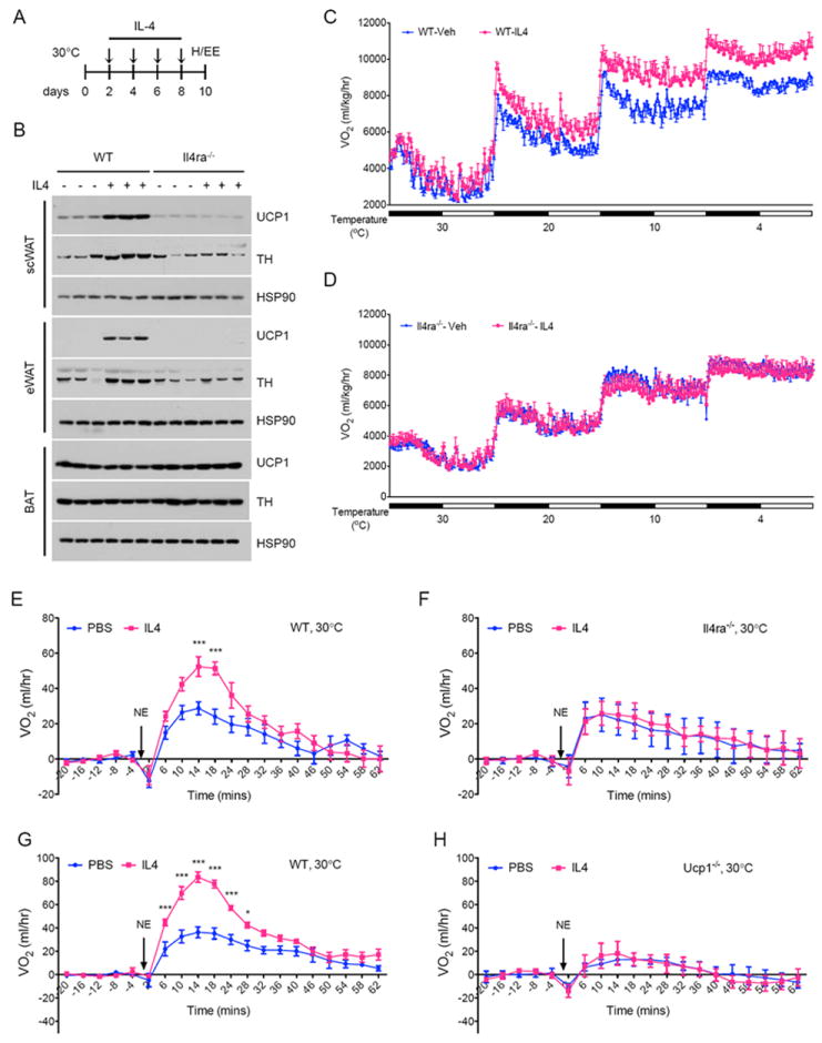
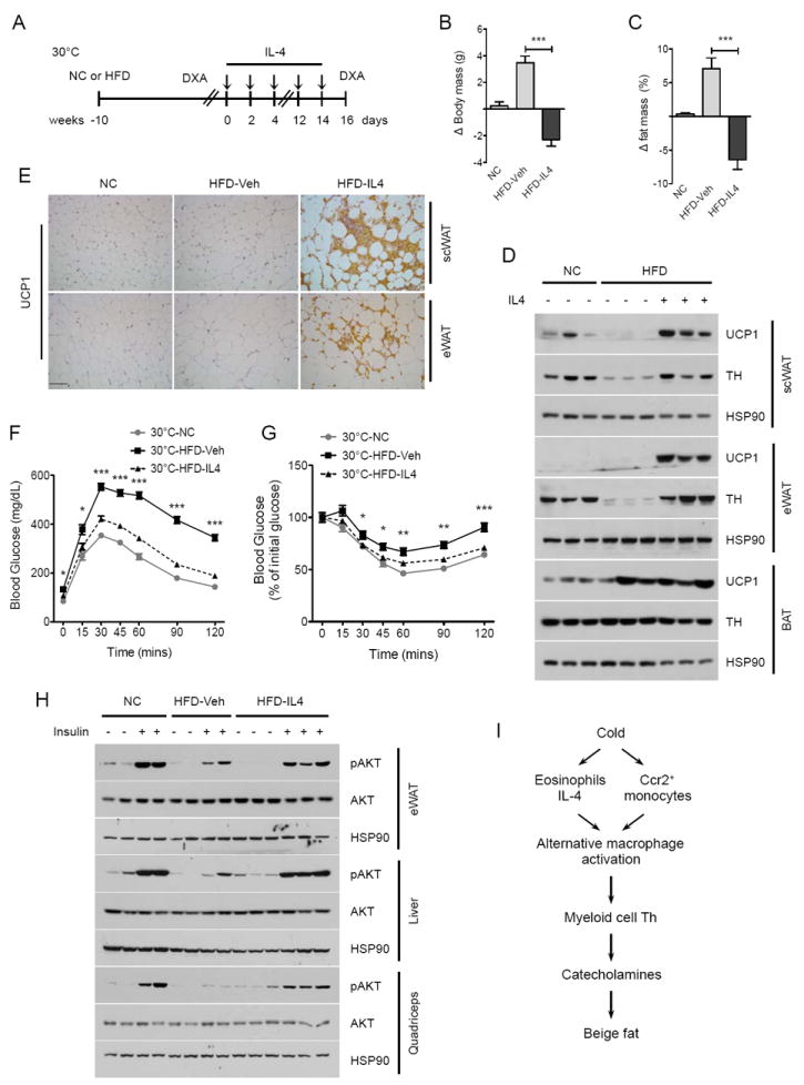
Beige fat, which expresses the thermogenic protein UCP1, provides a defense against cold and obesity. Although a cold environment is the physiologic stimulus for inducing beige fat in mice and humans, the events that lead from the sensing of cold to the development of beige fat remain poorly understood. Here, we identify the efferent beige fat thermogenic circuit, consisting of eosinophils, type 2 cytokines interleukin (IL)-4/13, and alternatively activated macrophages. Genetic loss of eosinophils or IL-4/13 signaling impairs cold-induced biogenesis of beige fat. Mechanistically, macrophages recruited to cold-stressed subcutaneous white adipose tissue (scWAT) undergo alternative activation to induce tyrosine hydroxylase expression and catecholamine production, factors required for browning of scWAT. Conversely, administration of IL-4 to thermoneutral mice increases beige fat mass and thermogenic capacity to ameliorate pre-established obesity. Together, our findings have uncovered the efferent circuit controlling biogenesis of beige fat and provide support for its targeting to treat obesity.


Conflict of interest statement

The authors declare that they have no competing financial interests.

Figures

Figure 1
Figure 1. Type 2 cytokines IL-4/13 are required for biogenesis of cold-induced beige fat
(A) Quantitative RT-PCR analysis of thermogenic genes in scWAT of WT and Il4/13-/- mice housed at 30°C, 22°C, or 5°C for 48 hours (n=5 per genotype and temperature). (B) Immunoblot analysis of UCP1 in scWAT and BAT of WT and Il4/13-/- mice housed at 22°C or 5°C for 48 hours (n=3 per genotype and temperature). (C, D) Representative scWAT sections of WT and Il4/13-/- mice housed at 30°C, 22°C, or 5°C for 48 hours were stained with hematoxylin and eosin (C) or for UCP1 (D), scale bar 100 μm. (E, F) Oxygen consumption in scWAT (E) and BAT (F) from WT and Il4/13-/- mice housed at 22°C or 5°C for 48 hours (n=7-8 per genotype and temperature). (G-I) Oxygen consumption (VO2) in WT and Il4/13-/- mice at different environmental temperatures: at thermoneutrality, 30°C (G) or at different ambient temperatures (H, I), n=5-8 per genotype. Data are represented as mean ± SEM. See also Figure S1.
Figure 2
Figure 2. Signaling via IL-4Rα and STAT6 controls growth of functional beige fat
(A, E) Immunoblot analysis for UCP1 protein in the scWAT and BAT of WT, Il4/13-/- (A) and Stat6-/-(E) mice housed at 22°C or 5°C for 48 hours (n=3 per genotype and temperature). (B, F) Representative sections of scWAT from WT, Il4ra-/- (B) and Stat6-/- (F) mice housed at various temperatures were stained for UCP1, scale bar 100 μm. (C-D, G-H) Oxygen consumption (VO2) in WT, Il4ra-/- (C) and Stat6-/- (F) mice at 22°C and 4°C (n=8-10 per genotype). (I) Core body temperature of WT, Il4/13-/-, Il4ra-/-, and Stat6-/- mice during a 48 hour cold challenge (n=4-5 per genotype). Data are represented as mean ± SEM. See also Figure S2.
Figure 3
Figure 3. IL-4 producing eosinophils are required for development of cold-induced functional beige fat
(A) Quantification of IL-4 producing cells in the scWAT of 4get mice housed at 30°C and 5°C. GFP expression marks cells competent for production of IL-4 (n=6 per temperature). (B-D) Quantification of eosinophils (B), GFP+ cells (C) and GFP+ SiglecF+ cells (eosinophils, D) in the scWAT of 4get and 4get-ΔdblGATA mice housed at 5°C (n=5-9 per genotype). (E) Quantitative RT-PCR analysis of thermogenic genes in scWAT of 4get and ΔdblGATA mice housed at 30°C, 22°C, or 5°C for 48 hours (n=5 per genotype and temperature). (F) Immunoblot analysis of UCP1 in scWAT and BAT of 4get and ΔdblGATA mice housed at 22°C or 5°C for 48 hours (n=3 per genotype and temperature). (G, H) Representative scWAT sections of 4get and ΔdblGATA mice housed at 22°C, or 5°C for 48 hours were stained with hematoxylin and eosin (G) or for UCP1 (H), scale bar 100 μm. (I, J) Oxygen consumption (VO2) in 4get and ΔdblGATA mice at different environmental temperatures (n=7-8 per genotype). Data are represented as mean ± SEM. See also Figure S3.
Figure 4
Figure 4. Macrophage recruitment via CCR2 and alternative activation via IL-4Rα is required for biogenesis of beige fat
(A) Ly6Chi macrophage content of scWAT in WT and Ccr2-/- mice housed at thermoneutrality (30°C) or at 5°C for 48 hours (n=5 per genotype and temperature). (B, F) UCP1 protein expression in WT and Ccr2-/- mice (B) or Il4raf/f and Il4raf/fLyz2Cre (F) mice at 22°C or 5°C (n=3 per genotype and temperature). (C, D) Cold-induced changes in oxygen consumption (VO2) in WT and Ccr2-/- mice (n=8 per genotype). Data are represented as mean ± SEM. (E) Expression of alternative activation marker Arginase 1 in scWAT macrophages of WT, Il4/13-/-, and Il4ra-/- mice housed at 30°C or 5°C (n=4-5 per genotype and temperature). (G, H) Oxygen consumption in scWAT (G) and BAT (H) from Il4raf/f and Il4raf/fLyz2Cre mice housed at 22°C or 5°C for 48 hours (n=6 per genotype and temperature). (I-K) Oxygen consumption (VO2) in Il4raf/f and Il4raf/fLyz2Cre mice at various environmental temperatures: thermoneutrality, 30°C (n=5 per genotype) or 22°C and 4°C (n=7-9 per genotype). (L) Adipose tissue weights of Il4raf/f and Il4raf/fLyz2Cre mice after cold challenge at 5°C for 48 hours (n=5 per genotype). (M) Core body temperature of Il4raf/f and Il4raf/fLyz2Cre mice during a 48 hour cold challenge (n=5 per genotype). Data are represented as mean ± SEM. See also Figures S3 and S4.
Figure 5
Figure 5. Myeloid cell tyrosine hydroxylase is required for biogenesis of functional beige fat
(A) Intracellular staining for TH expression in scWAT macrophages of WT, Il4/13-/- and Il4ra-/- mice housed at 30°C or 5°C. (B) Immunoblotting for TH protein in resident peritoneal macrophages of Thf/f and Thf/fLyz2Cre mice. (C, D) Norepinephrine content of scWAT (C) and BAT (D) of Thf/f and Thf/fLyz2Cre mice housed at 5°C (n=6-7 per genotype). (E) Representative sections of scWAT from Thf/f and Thf/fLyz2Cre mice stained for UCP1, scale bar 100 μm. (F) Immunoblot analysis for TH and UCP1 in scWAT, eWAT, and BAT of Thf/f and Thf/fLyz2Cre mice housed at 22°C or challenged with 5°C for 48 hours (n=3 genotype and temperature). (G) Serum concentration of free fatty acids in Thf/f and Thf/fLyz2Cre mice at various temperatures (n=6 per genotype and temperature). (H) Adipose tissue weights of Thf/f and Thf/fLyz2Cre mice after cold challenge at 5°C for 48 hours (n=5 per genotype). (I-K) Oxygen consumption (VO2) in Thf/f and Thf/fLyz2Cre mice at different environmental temperatures: at thermoneutrality, 30°C (I, n=5 per genotype) or during at different ambient temperatures (J, K), n=8 per genotype. (L) Core body temperature of Thf/f and Thf/fLyz2Cre mice during a 48 hour cold challenge (n=5 per genotype). Data are represented as mean ± SEM. See also Figure S5.
Figure 6
Figure 6. IL-4 induces beige fat in thermoneutral mice
(A) Schematic for IL-4 dosing and metabolic analysis in thermoneutral mice. (B) Immunoblotting for UCP1 and TH in scWAT, eWAT, and BAT of WT and Il4ra-/- thermoneutral mice administered IL-4 complex over 8 days (n=3 per genotype and treatment). (C, D) Cold-induced changes in oxygen consumption in WT (C) and Il4ra-/- (D) thermoneutral mice administered IL-4 complex over 8 days (n=4-5 per genotype and treatment). (E-H) Norepinephrine stimulated changes in oxygen consumption (VO2) in conscious, thermoneutral mice that were pretreated with vehicle (Veh) or IL-4 complexes (IL-4): BALB/cJ (E), Il4ra-/- (F), C57BL/6J (G) and Ucp1-/- (H). All metabolic and histological analyses were performed 2 days after the last dose of IL-4 complex. Data are represented as mean ± SEM. See also Figure S6.
Figure 7
Figure 7. Administration of IL-4 to obese thermoneutral mice reverses high fat diet-induced metabolic dysfunction
(A) Schematic for treatment of pre-established obesity with IL-4 complex in thermoneutral mice. (B, C) Changes in body mass (C) and fat mass (C) in obese C57BL/6J thermoneutral mice treated with vehicle (Veh) or IL-4 complex over 14 days (n=5 per treatment). (D) Immunoblot analysis for UCP1 and TH in scWAT, eWAT, and BAT of obese C57BL/6J thermoneutral mice administered Veh or IL-4 (n=3 per treatment). (E) Representative sections of scWAT and eWAT from thermoneutral mice treated with vehicle or IL-4 complex for 14 days were stained for UCP1, scale bar 100 μm. (F, G) Glucose (F) and insulin (G) tolerance tests in obese thermoneutral mice treated with Veh or IL-4 (n=5-7 per treatment). (H) Assessment of insulin signaling, as quantified by the phosphorylation of AKT, in obese thermoneutral mice treated with Veh or IL-4 (n=2-3 per treatment group). (I) Model for biogenesis and function of cold-induced beige fat. All metabolic and provocative testing was performed 2 days after the last dose of IL-4 complex. Data are represented as mean ± SEM. See also Figure S7.

Comment in

References

    1. Cannon B, Nedergaard J. Metabolic consequences of the presence or absence of the thermogenic capacity of brown adipose tissue in mice (and probably in humans) Int J Obes (Lond) 2010;34(Suppl 1):S7–16. - PubMed
    1. Cannon B, Nedergaard J. Nonshivering thermogenesis and its adequate measurement in metabolic studies. The Journal of experimental biology. 2011;214:242–253. - PubMed
    1. Charo IF, Ransohoff RM. The many roles of chemokines and chemokine receptors in inflammation. The New England journal of medicine. 2006;354:610–621. - PubMed
    1. Cypess AM, Lehman S, Williams G, Tal I, Rodman D, Goldfine AB, Kuo FC, Palmer EL, Tseng YH, Doria A, et al. Identification and importance of brown adipose tissue in adult humans. The New England journal of medicine. 2009;360:1509–1517. - PMC - PubMed
    1. Cypess AM, White AP, Vernochet C, Schulz TJ, Xue R, Sass CA, Huang TL, Roberts-Toler C, Weiner LS, Sze C, et al. Anatomical localization, gene expression profiling and functional characterization of adult human neck brown fat. Nat Med. 2013;19:635–639. - PMC - PubMed

Publication types

MeSH terms

LinkOut - more resources