. 2014 Jun 5;157(6):1324-1338.
doi: 10.1016/j.cell.2014.03.060.

Hippo pathway activity influences liver cell fate

Affiliations

Hippo pathway activity influences liver cell fate

Dean Yimlamai et al. Cell. .

Abstract

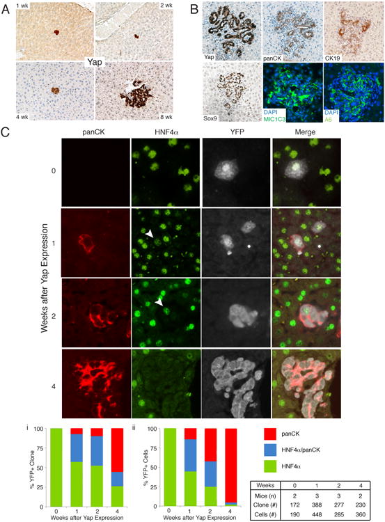
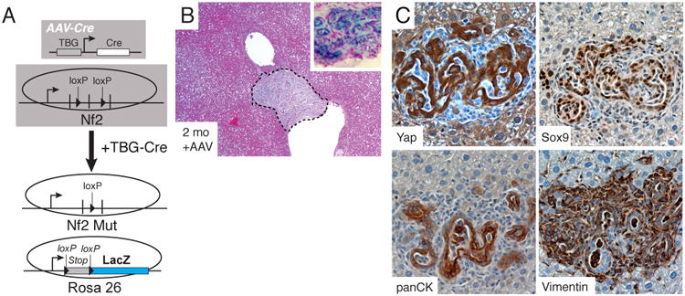
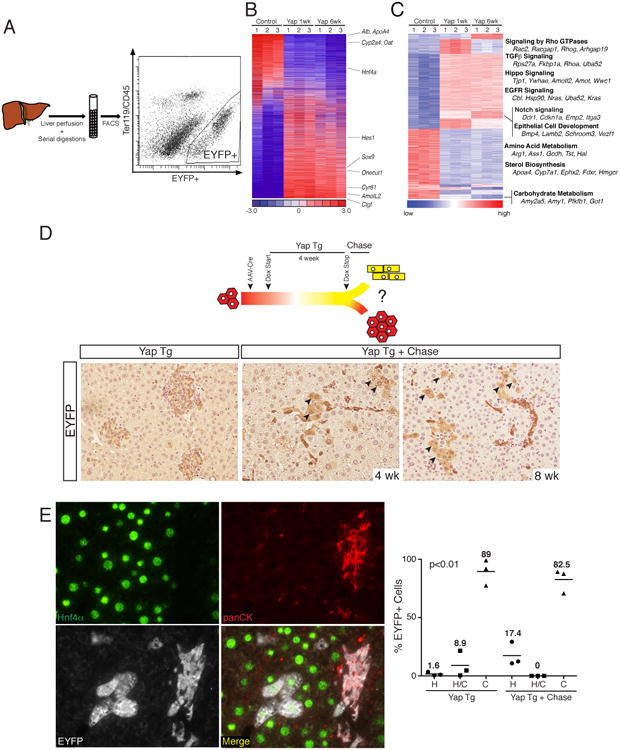
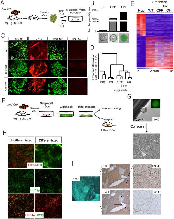
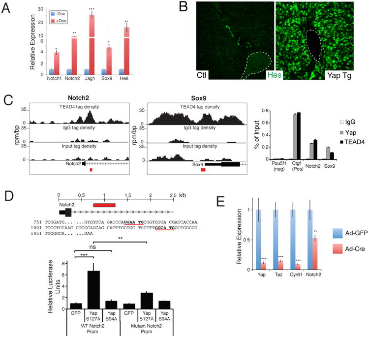
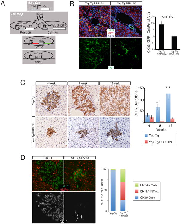
The Hippo-signaling pathway is an important regulator of cellular proliferation and organ size. However, little is known about the role of this cascade in the control of cell fate. Employing a combination of lineage tracing, clonal analysis, and organoid culture approaches, we demonstrate that Hippo pathway activity is essential for the maintenance of the differentiated hepatocyte state. Remarkably, acute inactivation of Hippo pathway signaling in vivo is sufficient to dedifferentiate, at very high efficiencies, adult hepatocytes into cells bearing progenitor characteristics. These hepatocyte-derived progenitor cells demonstrate self-renewal and engraftment capacity at the single-cell level. We also identify the NOTCH-signaling pathway as a functional important effector downstream of the Hippo transducer YAP. Our findings uncover a potent role for Hippo/YAP signaling in controlling liver cell fate and reveal an unprecedented level of phenotypic plasticity in mature hepatocytes, which has implications for the understanding and manipulation of liver regeneration.


Figures

Figure 1
Figure 1. Hepatocyte-specific YAP expression results in ductal/progenitor marker expression
A. YAP protein/activity is enriched in a subset of ductal cells. Control (Ctl) liver YAP staining (400×) shows prominent signaling in bile ductules. Arrowheads indicate ductal cells with nuclear YAP (inset shows magnified view). Yapfl/fl mice given Ad-Cre recombinase (Ad-cre Yapfl/fl) demonstrates patchy YAP staining in hepatocytes (100×). B. Immunoblots of human hepatocyte (Hep) and biliary (Bil) lysates for YAP, pYAP, CTGF and β-ACTIN. C. qPCR comparing relative levels of YAP and its targets in hepatocytes (Hep) and biliary cells (BC). N=3, Mean +/- SEM. D. Ctgf-EGFP mice shows EGFP co-staining (Ctgf) in a subset of CK19, SOX9 and A6-expressing cells. E. Experimental design for hepatocyte-specific YAP overexpression. F. H&E of uninduced (-Dox) or YAP Tg mouse (+Dox) for 21 days following injection with 1011 PFU of AAV-Cre. Right image displays a magnification of inset. G. Representative immunohistochimical stains of portal areas for YAP, panCK, SOX9 and Hnf1β for the mice displayed in F. *, p<0.05; ***, p<0.001. See also Figure S1.
Figure 2
Figure 2. Clonal analysis of YAP-mediated de-differentiation
A. Low dose (108 PFU) AAV-Cre and Dox administration allows clonal tracking of hepatocytes expressing YAP for several weeks. Representative images of clonal events at 1, 2, 4 and 8 weeks post Dox (100×). B. 8-weeks post Dox single hepatocytes give rise to ectopic ductal structures showing expression of multiple progenitor/biliary markers. C. Clonal and dynamic analysis of fate change driven by YAP. Representative images and quantitation of hepatocyte to progenitor/ductal cell de-differentiation following YAP expression. Arrowhead indicates weak HNF4α staining. Bar graphs represent measurements of cellular fates as examined by the presence of HNF4α only, HNF4α/panCK or panCK only in clones or indivisual cells within clones. Table indicates number of mice, clones and cells examined for the associated analysis. See also Figure S2.
Figure 3
Figure 3. Hepatocyte-specific Nf2-loss results in progenitor/ductal de-dedifferentiation
A. Experimental design for generating hepatocyte-specific Nf2 loss. B. Representative H&E stains of Nf2-deficient (Mut) livers 2 months after AAV-Cre administration. Inset shows a LacZ stained nodule from an Nf2-mutant mouse. C. Stained serial section of a biliary malformation for YAP, SOX9, panCK and Vimentin from an Nf2-mutant mouse, 2 months after AAV-Cre.
Figure 4
Figure 4. Molecular characterization of de-differentiated hepatocytes and consequences of restoring endogenous Hippo-pathway signaling
A. Schematic representation of EYFP+ cell isolation from induced TetOYAP mice. Representative FACS plot 1-week after Dox administration is shown. B. Heat map of 6,536 rank-ordered differentially expressed genes from microarray experiments from hepatocytes (control), and sorted EYFP+ hepatocytes expressing YAP for 1 or 6 weeks. Hepatocytic, biliary and YAP target genes are indicated to the right. C. Heat map of 1762 genes grouped by transcriptional gene program using Mclust. Annotated transcriptional programs of interest are noted to the right. D. Experimental design for the evaluation of fate outcomes following Dox removal in hepatocytes exposed to Dox for 4 weeks. EYFP stains of representative slides from TetOYAP mice given AAV-cre, placed on Dox for 4 weeks (YAP Tg) and following a 4 or 8 week Dox wash period (YAP Tg + Chase, 200×). Arrowheads indicate EYFP+ cells with hepatocyte morphology. F. Triple stain of a representative image (200×) (4 week on, 4 week chase) showing HNF4α (green), panCK (red), EYFP (white) and merge picture. Dot plot of average number of EYFP+ cells for the indicated staining patterns and representative treatments. Horizontal line and number represents the mean. One-way ANOVA was performed using the Kruskal-Wallis test. See also Figure S4.
Figure 5
Figure 5. YAP-reprogrammed progenitors are clonogenic and produce hepatocyte progeny
A. Schematic representation of liver organoid generation from YAP Tg mice and expansion procedure. B. Analysis of the number of organoids derived from livers from AAV-Cre infected Dox-uninduced (UI) and 3-week induced YAP Tg mice. YAP Tg results in dramatic increase of the liver organoid generation both in the presence (ON) and absence (OFF) of Dox in culture. Bottom shows representative immunofluorescent (IF) images of organoids in each category. Bars represent value of N=3. C. IF of wild-type (WT), ON Dox YAP Tg and OFF Dox YAP Tg organoids for biliary (SOX9, CK19, Hnf1β) and hepatocyte (HNF4α) markers. D. Hierarchical clustering analysis of primary hepatocytes, WT and hepatocyte-derived YAP Tg organoids demonstrates close clustering of all organoid groups. E. Differential expression analysis of hepatocytes compared to distinct organoid populations. Heat map demonstrates all differentially expressed genes with ≥ 2.5 fold change. F. Experimental design for the isolation, expansion and characterization of single-cell hepatocyte-derived organoids. G. Representative image of organoid derived from single sorted EYFP+ YAP Tg cell, followed by monolayer expansion. H. Differentiation of hepatocyte-derived organoid clone in the presence of γ-secretase inhibitor and in the absence of Dox. Day 15 differentiated cells demonstrate downregulation of biliary (CK19, SOX9, Hnf1β) and increase of hepatocyte markers (ALB, HNF4α). I. Representative liver images 5 months after transplantation of differentiated clonal YAP-Tg cells into Fah-/- mice. Engrafted cells are positive for EYFP (5×), FAH (5×) and HNF4α (hepatocyte marker), and negative for CK19 (biliary marker). See also Figure S5.
Figure 6
Figure 6. YAP and TEAD regulates Notch2 transcription
A. qRT-PCR analysis of NOTCH pathway genes from EYFP+ sorted uninduced and 1-week Dox YAP Tg liver cells post AAV-Cre infection. B. Immunofluorescent analysis for HES1 in an uninduced (Ctl) and a 2-week YAP Tg mouse. Dotted line outlines portal vein. C. ChIP-Seq binding profiles (reads per million per base pair) for TEAD4 at the Notch2 and Sox9 loci in trophoblast stem cells. Graph on the right shows ChIP-PCR assays for the indicated validation sites (red boxes) performed in liver cells isolated from YAP Tg mice 2 weeks post Dox . D. Localization and sequence of TEAD binding sites (bold and underlined) present in the NOTCH2 promoter. Red box indicates area of genomic sequence (WT Notch2 prom) that was cloned into a luciferase expression construct for functional analyses in CCLP1 cells (bottom). Mutant Notch2 promoter construct contains 3 mutated base pairs at each of the TEAD binding sites. n=3, mean +/- SEM. E. qPCR analysis of the indicated target genes in Yapfl/fl Tazfl/fl liver organoids given either Adenovirus-EGFP or Ad–Cre:EGFP. mRNA analysis of sorted infected cells was done 48 hours following infection. n=3, mean +/- SD. *, p<0.05; **, p<0.01; ***, p<0.001. See also Figure S6.
Figure 7
Figure 7. NOTCH signaling is a functional target of YAP/TEAD in vivo
A. Experimental design for hepatocyte-specific YAP overexpression with concomitant loss of NOTCH signaling. B. CK19 (Green), GFP (red) and DAPI (blue) or JAG1 immunofluorescence (IF) in TetOYAP:Rbpjfl/+ and TetOYAP:Rbpjfl/fl mice infected with high-dose AAV-Cre and treated with Dox for 2 weeks. Bar graph shows quantitation of GFP+CK19+ cells. C. GFP stain of representative animals infected with low-dose AAV-Cre and treated with Dox for the indicated times. Graph on the right depicts the number of GFP+ cells per clone analyzed. D. Representative IF triple stain (GFP, HNF4α, Ck19) of single cell derived clones with the noted genotypes, 12 weeks after Dox induction. Bar graph represents proportion of clones displaying indicated markers. ***p<0.001. See also Figure S7.

Comment in

  • Prometheus and progenitors.
    Diehl AM. Diehl AM. Hepatology. 2015 Apr;61(4):1427-9. doi: 10.1002/hep.27676. Epub 2015 Feb 13. Hepatology. 2015. PMID: 25545250 Free PMC article. No abstract available.

Similar articles

Cited by

References

    1. Benhamouche S, Curto M, Saotome I, Gladden AB, Liu CH, Giovannini M, Mcclatchey AI. Nf2/Merlin controls progenitor homeostasis and tumorigenesis in the liver. Genes Dev. 2010;24:1718–1730. - PMC - PubMed
    1. Camargo FD, Gokhale S, Johnnidis JB, Fu D, Bell GW, Jaenisch R, Brummelkamp TR. YAP1 increases organ size and expands undifferentiated progenitor cells. Current Biology. 2007;17:2054–2060. - PubMed
    1. Demetris AJ, Seaberg EC, Wennerberg A, Ionellie J, Michalopoulos G. Ductular reaction after submassive necrosis in humans. Special emphasis on analysis of ductular hepatocytes. Am J Pathol. 1996;149:439–448. - PMC - PubMed
    1. Dong J, Feldmann G, Huang J, Wu S, Zhang N, Comerford SA, Gayyed MF, Anders RA, Maitra A, Pan D. Elucidation of a universal size-control mechanism in Drosophila and mammals. Cell. 2007;130:1120–1133. - PMC - PubMed
    1. Dorrell C, Erker L, Schug J, Kopp JL, Canaday PS, Fox AJ, Smirnova O, Duncan AW, Finegold MJ, Sander M, et al. Prospective isolation of a bipotential clonogenic liver progenitor cell in adult mice. Genes Dev. 2011;25:1193–1203. - PMC - PubMed

Publication types