. 2014 Jun 5;157(6):1380-1392.
doi: 10.1016/j.cell.2014.05.009.

Bromine is an essential trace element for assembly of collagen IV scaffolds in tissue development and architecture

Affiliations

Bromine is an essential trace element for assembly of collagen IV scaffolds in tissue development and architecture

A Scott McCall et al. Cell. .

Abstract

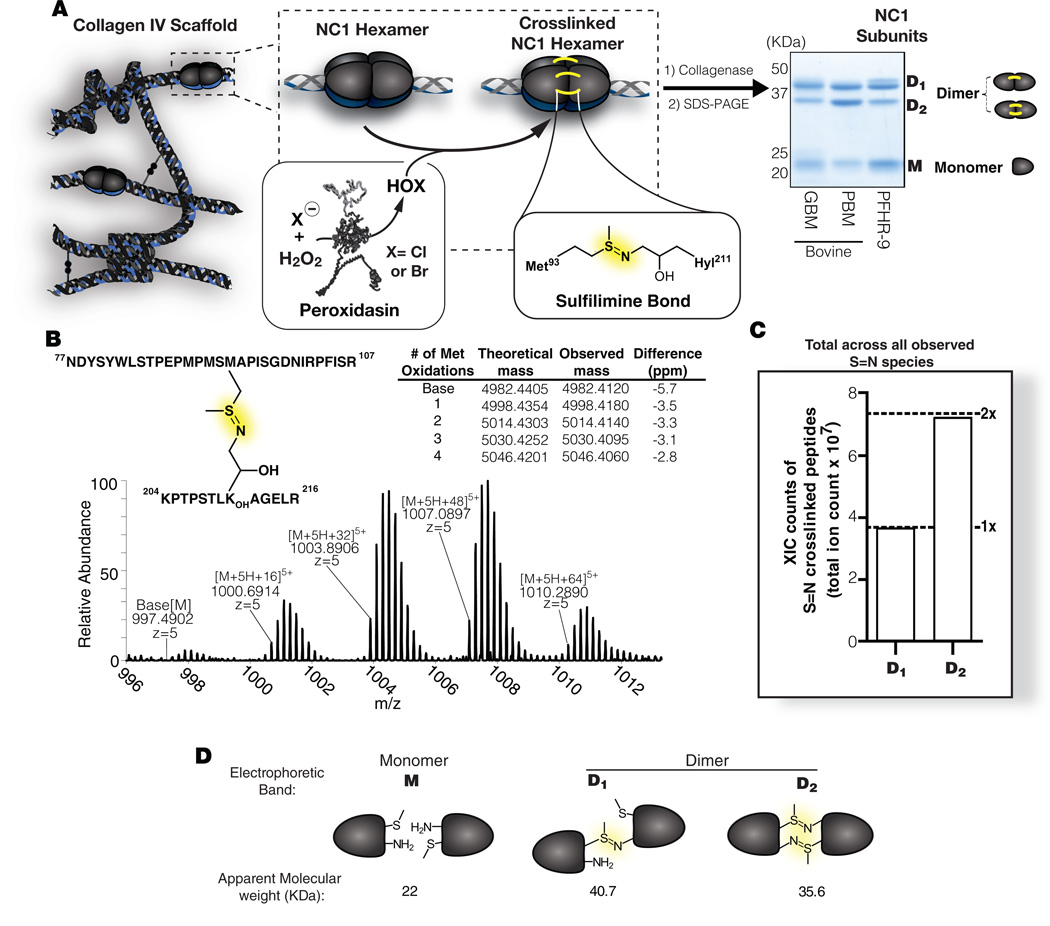
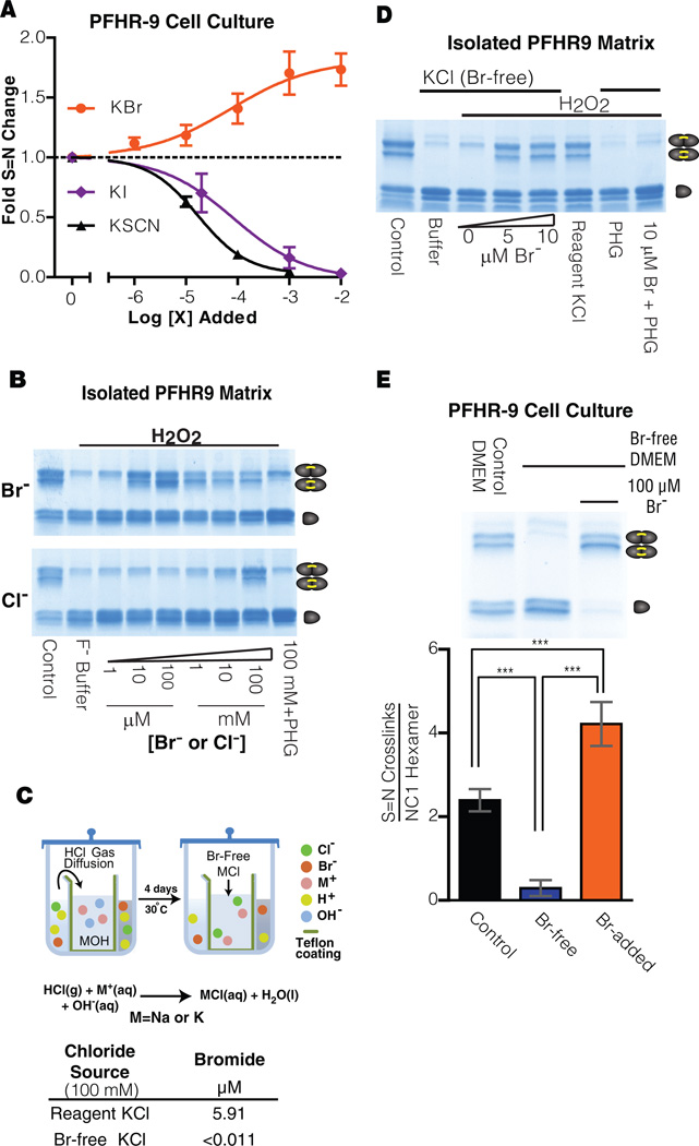
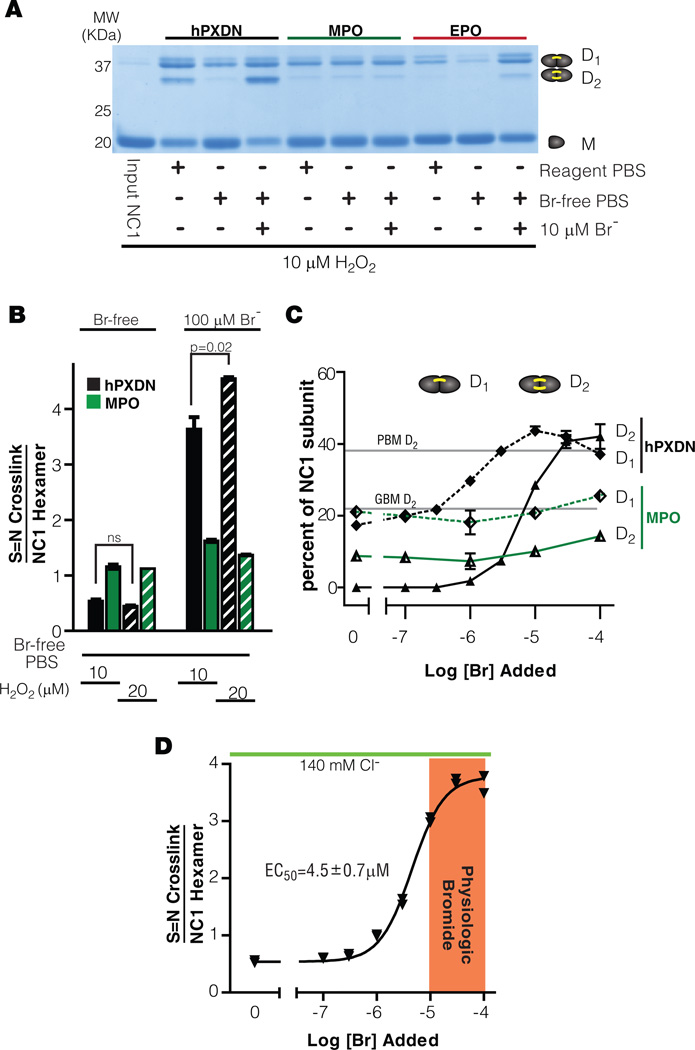
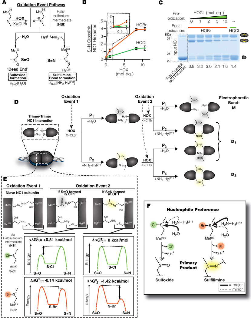
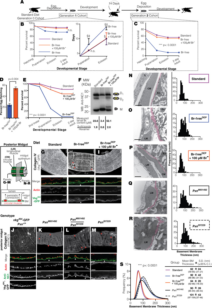
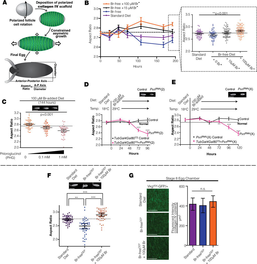
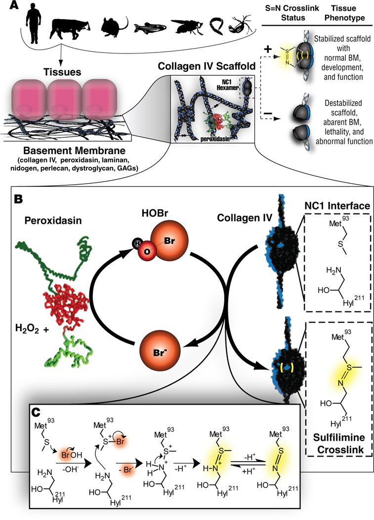
Bromine is ubiquitously present in animals as ionic bromide (Br(-)) yet has no known essential function. Herein, we demonstrate that Br(-) is a required cofactor for peroxidasin-catalyzed formation of sulfilimine crosslinks, a posttranslational modification essential for tissue development and architecture found within the collagen IV scaffold of basement membranes (BMs). Bromide, converted to hypobromous acid, forms a bromosulfonium-ion intermediate that energetically selects for sulfilimine formation. Dietary Br deficiency is lethal in Drosophila, whereas Br replenishment restores viability, demonstrating its physiologic requirement. Importantly, Br-deficient flies phenocopy the developmental and BM defects observed in peroxidasin mutants and indicate a functional connection between Br(-), collagen IV, and peroxidasin. We establish that Br(-) is required for sulfilimine formation within collagen IV, an event critical for BM assembly and tissue development. Thus, bromine is an essential trace element for all animals, and its deficiency may be relevant to BM alterations observed in nutritional and smoking-related disease. PAPERFLICK:


Figures

Fig 1
Fig 1. Measurement of sulfilimine crosslink content within NC1 domains of collagen IV scaffolds
(A) Diagram of the collagen IV scaffold, showing the relationship of NC1 hexamers sulfilimine crosslinks, peroxidasin (PXDN), and hypohalous acids (HOX). Inset shows resolution of dimeric (D1 and D2) and monomeric (M) NC1 domains by SDS-PAGE. Representative NC1 domains are shown from bovine placental BM (PBM), bovine glomerular BM (GBM), and murine collagen IV matrix produced in PFHR-9 cell culture (B) High-resolution mass spectrum depicting the multiple oxidation states of tryptic peptides containing the sulfilmine (S=N) crosslink (C) Extracted ion current (XIC) based quantitation of S=N crosslinked peptides from D1 and D2. Full data appears in Figure S1. (D) Diagram showing the crosslinking status of observed NC1 banding in SDS-PAGE, where D1 is singly crosslinked and D2 is doubly crosslinked with a resultant higher electrophoretic mobility.
Fig 2
Fig 2. Bromide is the required cofactor for sulfilimine crosslink formation
(A) The effect of halide ions on sulfilimine crosslink formation is examined in PFHR-9 matrix. Inhibition values were calculated from non-linear curve fitting: KI (IC50= 84µM 95%CI[30–241µM]), KSCN (IC50=17µM 95%CI[3-24µM]). Contrasting with these effects, exogenous potassium bromide (KBr) enhanced the reaction. Points represent mean ± S.D. (n=3). See also Fig. S2. (B) Uncrosslinked PFHR9 matrix was crosslinked in vitro in the presence of KCl and KBr. Reacted for 1 hr. at 37°C with 1 mM H2O2 100 mM KF used as ionic strength control. Collagenase digest analyzed by SDS-PAGE and Coosmassie staining. (C) Schematic of Br-free Cl- salt purification apparatus and setup. Resulting salt was analyzed by ICP-MS for bromide content. Further analysis of salt reagents appears in Table S1 and Figure S3 (D) Crosslink formation in PFHR-9 matrix with Br-free KCl. Reaction buffer contained 10 mM phosphate buffer (pH 7.4), 100 mM Br-free or reagent grade KCl, and 1 mM H2O2 and 200 µM PHG where appropriate. Displayed SDS-PAGE gels were stained with Coomassie blue. (E) Sulfilimine (S=N) crosslink formation in PFHR-9 cell-culture tested under Br-free conditions. Culture conditions and media formulations are presented in detail in the supplementary methods. NC1 hexamers were isolated via collagenase treatment and analyzed by SDS-PAGE. The amount of crosslinks per hexamer is graphed as the mean±95% C.I. (n=3). All sample groups had equal variance, one-way ANOVA was performed (p<0.001) and differences between groups was tested using Tukeys post-hoc analyisis (***p<0.001)
Fig 3
Fig 3. Peroxidasin uses physiologic Br levels to form sulfilimine crosslinks
(A–D) Mammalian peroxidases are compared for ability to crosslink collagen IV NC1 domains in Br-free 1x PBS, (recombinant human peroxidasin (hPXDN), myeloperoxidase (MPO), eosinophilperoxidase (EPO).Uncrosslinked NC1 domains were isolated from PHG-treated PFHR-9 cultures. All peroxidase activity enzymes was nominalized by TMB assay prior to assay (Bozeman et al., 1990). Reactions proceeded for 10 minutes at 37° C after initiation by the addition of H2O2 and quenched with 5 mM PHG, 0.2 mg/ml bovine catalase, and 10 mM methionine. Gel representative of 3 experiments. (A) Coomassie stained gel of enzymatic crosslink formation under reagent grade and Br-free conditions. (B) Quantitative analysis of crosslinks formed per NC1 hexamer by MPO and hPXDN under Br-free and Br-added (100 µM) conditions. Data shown as mean ± 1 S.D. n=2. Student’s T-Test was performed (n=2) due to equal variance between groups. (C) Effect of bromide titration on the proportion of D1 (1 crosslink) and D2 (2 crosslinks) NC1 populations following reaction with MPO and hPXDN. For reference, the proportions of D2 found in PBM and GBM are denoted on the graph. Data shown as mean ± 1 S.D. (n=2). (D) Crosslinking efficacy of peroxidasin measured as crosslink formed per hexamer upon Br titration. EC50 value ± 95% C.I. (n=2).
Fig 4
Fig 4. Chemical mechanism of sulfilimine (S=N) formation within the NC1 hexamer
(A) Working model of the oxidative formation of either sulfilimine crosslinks or methionine sulfoxide. kS=O and kS=N refer to rate constants in the formation of sulfoxides and sulfilimines, respectively. (B) Uncrosslinked NC1 hexamers (5 µM) were reacted with hypohalous acids for 5 minutes at 37˚ C and the products analyzed by SDS-PAGE. Values represent mean ± 95%CI. (n=3). (C) Uncrosslinked NC1 hexamer (1.3 µM) was reacted with indicated amounts of HOCl for 1 minute at 37° C in Br-free 1x PBS, followed by subsequent treatment with of 8 mol eq. HOBr (or HOCl as a control) and reacted for an additional minute at 37° C. Reactions were quenched with 20 mM methionine. Gel is representative of two experiments. (D) The sequential model for D1 and D2 formation within the NC1 hexamer following complete stoichiometric oxidation of Met93. P1-P4 indicate the proportional probabilities of forming the observed products. Calculations are presented in Extended Experimental Methods. (E) Free energy landscape for S=N formation within the NC1 hexamer based on the model outlined in panel D and Fig S5–6. (F) Outline of overall chemical pathway governing the intrinsic chemical reactivity of S-Br and S-Cl at Met93.
Fig 5
Fig 5. Bromide is essential for development and basement membrane architecture in Drosophila
(A) Generational Br-depletion scheme (B) Generation 1 survival and time-to-development curves for w1118 flies on the standard diet vs experimental diets. Embryos from mothers fed a standard diet were placed on the indicated diet, and progeny were scored every 24 hours. There was not a signifigant difference in survival between groups by Log-Rank test (Left panel). The Br-free+ 100µM Br diet supported the same timing of development as the standard diet, whereas the Br-free diet caused a significant delay (p<0.001 compared to both standard diet and Br-free +100 µM Br) prior to pupariation (8 days) and eclosion (14 days) (Right panel). Data plotted as the group median ± interquartile range. N=30 for each group. 2 way ANOVA test showed a significant difference for pupariation and eclosion (p<0.001 ) §=different from standard, ‡=different from Br-free+100µM Br. (C) Generation 2 developmental survival on experimental diets. n>40 for each cohort. Tested by Log-Rank test. (D) Percentage of eggs (mean +/− 95% C.I.) completing embryogenesis from mothers reared on Br-freeDEP or Br-freeDEP +100 µM Br diets for 5 days. In the Br-freeDEP experimental group, mothers were fed Br-free synthetic diet containing 80 mM total NaCl (Br-freeDEP) for 3 days prior to egg collection. The Br-freeDEP+100µM Br was treated in the same manner except that 100 µM NaBr was added to all food components of the Br-freeDEP synthetic diet. Hatching rate differences were observed for eggs collected 3–7 days after maternal diet implementation. n=300 eggs. Analyzed by the Mann-Whitney U test. (E) Survival curve for w1118 flies under Standard, Br-freeDEP , and Br-freeDEP+100µM Br dietary conditions. The survival difference between groups was highly significant (log rank test, n>40 for each group). (F) Western blot of isolated NC1 domain from larvae treated as in (E), probed with an anti-Drosophila NC1 polyclonal antibody (Extended Experimental Methods). Associated larval Br-content was measured by EINAA (additional data in Table S2). Bonds/hexamer were calculated from the Western blot. (G–I) Representative images of vkg454-GFP homozygous larvae reared under the conditions tested in (E) demonstrating holes in the BM (indicated by orange arrows) in the distal posterior midgut of Br-freeDEP larvae. Optical sections of mid-lateral gut plane visualizing the circular muscles in cross-section (f-actin stained with phalloidin) surrounded by a collagen IV (Vkg454 -GFP) scaffold and the gut epithelial BM. Gut lumen is oriented at the top of the image, anterior-posterior axis is horizontal. *= BM defect. Whole gut images, scale bar = 20 µ, mid-lateral plane optical sections, scale bar = 10 µ. (J–M) Representative images of posterior midgut of 4 day old larvae with indicated peroxidasin genotype on standard food. Collagen IV anti-NC1 staining on non-permeablized samples, demonstrating similar BM staining as Vkg454-GFP (J). F-actin was stained with phalliodin. No muscle actin staining is visible in (M) due to muscle death, directly visualized by EM in (R). *= BM defect. Whole gut images, scale bar = 20 µ, mid-lateral plane optical sections, scale bar = 10 µ. (N–R) Electron micrographs of circular sections through the posterior midgut, focusing on the BM (magenta psuedocolor) beneath the enterocyte (En) near a longitudinal muscle belly (LM). Trachioles (Tr) are occasionally visualized. Standard diet control (N) has a compact, normal BM. BMs are thickened and irregular in Br-freeDEP (O), PxnMI01492(Q), and Pxnf07229 (R); BM is similar to control in Br-freeDEP+100µM Br (P). BMs from 15 independent sections for each group were evaluated for thickness and the histograms plotted. Scale bar = 0.5 µ. (S) Distribution curves and parameters from fitted distributions of each experimental group. Optimal Box-Cox transformations were performed for normality and the transformed distributions were fitted. The curves to fit the data were significantly different by the F test. Pairwise comparison revealed no significant difference between standard and Br-freeDEP+100µM Br while both curves differed significantly from Br-freeDEP, PxnMI01492, and Pxnf07229. Bootstrapping was performed to obtain the 95% C.I.s for the mean and S.D. for each group, revealing that the variance in BM thickness in Br-freeDEP, PxnMI01492, and Pxnf07229 is substantially higher. (See also Fig. S7)
Fig 6
Fig 6. Br and peroxidasin interact in vivo to strengthen collagen IV scaffolds
(A) Schematic overview of polarized collagen IV scaffolds (molecular corset, green) which determines aspect ratio in Drosophila eggs. (B) Br concentration effect on egg aspect ratio, in single age-matched cohort of w1118 flies, over time. Vertical axis represents mean aspect ratio (±S.E.M). At 192 hours (inset), egg aspect ratio had increased proportionally to Br concentration, with similar aspect ratios in 15 µM added NaBr and standard diet (measured as 15 µM Br by NAA). Inset plotted as mean±95% C.I. and significance calculated with the Kruskall-Wallis test. Dotted line indicates egg aspect ratio reported by (Haigo and Bilder, 2011). (C) An irreversible peroxidasin inhibitor, PHG, causes a dose-dependent reduction in the exaggerated egg elongation caused by excess dietary (100µM) Br. PHG was administered in the food. All wild-type (w1118) mothers were from the same cohort and reared identically, then divided into sub-cohorts for exposure to the indicated experimental diet. Significance among the conditions calculated using the Kruskal-Wallis test. Data plotted as mean±95% C.I. (image; scale bar = 500 µm). Dotted line indicates reported value for egg aspect ratio (Haigo and Bilder, 2011). All groups also differed significantly when compared individually using Dunn’s multiple Comparison testing (p<0.05). (D–E) Pxn is required for Br-induced egg elongation. Two independent temperature-inducible RNAi constructs targeting Pxn were expressed in adult females fed 100 µM added Br. Aspect ratios from maternally expressed PxnRNAi were significantly different than sibling-matched controls after induction for 72 hours (29° C) by Mann-Whitney U test. *p<0.05, ***p<0.001. Data plotted as mean as mean±95% C.I., (inset image scale bar = 500 µm). Dotted line indicates reported value for egg aspect ratio (Haigo and Bilder, 2011). (F) Egg aspect ratio on standard diet and synthetic Br-freeDEP and Br-freeDEP + 100µM Br diets. Eggs were collected after mothers were fed indicated diet for 7 days. Differences in egg aspect ratio were observed in eggs collected after 5–7 days of experimental diets. Representative pictures of eggs are shown (scale bar = 500 µm). Aspect ratio plotted as mean +/− 95% C.I. (graph; Mann Whitney U Test **p<0.01 ***p<0.001). Dotted line indicates reported value for egg aspect ratio (Haigo and Bilder, 2011). (G) Collagen IV density appears normal in eggs from Br-depleted mothers. BM of stage 8 egg chambers from mothers expressing Vkg-GFP and fed the indicated diet (confocal images). For quantitation, fluorescence intensities of z-stack projections were summed in areas where the whole thickness of the BM had been observed, and normalized to the observational area. n=9 for each group, There was no difference in the medians between the groups by the Kruskal-Wallis test. Data plotted as mean ±95% C.I.., (image; scale bar = 20 µm). (See also Fig. S7)
Fig 7
Fig 7. Model of the essentiality of bromine in forming collagen IV sulfilimine crosslinks
(A) Diagrammatic relationship between collagen IV sulfilimine formation and tissue phenotype. (B) Schematic representation of role of bromide in oxidative formation of sulfilimine crosslinks. (C) Proposed chemical mechanism of sulfilimine formation by HOBr.

References

    1. Anke M, Regius Á, Groppel B, Arnhold W. Essentiality of the trace element bromine. Acta Agronomica Hung. 1990;39:297–303.
    1. Armesto XL, Canle LM, Fernandez MI, Garca MV, Santaballa JA. First Steps in the Oxidation of Sulfur-Containing Amino Acids by Hypohalogenation: Very Fast Generation of Intermediate Sulfenyl Halides and Halosulfonium Cations. Tetrahedron. 2000;56:1103–1109.
    1. Asmussen I. Fetal cardiovascular system as influenced by maternal smoking. Clin Cardiol. 1979;2:246–256. - PubMed
    1. Barratt TM, Walser M. Extracellular fluid in individual tissues and in whole animals: the distribution of radiosulfate and radiobromide. The Journal of Clinical Investigation. 1969;48:56–66. - PMC - PubMed
    1. Bhave G, Cummings CF, Vanacore RM, Kumagai-Cresse C, Ero-Tolliver IA, Rafi M, Kang JS, Pedchenko V, Fessler LI, Fessler JH, et al. Peroxidasin forms sulfilimine chemical bonds using hypohalous acids in tissue genesis. Nat Chem Biol. 2012;8:784–790. - PMC - PubMed

Publication types