. 2014 Jun 5;157(6):1405-1415.
doi: 10.1016/j.cell.2014.03.061.

Molecular basis for age-dependent microtubule acetylation by tubulin acetyltransferase

Affiliations

Molecular basis for age-dependent microtubule acetylation by tubulin acetyltransferase

Agnieszka Szyk et al. Cell. .

Abstract

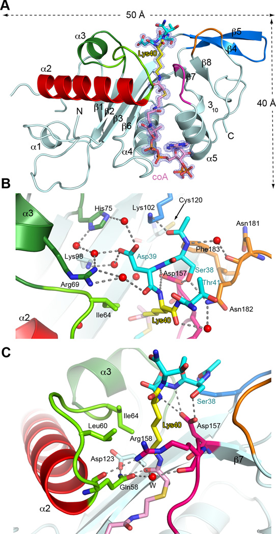
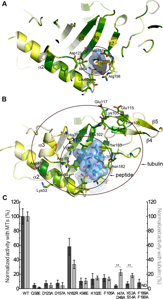
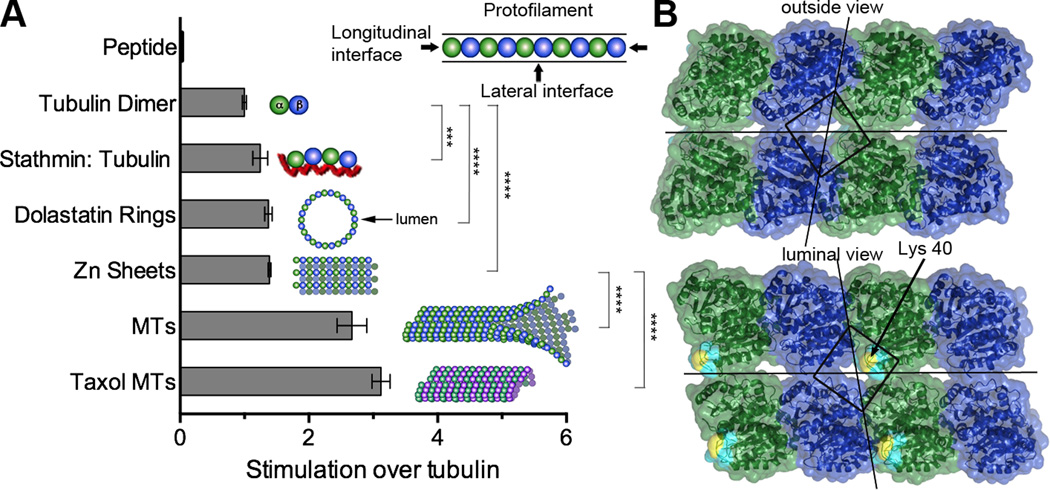
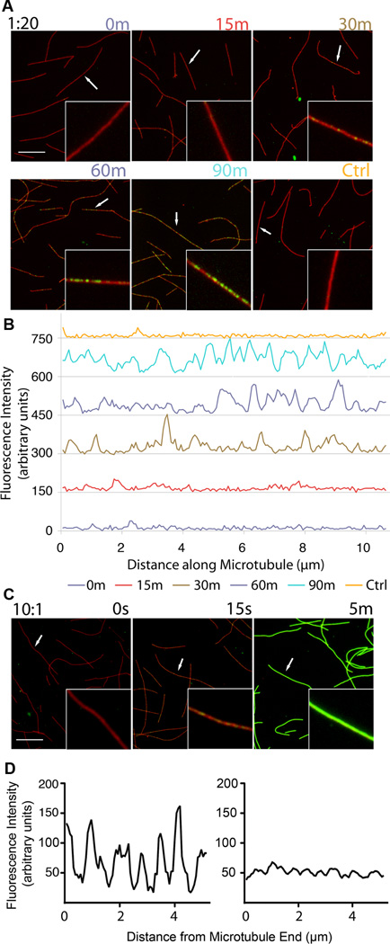
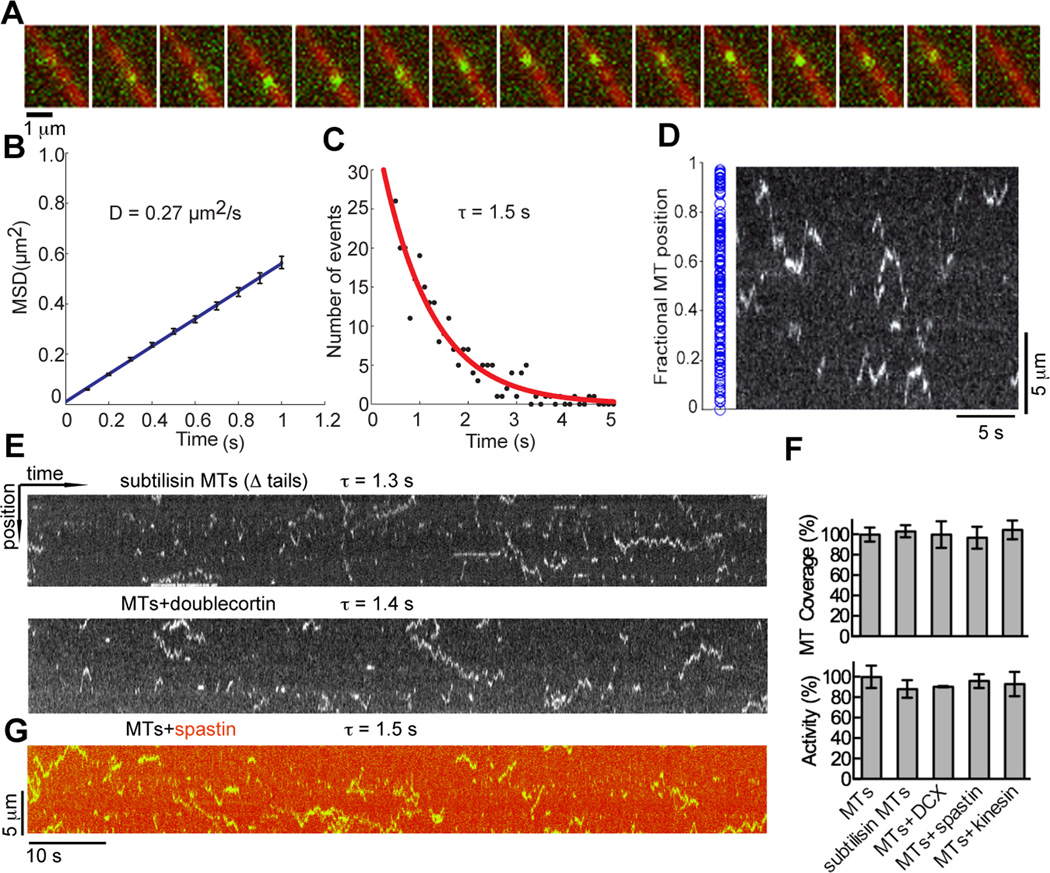
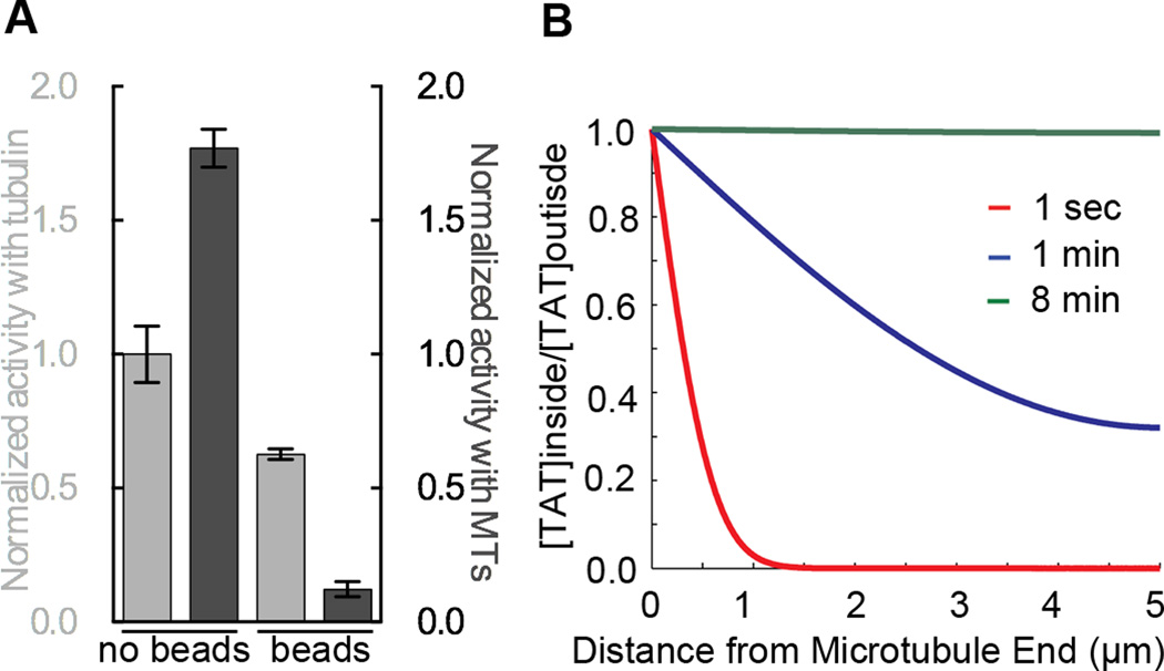
Acetylation of α-tubulin Lys40 by tubulin acetyltransferase (TAT) is the only known posttranslational modification in the microtubule lumen. It marks stable microtubules and is required for polarity establishment and directional migration. Here, we elucidate the mechanistic underpinnings for TAT activity and its preference for microtubules with slow turnover. 1.35 Å TAT cocrystal structures with bisubstrate analogs constrain TAT action to the microtubule lumen and reveal Lys40 engaged in a suboptimal active site. Assays with diverse tubulin polymers show that TAT is stimulated by microtubule interprotofilament contacts. Unexpectedly, despite the confined intraluminal location of Lys40, TAT efficiently scans the microtubule bidirectionally and acetylates stochastically without preference for ends. First-principles modeling and single-molecule measurements demonstrate that TAT catalytic activity, not constrained luminal diffusion, is rate limiting for acetylation. Thus, because of its preference for microtubules over free tubulin and its modest catalytic rate, TAT can function as a slow clock for microtubule lifetimes.


Figures

Figure 1
Figure 1
Active site architecture and α−tubulin Lys40 loop recognition (A) Ribbon representation of TAT bound to peptide-coA bisubstrate analog 1 (IC50 ∼ 100 µM; Figures S1C and S1D); analog, shown as a stick model with Lys40 yellow, the rest of the peptide moiety, cyan, coA and linker, pink in the same scheme as Figure S1C; the |Fo|-|Fc| density (prior to modeling the ligand) is contoured at 3.5σ (blue); Regions engaged in substrate binding and catalysis colored green, red, magenta, orange and blue (B) Close-up of the active site showing residues engaged in tubulin peptide recognition; red spheres and dashed lines denote water molecules and hydrogen bonds, respectively; color scheme as in A; TAT residues labeled in black, peptide residues in color (C) Close-up of the active site showing residues important for Lys40 recognition and catalysis; colored as in A. Water molecule depicted as a red sphere labeled W. See also Figure S1 and Table S1.
Figure 2
Figure 2
Molecular determinants for Lys40 loop and tubulin recognition (A) A slice through TAT shown in ribbon representation color-coded for conservation on a gradient from white (40% identity) to green (100% identity). The tubulin peptide bisubstrate analog is shown as a stick model encased in a transparent molecular surface with α-tubulin Lys40 yellow (B) Peptide and tubulin binding interface. Inner and outer circles denote interaction surfaces with the Lys40 loop and tubulin, respectively. TAT represented as in A, TAT residues labeled in black, peptide residues in blue. The Lys40 side chain projects into the plane of the figure and is not visible in this view. (C) Normalized acetylation activity of structure-guided TAT mutants with microtubules (dark grey) and tubulin (light grey). Error bars indicate standard error of the mean (S.E.M.; N≥3). **, p < 0.01, t-test. See also Figure S2.
Figure 3
Figure 3
TAT activity is enhanced predominantly by lateral lattice interactions (A) TAT activity with tubulin substrates of diverse geometries schematically represented with α-and β-tubulin as green and blue spheres, respectively. Stathmin, shown as red line. MTs, non-taxol stabilized glycerol microtubules; taxol MTs, microtubules stabilized with 20 µM taxol. The presence of dolastatin-10 rings, Zn sheets and microtubules under the reaction conditions was confirmed by negative stain EM (Figure S3A). Error bars indicate S.E.M. (N≥3). ***, p < 0.001, ****, p < 0.0001, t-test (B) Outer and luminal surface of two microtubule protofilaments; α-and β-tubulin, colored green and blue, respectively; the modeled Lys40 loop (visible only in the luminal view) is shown in cyan with Lys40 in yellow. Lines bisecting the surface indicate longitudinal and lateral microtubule lattice interfaces. The square surrounds the vertex of four tubulin subunits that overlies the Lys40 loop.
Figure 4
Figure 4
Acetylation occurs stochastically along the microtubule and is not biased for microtubule ends (A) Time-course of taxol stabilized microtubules stained for tubulin (red) and acetylated tubulin (green) after exposure to TAT at 1:20 TAT: tubulin molar ratio; insets, close-ups of selected microtubules (arrows); scale bar 5 µm. (B) Line-scans of selected microtubules (arrows) from panel A in the acetylated tubulin channel. Line-scans from successive time points are staggered vertically by 150 units for clarity. (C) Time-course of taxol microtubules exposed to excess TAT at 10:1 TAT:tubulin molar ratio; insets, close-ups of selected microtubules (arrows); scale bar 5 µm. (D) Analysis of acetylation near microtubule ends with excess TAT 15 seconds into the reaction. Left, representative scan of the terminal 5 µm of a microtubule; right, average of 25 line-scans of microtubule ends from the tip to 5µm into the microtubule. Scanned microtubules had 10 µm minimum length. See also Figure S4.
Figure 5
Figure 5
TAT-GFP scans the microtubule (A) Sequential frames of a TAT-GFP (green) movie. 15 frames of a continuous TIRF recording (100 ms frames) were overlaid on one microtubule image (red). (B) Mean-squared displacement (MSD) of TAT-GFP plotted against time. A linear curve fitted to the shown time interval yields a diffusion coefficient of 0.27 ± 0.01 µm2/s. Error bars represent S.E.M. (C) Distribution of durations of TAT-GFP interactions with the microtubule. An exponential curve fitted to the histogram and corrected for photobleaching (Figure S5A) yields a mean lifetime of interaction (τ) of 1.5 ± 0.3 s (R2 = 0.91; N=269). (D) Initial positions (blue dots) of scanning TAT-GFP along microtubules, N=163 (left) (Figure S5C); kymograph depicting TAT-GFP motion on a microtubule showing random initial positions along the microtubule (right). (E) Kymographs depicting the motion of TAT-GFP on a subtilisin treated microtubule missing C-terminal tails (top) and microtubule in the presence of saturating amounts of doublecortin (bottom) (see also Figure S5K). (F) Normalized total microtubule length scanned by TAT-GFP molecules (top) and acetylation activity (bottom) with subtilisin microtubules and microtubules coated with doublecortin, spastin and kinesin-5 motor domain (see also Figure S5J). (G) Kymograph depicting the motion of TAT-GFP on microtubules decorated with DyLight 550 labeled spastin in the presence of ATPγS, which inhibits severing, but not microtubule binding (Roll-Mecak and Vale, 2008). τ, defined as in C.
Figure 6
Figure 6
Substrate access by TAT is spatially constrained but not rate limiting for microtubule acetylation (A) Normalized activity of biotinylated TAT, free in solution or immobilized to 1 µm diameter streptavidin beads, with tubulin (light grey) and microtubules (dark grey). (B) Calculated profiles of TAT diffusion into a microtubule (or probability of residence) at positions along the microtubule at indicated time points (Extended Experimental Procedures). At 8 min, TAT is completely equilibrated between the lumen of a 10 µm long microtubule and the outside volume. See also Figure S6.

Comment in

References

    1. Akella JS, Wloga D, Kim J, Starostina NG, Lyons-Abbott S, Morrissette NS, Dougan ST, Kipreos ET, Gaertig J. MEC-17 is an alpha-tubulin acetyltransferase. Nature. 2010;467:218–222. - PMC - PubMed
    1. Bechstedt S, Brouhard GJ. Doublecortin recognizes the 13-protofilament microtubule cooperatively and tracks microtubule ends. Dev Cell. 2012;23:181–192. - PMC - PubMed
    1. Black MM, Baas PW, Humphries S. Dynamics of alpha-tubulin deacetylation in intact neurons. J Neurosci. 1989;9:358–368. - PMC - PubMed
    1. Bungay PM, Brenner H. The motion of a closely-fitting sphere in a fluid-filled tube. Internaltional Jounral of Multiphase Flow. 1973;1:25–56.
    1. Burton PR. Luminal material in microtubules of frog olfactory axons: structure and distribution. J Cell Biol. 1984;99:520–528. - PMC - PubMed

Publication types