. 2014 Jun 5;157(6):1416-1429.
doi: 10.1016/j.cell.2014.03.063.

A "push and slide" mechanism allows sequence-insensitive translocation of secretory proteins by the SecA ATPase

Affiliations

A "push and slide" mechanism allows sequence-insensitive translocation of secretory proteins by the SecA ATPase

Benedikt W Bauer et al. Cell. .

Abstract

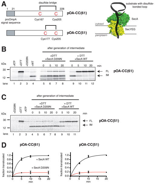
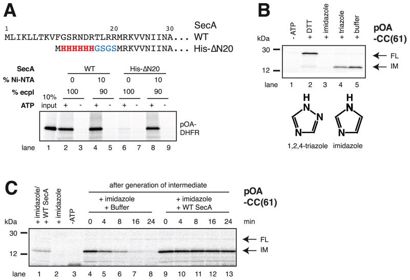
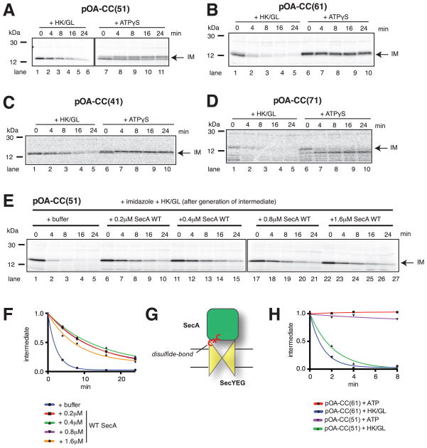
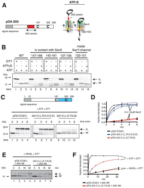
In bacteria, most secretory proteins are translocated across the plasma membrane by the interplay of the SecA ATPase and the SecY channel. How SecA moves a broad range of polypeptide substrates is only poorly understood. Here we show that SecA moves polypeptides through the SecY channel by a "push and slide" mechanism. In its ATP-bound state, SecA interacts through a two-helix finger with a subset of amino acids in a substrate, pushing them into the channel. A polypeptide can also passively slide back and forth when SecA is in the predominant ADP-bound state or when SecA encounters a poorly interacting amino acid in its ATP-bound state. SecA performs multiple rounds of ATP hydrolysis before dissociating from SecY. The proposed push and slide mechanism is supported by a mathematical model and explains how SecA allows translocation of a wide range of polypeptides. This mechanism may also apply to hexameric polypeptide-translocating ATPases.


Figures

Figure 1
Figure 1. SecA is moderately processive
(A) Generation of translocation intermediates. The substrates pOA-CC(61) and pOA-CC(51) contain a signal sequence (grey box) and cysteines (C) at the indicated positions, which form a disulfide-bonded loop that prevents complete transport through the complex of SecA and SecYEG (shown in green and yellow, respectively). (B) A translocation intermediate was generated with pOA-CC(61), wild type (WT) SecA, ATP, and proteoliposomes containing SecYEG. Then, a 64fold molar excess of either the dominant-negative mutant SecA D209N or WT SecA was added together with DTT. Samples were taken at the indicated time points, treated with proteinase K, and analyzed by SDS-PAGE and autoradiography. In lanes 1–4, the indicated components were added or omitted before generation of the intermediate. IM and FL indicate protease-protected fragments, corresponding to the intermediate and fully translocated pOA-CC(61), respectively. Molecular weight markers are indicated (in kDa). (C) As in (B), but with pOA-CC(51). (D) Quantification of the experiments in (B) and (C). Plotted is the amount of FL relative to the final levels of FL seen with WT SecA (means and standard deviations of three experiments).
Figure 2
Figure 2. SecA rebinds to the translocating SecY channel through prior interaction with lipids
(A) N-terminal sequences of wild type (WT) SecA and of the His-ΔN20 mutant. These proteins were incubated with radiolabeled substrate (a fusion of proOmpA and DHFR; pOA-DHFR), ATP, and proteoliposomes containing SecYEG and either 100% E. coli polar lipids (ecpl) or 90% ecpl and 10% Ni-NTA lipids. Translocation of pOA-DHFR was assessed by proteinase K treatment followed by SDS-PAGE and autoradiography. 10% of the input material was loaded in lane 1. (B) SecA His-ΔN20 was incubated with radiolabeled pOA-CC(61), ATP, and Ni-NTA proteoliposomes containing SecYEG. Hexokinase/glucose (-ATP), DTT, imidazole, or triazole were added, as indicated, before the start of the translocation reaction. The samples were analyzed as in (A). IM and FL indicate protease-protected fragments, corresponding to the intermediate and fully translocated pOA-CC(61), respectively. Molecular weight markers are indicated (in kDa). The lower panel shows the structures of imidazole and triazole. (C) As in (B), but imidazole was added after generation of the translocation intermediate, together with buffer or WT SecA. In lanes 1–3, the indicated components were added before generation of the intermediate.
Figure 3
Figure 3. Dynamic interaction between SecA and SecY channel
(A) A translocation intermediate was generated by fusing proOmpA with dehydrofolate reductase (DHFR) (pOA-DHFR). The DHFR domain folds in the presence of methotrexate (MTX), and stalls translocation. A FRET assay was used to follow the interaction of SecA (in green), labeled with the FRET-acceptor Cy5 (in blue), with SecYEG (in yellow), labeled with the FRET-donor tetramethyl rhodamine (TMR; in red). (B) SecA-Cy5 was mixed with ATP, MTX, and proteoliposomes containing SecYEG-TMR in the presence or absence of pOA-DHFR. The translocation intermediate was then incubated with a 25fold molar excess of unlabeled SecA, and the resulting decay of the FRET signal measured over time. The curves represent exponential fits to the data points. (C) As in (B), but in the presence or absence of MTX. (D) As in (B), but with ATP, ATPγS, or hexokinase/glucose (HK/GL) added to the reactions. (E) As in (D), but without substrate.
Figure 4
Figure 4. Nucleotide-dependent interaction of SecA with a translocation substrate
(A) Radiolabeled pOA-CC(51) was mixed with ATP, wild type (WT) SecA, and proteoliposomes containing SecYEG. After generation of the intermediate, DTT was added together with hexokinase/glucose (HK/GL) or ATPγS. Samples were taken at different times, treated with proteinase K, and subjected to SDS-PAGE followed by autoradiography. IM indicates the position of the translocation intermediate. Molecular weight markers are indicated (in kDa). (B) As in (A), but with pOA-CC(61). (C) As in (A), but with pOA-CC(41). (D) As in (A), but with pOA-CC(71). (E) Radiolabeled pOA-CC(51) was mixed with ATP, SecA His-ΔN20, and Ni-NTA proteoliposomes containing SecYEG. After generation of a translocation intermediate, imidazole was added together with HK/GL and either buffer or WT SecA at various concentrations. The samples were analyzed as in (A). (F) Quantification of the experiments shown in (E). The amount of IM, relative to the initial level, was plotted over time. (G) Scheme showing a translocation intermediate generated with SecA disulfide-crosslinked to the SecY complex. (H) A translocation intermediate was generated with a covalent SecA-SecY complex (see (G)) and either pOA-CC(61) or pOA-CC(51). The intermediates were incubated with either HK/GL or ATP, and backsliding determined as in (A).
Figure 5
Figure 5. SecA interacts with amino acid side chains of the polypeptide substrate
(A) Stretches of 20 consecutive glycines (G) were inserted into the translocation substrate pOA-CC(61) at various distances in front of Cys167. Backsliding in the presence of ATPγS arrests polypeptide movements at interacting amino acids in the flanking regions, giving rise to two defined proteolytically protected fragments (arrows in right scheme). (B) pOA-CC(61) and derivatives with glycine stretches inserted at the indicated positions were mixed with ATP, wild type SecA, and proteoliposomes containing SecYEG. After generation of a translocation intermediate, ATP or ATPγS were added. The samples were analyzed at different times by treatment with proteinase K, followed by SDS-PAGE and autoradiography. Where indicated, DTT was added at the beginning of the translocation reaction. IM and FL indicate protease-protected fragments, corresponding to the intermediate and fully translocated products, respectively. Molecular weight markers are indicated (in kDa). (C) Translocation of the C-terminal tail (in blue) was tested with pOA-CC(61) or derivatives in which several amino acids in the segment following the first cysteine were replaced with glycines (the substituted amino acids are given in one-letter code). The radiolabeled substrate was mixed with ATP, SecA His-Δ N20, and Ni-NTA proteoliposomes containing SecYEG. The generated translocation intermediate was incubated with imidazole and wild type (WT) SecA, before addition of DTT. The samples were analyzed as in (B). (D) Quantification of the experiments in (C). The experiments were performed at different concentrations of SecA. Plotted is the amount of FL relative to the initial level of IM, divided by the ratio determined for wild type substrate at the last time point. A correction was made for the different numbers of methionines in IM and FL (8 versus 9). (E) A comparison of translocation and passive forward sliding was performed with substrates in which the segment in pOA-CC(61) following the second cysteine was replaced with four methionines (pOA-CC(61) 1-205 4M). The segment in the disulfide-bonded loop was either the wild type sequence or lacked hydrophobic and polar amino acids (d(A,V,I,L,S,T,N,Q)). Translocation intermediates were incubated with hexokinase/glucose (HK/GL), followed by addition of DTT. The appearance of FL was followed by protease protection. (F) Quantification of the experiment in (E), done as in (D). A correction was made for the different numbers of methionines in IM and FL (8 versus 13). Shown are the means and standard deviation of three experiments. Also shown is translocation in the presence of ATP, determined in parallel.
Figure 6
Figure 6. Substrate interaction of SecA’s two-helix finger
(A) The two-helix finger (THF) of SecA has a loop with the indicated sequence between helices 1 and 2. Residues R792/K797 and Y794 (in red) were replaced by glycines. (B) Radiolabeled pOA-CC(61) was mixed with ATP, SecA His-ΔN20, and Ni-NTA proteoliposomes containing SecYEG. After generation of a translocation intermediate, imidazole was added together with either wild type (WT) SecA or fingertip SecA mutants. After 20 min, DTT was added, and the conversion of the intermediate (IM) to fully translocated product (FL) was determined by protease protection. (C) As in (B), but with pOA-CC(61) in which the indicated amino acids following the first cysteine were replaced by glycines. (D) Quantification of the experiments in (B). Plotted is the amount of FL relative to the final levels of FL seen with WT SecA (means and standard deviations). (E) As in (D), but for the experiments in (C). (F) As in (B) with SecA Y794G added at the indicated concentrations, but without DTT and with either buffer (BF), ATP, ATPγS, or hexokinase/glucose (HK/GL) added. The amount of IM, relative to the initial level, was plotted over time.
Figure 7
Figure 7. A mathematical model for the “push and slide” mechanism of SecA
(A) Push and slide model. Amino acids of the translocating polypeptide are shown as circles. Upon ATP binding by SecA (rate constant Kbind), the two-helix finger (in blue) undergoes a power stroke (curved arrow). If it interacts with an amino acid (green), it pushes it forward (green arrow). Otherwise, the amino acid (in red) can diffuse forward or backward (diffusion constant DATP). SecA-ATP is converted into SecA-ADP with the rate constant Khyd, resulting in resetting of the two-helix finger (purple; curved arrow). In the ADP-state, SecA allows passive sliding of any amino acid (diffusion constant DADP). The translocation direction is indicated with a blue arrow. (B) Modeling of the experiment in Figure 5D. The translocated last 61 residues of the substrates correspond to either wild type (black curve) or mutant sequences (blue and red). The best fit was obtained with the percentages of interacting amino acids indicated in the inset, distributed evenly throughout the segment. Parameters: DATP=DADP=0.04 nm2/sec; Khydr=107/sec; Kbind=11/sec. (C) Translocation of a 40-residue polypeptide containing interacting (green) and non-interacting (red) amino acids. SecA was assumed to be 90% of the time in its ADP-bound state. The fraction of polypeptide chains translocated to different positions is given at different time points (blue columns; time in sec). Position 40 corresponds to complete translocation. The parameters were as in (B). (D) As in (C), but with SecA being 10% of its time in the ADP-bound state. (E) The translocation time (the time its takes to fully translocate 95% of all chains) was plotted over the ATP consumption rate (the average of ATP molecules hydrolyzed per sec during translocation of the entire chain). SecA was assumed to spend different fractions of time in the ADP-bound state (inset). The substrate (50 amino acids) contained 90% or 10% of evenly spaced interacting amino acids. (F) As in (E), but with translocation rates (inverse of translocation time) expressed as percentage relative to the maximal rate at infinite ATP consumption rate. The calculations were performed for different percentages of evenly spaced interacting amino acids (inset), assuming SecA to be 90% in its ADP-bound state. The broken line indicates the experimentally determined ATP consumption rate (7.6/sec). This corresponds to an optimal situation, as translocation would be slow towards the left, and ATP consumption excessive towards the right.

References

    1. Alami M, Dalal K, Lelj-Garolla B, Sligar SG, Duong F. Nanodiscs unravel the interaction between the SecYEG channel and its cytosolic partner SecA. EMBO J. 2007;26:1995–2004. - PMC - PubMed
    1. Barkow SR, Levchenko I, Baker TA, Sauer RT. Polypeptide Translocation by the AAA+ ClpXP Protease Machine. Chem Biol. 2009;16:605–612. - PMC - PubMed
    1. Bauer BW, Rapoport TA. Mapping polypeptide interactions of the SecA ATPase during translocation. Proc Natl Acad Sci USA. 2009;106:20800–20805. - PMC - PubMed
    1. DeLaBarre B, Christianson JC, Kopito RR, Brunger AT. Central Pore Residues Mediate the p97/VCP Activity Required for ERAD. Mol Cell. 2006;22:451–462. - PubMed
    1. Economou A, Pogliano JA, Beckwith J, Oliver DB, Wickner W. SecA membrane cycling at SecYEG is driven by distinct ATP binding and hydrolysis events and is regulated by SecD and SecF. Cell. 1995;83:1171–1181. - PubMed

Publication types

MeSH terms