Review
. 2014 Aug;35(26):7308-25.
doi: 10.1016/j.biomaterials.2014.04.091. Epub 2014 Jun 3.

Microfluidic techniques for development of 3D vascularized tissue

Affiliations
Review

Microfluidic techniques for development of 3D vascularized tissue

Anwarul Hasan et al. Biomaterials. 2014 Aug.

Abstract

Development of a vascularized tissue is one of the key challenges for the successful clinical application of tissue engineered constructs. Despite the significant efforts over the last few decades, establishing a gold standard to develop three dimensional (3D) vascularized tissues has still remained far from reality. Recent advances in the application of microfluidic platforms to the field of tissue engineering have greatly accelerated the progress toward the development of viable vascularized tissue constructs. Numerous techniques have emerged to induce the formation of vascular structure within tissues which can be broadly classified into two distinct categories, namely (1) prevascularization-based techniques and (2) vasculogenesis and angiogenesis-based techniques. This review presents an overview of the recent advancements in the vascularization techniques using both approaches for generating 3D vascular structure on microfluidic platforms.

Keywords: Angiogenesis; Microfluidics; Micromolding; Tissue engineering; Vascularization; Vasculogenesis.


Figures

Figure 1
Figure 1
Schematic representation for mechanisms of in vivo blood vessels formation and their structure, (A) vasculogenesis and angiogenesis: two predominant mechanisms of blood vessel formation. Vasculogenesis gives rise to the primitive vascular plexus during embryogenesis. Angiogenesis remodels and expands the vascular network through biochemical cues including growth factor gradient, budding tumors, and hypoxia conditions [30]. (B) Schematic diagrams for structure of blood vessels: Cross-section of arteries with their major components and maturation steps [39]. Figures reproduced from [30] and [39] with permissions from Nature publishing group.
Figure 2
Figure 2
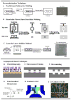
Schematic diagrams of different types of in vitro vascularization techniques: (A–D) various prevascularization techniques, (A–B) subtractive methods, (A) stainless steel needle- based molding, (B) dissolvable-network-based sacrificial molding, (C) additive method - soft lithography/PDMS stamping-based micromolding and layer-by-layer stacking, (D) hybrid method – Bioprinting, (E–I) various vasculogenesis and angiogenesis-based techniques, (E) photolithography, (F) microcontact printing, (G) functionalization of scaffold material, (H) gradient of growth factor in a microfluidic device, and (I) co-culture of multiple cells. Figures adopted and modified from [83, 85, 87, 98, 160, 161] with permissions from American Chemical Society, Royal Society of Chemistry and Elsevier Science.
Figure 3
Figure 3
Fabrication of single vascular-like structures in hydrogels using needle-based subtractive prevascularization technique. (A) Microvascular tubes were formed in type I collagen hydrogel by extracting a pre-inserted 120-μm-diameter stainless steel needle. Removal of the needle generated cylindrical channels that were seeded with microvascular ECs. The EC monolayer exhibited strong cell-cell junctions as shown by expression of Prox-1, junctional markers VE-cadherin and PECAM [162]. (B) A cylindrical channel was formed in a cell-laden hydrogel using extraction of a stainless steel microneedle [89]. (C) A 300 μm microchannel in cell-laden GelMA hydrogels seeded in the lumen with GFP-labeled-HUVECs [86]. (D) Fabrication of a vascular-like structure in cell-laden GelMA using cell transfer technique. A peptide chemically adsorbed on a gold wire was used to adsorb/capture cells that were transferred to a hydrogel construct by embedding the rod in hydrogel and applying an electrical voltage. The gold was then removed leaving a channel seeded with HUVECs. The multilayer structure was generated by an additional dip-coating step in GelMA solution enriched with 3T3 cells, (i) schematic representation of the dip-coating-cell-transfer technique, (ii) a 3D continuous monolayer of ECs in the microvasculature, (iii) the bilayer structure of 3T3-HUVECs, (iv–vi) confocal fluorescence image of the cross section, red: 3T3 cells, green: HUVECs [90]. (E) A multilayered capillary structure fabricated using a hierarchical cell deposition technique in a prevascularized polyglycolic acid (PGA) scaffold. A uniaxial microchannel was formed inside the scaffold by extraction of a silica tube. Layers of SMCs and ECs were formed sequentially inside the channel using a hierarchical cell deposition [91], (i) confocal fluorescence image of the fabricated bilayer capillary structure, green: SMCs, red: HUVECs, (ii) a magnified image of (i), (iii) schematic representation of the bilayer structure. Scale bar: 200 μm in C(ii), 400 μm in E (i), and 100 μm in E (ii). Figures modified from [87] [89] [86] [90] and [91] with permissions from Elsevier Ltd. Wiley Periodicals Inc. and Wiley-VCH Verlag GMbh & Co. respectively.
Figure 4
Figure 4
Vascularization using various prevascularization techniques: (A) Sacrificial molding by dissolving a gelatin mesh in fibrin and collagen hydrogels [85], (i) overlaid phase contrast images of a hexagonal vascular network in fibrin gel perfused with fluorescent microspheres, (ii) perfusable collagen gel with multiplanar networks shown with an overlay of suspensions of green and red fluorescent beads, respectively; (B) Prevascularization using omnidirectional printing of 3D microvascular networks with a fugitive ink into a physical gel reservoir. The technique allows full control of channel hierarchical and branching patterns. Post photocrosslinking the ink (shown in red) is liquefied and removed slowly under pressure exposing the microvascular channels [96]. (C) A rapid casting-based prevascularization technique (i) schematic overview of generating 3D vascular network: an open, perfusable vascular network was generated using 3D printing of an interconnected sacrificial carbohydrate network able to support cell-laden hydrogels [96], (ii) a single carbohydrate-glass fiber approximately 200 μm is encapsulated in a fibrin gel showing the dissolution of the carbohydrates within hydrogel post-crosslinking, (iii) viable cells constitutively expressing enhanced green fluorescent protein (eGFP) imaged with confocal microscopy z-stack to visualize two intersecting channels demonstrating endothelialization of channel walls and across the intervessel junctions. (D) A microfluidic vessel networks (μVNs) fabricated using micromolding method followed by additive stacking and bonding of two hydrogel slabs [99]: (i) schematic cross-sectional view of the microfluidic collagen construct after fabrication, (ii) cell-cell junction of EC seeded inside the channels, (iii) Z-stack projection of horizontal confocal sections of endothelialized microfluidic vessels, (iv) view of xz plane and (v) view of a corner. Red: CD31, blue: nuclei. (Scale bar: 100 μm) [99]. (E) Vascular tubes fabricated using bioprinting technique [110], (i) the tubular constructs after fusing the cylindrical bioink, (ii) a bilayer tubular construct fabricated using spheroids of ECs (green) and SMCs (red), (iii) a bilayer construct fabricated using building blocks of fibroblast cells (outer layer) and SMCs (inner layer) representing the adventitia and media of a blood vessel. Figures reproduced and modified from [85, 92, 96, 99, 110] with permissions from the Royal Society of Chemistry, Wiley-VCH Verlag GMbh & Co. Nature Publishing, PNAS and IOP Publishing respectively.
Figure 5
Figure 5
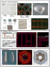
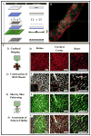
Formation of vascular structure using photolithography-based micropatterning technique: (A) schematic representation of the micropatterning process. Cell encapsulated photosensitive hydrogel prepolymer suspension placed between two spacers and covered with a glass is exposed to UV light through a photomask. The areas exposed to UV light gets crosslinked while the uncrosslinked region is washed out following the crosslinking step. (B) Confocal fluorescence image of a microconstruct showing the organization of cytoskeletal actin fibers (red) of HUVECs, encapsulated in GelMa hydrogel, leading to the formation of an endothelial cord. The cord was imaged after 5 days of culture [113]. Scale bar represents 100 μm. (C) Micropatterning using confocal-imaging-of-native-tissue-based templates and two- photon laser lithography technique: Confocal microscopy was used to image labeled tissues in 3D. Each optical section was then processed to reconstruct the cross-sectional structure of the tissue using a mosaic of regions of interest (ROIs). Next the ROIs were used to control precise scanning of a laser scanning microscope. To pattern a 3D structure, the mosaic of ROIs for each axial cross section was utilized to sequentially pattern each corresponding plane of the hydrogel [123]. (D) Projections of imaged vasculature from the retina, cerebral cortex, and heart. (E) ROI mosaics reconstructing the vasculature of various tissues at individual cross-sectional planes. (F) 3D projections of hydrogels with fluorescently labeled PEG-RGD patterned to mimic the vasculature from various tissues. (G) A merge of the imaged vasculature with the imaged hydrogels, with yellow indicating excellent overlap between vessels and patterns. Scale bars = 100 μm (5 μm for insets). Figures reproduced from [113] and [123] with permissions from Elsevier Ltd. and Wiley-VCH Verlag GMbh & Co. respectively.
Figure 6
Figure 6
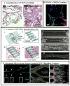
Vasculogenesis and angiogenesis-based approaches for vascularization: (A) promoting vascularization using functionalization of collagen hydrogel with covalently immobilized VEGF [135]. The organization and morphology of cells after a 7- day in vitro cultivation are shown using hematoxylin and eosin staining (arrowheads indicate vascular like structures). (B) Formation of microvascular network using coculture of HUVECs and NHLFS [149]. (C) Formation of vascular capillaries using co-culture of EC and fibroblast cells in a microfluidic device [150], (i) schematic of the microfluidic device consisting of two main channels connected with a group of bridge channels arranged in a ladder like structure. (ii) Enlarged view of ladder structure filled with fibrin gel forming concave sidewalls. (iii) HUVECs loaded and attached on the concave sidewalls and fibroblasts loaded through the two main inlet channels after attachment of HUVECs to the fibrin gel. (iv) Formation of self-organized capillaries by HUVECs grown from both sides. (v–vii) Image of HUVECs sprouts from both sides and formation of capillaries through fusion of the sprouts’ tips over time. (viii) Imunostaining of perfusable capillaries. Blood vessels were stained with Hoechst 33342 for the nucleus in blue and Phalloidin for F-actin in green and ZO-1 for tight junction in red. (D) Formation of a perfusable, functional microvascular network in a versatile microfluidic device [151]: (i) a representative microvessel formed, (ii) The cells were able to form tight cell-junction connections visualized by VE cadherin and β-catenin. (iii) aberrant morphology and frequent branching of immature tubules within 48h of culture as shown by multiple tip cells visualized by F-actin and CD31 staining. Figures adapted and modified from [, –151] with permissions from Elsevier Ltd., Federation of American Society for Experimental Biology and Royal Society of Chemistry.

References

    1. Bae H, Puranik AS, Gauvin R, Edalat F, Carrillo-Conde B, Peppas NA, et al. Building vascular networks. Sci Trans Med. 2012;4:1–4. - PMC - PubMed
    1. Du Y, Cropek D, Mofrad MRK, Weinberg EJ, Khademhosseinil A, Borenstein J. Microfluidic systems for engineering vascularized tissue constructs. In: Tian W-C, Finehout E, editors. Microfluidics for Biological Applications. New York: Springer; 2008.
    1. Kaully T, Kaufman-Francis K, Lesman A, Levenberg S. Vascularization - the conduit to viable engineered tissues. Tissue Eng Pt B: Rev. 2009;15:159–69. - PubMed
    1. Hasan A, Ragaert K, Swieszkowski W, Selimović Š, Paul A, Camci-Unal G, et al. Biomechanical properties of native and tissue engineered heart valve constructs. J Biomech. 2013 In press. - PubMed
    1. Du Y, Lo E, Ali S, Khademhosseini A. Directed assembly of cell-laden microgels for fabrication of 3D tissue constructs. Proc Natl Acad Sci U S A. 2008;105:9522–7. - PMC - PubMed

Publication types

MeSH terms