

Far-red tracer analysis of traumatic cerebrovascular permeability
- PMID: 24906578
- PMCID: PMC4096579
- DOI: 10.1016/j.jss.2014.05.011
Far-red tracer analysis of traumatic cerebrovascular permeability
Abstract
Background: Blood brain barrier (BBB) compromise is a key pathophysiological component of secondary traumatic brain injury characterized by edema and neuroinflammation in a previously immune-privileged environment. Current assays for BBB permeability are limited by working size, harsh extraction processes, suboptimal detection via absorbance, and wide excitation fluorescence spectra. In this study, we evaluate the feasibility of Alexa Fluor 680, a far-red dye bioconjugated to dextran, as an alternative assay to improve resolution and sensitivity.
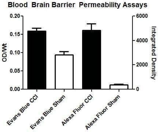
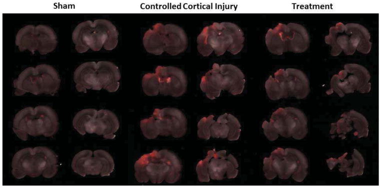
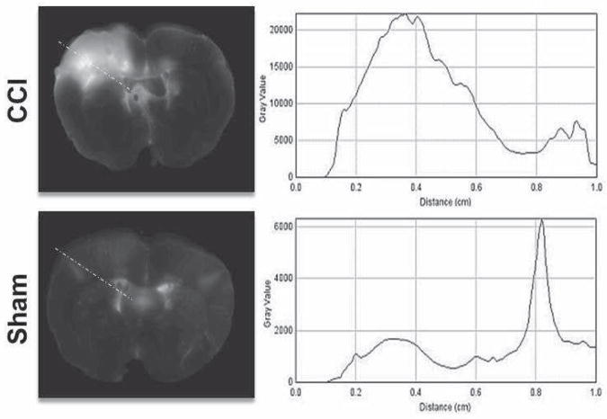
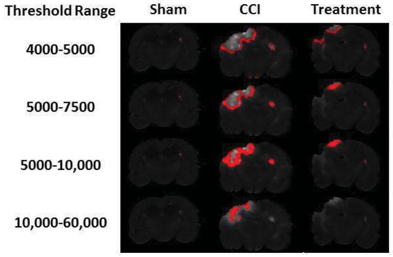
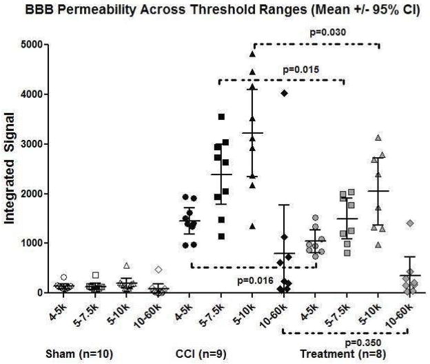
Methods: Alexa Fluor was introduced intravenously on the day of sacrifice to three groups: sham, controlled cortical impact (CCI), and CCI treated with a cell based therapy known to reduce BBB permeability. The brains were sectioned coronally and imaged using an infrared laser scanner to generate intensity plot profiles as well as signal threshold images to distinguish regions with varying degrees of permeability.
Results: Linear plot profile analysis demonstrated greater signal intensity from CCI than treated rats at corresponding injury depths. Threshold analysis identified rims of signal at low + narrow threshold ranges. The integrated signals from a treatment group known to preserve the BBB were significantly less than the groups with CCI injury alone. There was no significant difference at high + wide signal intensity threshold ranges.
Conclusions: Alexa Fluor 680 infrared photodetection and image analysis can aid in detecting differential degrees of BBB permeability after traumatic brain injury and maybe particularly useful in demonstrating BBB preservation of at-risk regions in response to therapeutic agents.
Keywords: Blood brain barrier; Edema; Fluorescence; Permeability; Traumatic brain injury.
Copyright © 2014 Elsevier Inc. All rights reserved.
Conflict of interest statement
Conflict of Interest: none
Figures
References
-
- Barzo P, Marmarou A, Fatouros P, Hayasaki K, Corwin F. Contribution of vasogenic and cellular edema to traumatic brain swelling measured by diffusion-weighted imaging. Journal of neurosurgery. 1997 Dec;87(6):900–907. - PubMed
-
- Saw MM, Chamberlain J, Barr M, Morgan MP, Burnett JR, Ho KM. Differential Disruption of Blood-Brain Barrier in Severe Traumatic Brain Injury. Neurocritical care. 2013 Nov 14; - PubMed
-
- van den Brink WA, Marmarou A, Avezaat CJ. Brain oedema in experimental closed head injury in the rat. Acta neurochirurgica Supplementum. 1990;51:261–262. - PubMed
MeSH terms
Substances
Grants and funding
LinkOut - more resources
Full Text Sources
Other Literature Sources
