

Case Reports
doi: 10.1186/1477-7819-12-180.
Splenic hamartomas in two children
Affiliations
- PMID: 24906658
- PMCID: PMC4068870
- DOI: 10.1186/1477-7819-12-180
Item in Clipboard
Case Reports
Splenic hamartomas in two children
World J Surg Oncol.
.
Abstract
Hamartomas are extremely rare splenic benign tumours in children. We present two cases, both in boys (6 and 8 years old), with left upper quadrant abdominal pain that were otherwise asymptomatic. Both patients showed a splenic mass on preoperative ultrasonography and magnetic resonance imaging (MRI). One patient had a focal splenic mass that was identified preoperatively with contrasted computed tomography (CT) scans. Both patients underwent a total splenectomy. Although multi-modality imaging findings were described preoperatively, the final diagnosis in each case was splenic hamartoma based on histology and immunohistochemistry. The postoperative courses were uneventful.
Figures
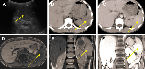
Splenic hamartoma in an 8-year-old boy was found during examination. (A) Abdominal ultrasonography showed an isoechoic oval-shaped mass (arrow) in the mid-portion of the spleen; (B) Non-enhanced CT showed a slightly lower-density mass (arrow) in the spleen. (C) Contrast-enhanced CT scan shows mild diffuse heterogeneous enhancement in the arterial phase. Magnetic resonance imaging (MRI) showed a hypointense mass (arrow) on the T1WI image (D) and a slightly hyperintense mass in the T2WI image (E). (F) The lesion (arrow) shows moderate enhancement on gadolinium-enhanced MRI.
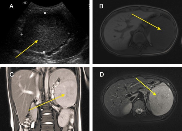
Splenic hamartoma in a 6-year-old boy with left upper quadrant abdominal pain. (A) Abdominal ultrasonography showed a hypoechoic mass (arrow) with well-defined borders in the lower pole of spleen. Magnetic resonance imaging (MRI) showed slightly hyperintense signal in the T1WI (B), T2WI images (C), and T2WI + Fatsat images (D), and a non-homogeneous signal within the lesion (arrow).
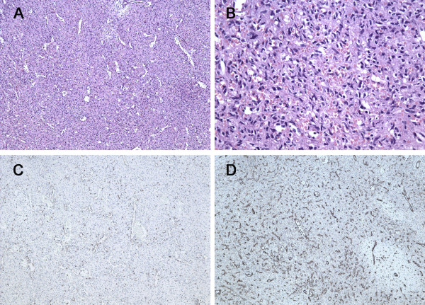
Pathology of splenic hamartoma. (A) Microscopic image of the splenic hamartoma showed the lesion containing a mixture of unorganised vascular channels and fibrotic cords of splenic red pulp-like area (hematoxylin-eosin, original magnification × 100). (B) Higher magnification of the lesion reveals no cytologic atypia and mitosis (hematoxylin-eosin, original magnification × 400). (C) CD8 immunostaining was positive in the lining cells and scattered lymphocytes (original magnification × 100). (D) Immunohistochemistry for CD31 is positive in vascular lining cells (original magnification × 100).
References
-
- Basso SM, Sulfaro S, Marzano B, Fanti G, Chiara GB, Lumachi F. Incidentally discovered asymptomatic splenic hamartoma with rapidly expansive growth: a case report. In Vivo. 2012;26(6):1049–1052. - PubMed
-
- Abramowsky C, Alvarado C, Wyly JB, Ricketts R. “Hamartoma” of the spleen (splenoma) in children. Pediatr Dev Pathol. 2004;7(3):231–236. - PubMed
Publication types
MeSH terms
LinkOut - more resources
Full Text Sources
Other Literature Sources
Medical
