

Case Reports
doi: 10.1136/bcr-2014-204122.
Hepatic PEComa: a potential pitfall in the evaluation of hepatic neoplasms
Affiliations
- PMID: 24907216
- PMCID: PMC4054585
- DOI: 10.1136/bcr-2014-204122
Item in Clipboard
Case Reports
Hepatic PEComa: a potential pitfall in the evaluation of hepatic neoplasms
BMJ Case Rep.
.
Abstract
Perivascular epithelioid cell tumour (PEComa) of the liver is very uncommon and may be overlooked in the clinical and histological differential diagnosis of a liver tumour. We report the case of an incidentally discovered liver mass suspicious for hepatocellular carcinoma, which on biopsy was suggestive of a pseudocyst but after resection was found to be hepatic PEComa with some of the usual characteristics of this neoplasm as well as several less familiar features. We have also reviewed cases of hepatic PEComa from the literature in order to provide insight into recognising possible PEComa preoperatively and assessing its risk of malignancy after diagnosis.
2014 BMJ Publishing Group Ltd.
Figures
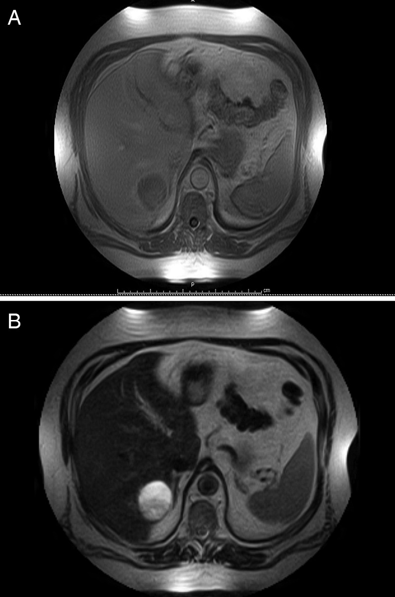
(A) MRI T1-weighted image showing hypointense, complex heterogeneous mass in segment 7/8. (B) MRI T2-weighted image showing mildly heterogeneous high signal intensity with well demarcated margins.
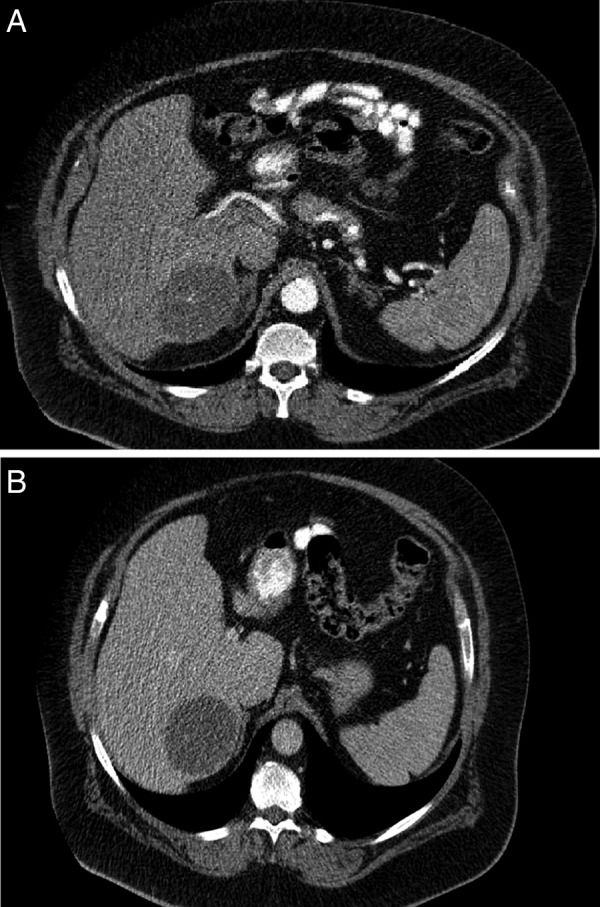
(A) CT scan (arterial phase) demonstrating partially enhancing mass in segment 7/8. (B) CT scan (venous phase) demonstrating decreased enhancement in the segment 7/8 mass.
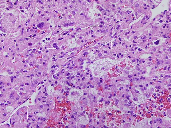
Appearance of the solid portion of the tumour demonstrating trabecular arrangement of large cells with abundant pale to clear cytoplasm, large vesicular pleomorphic nuclei and prominent nucleoli in a vascular network resembling clear-cell tumours of several different types. H&E ×40.
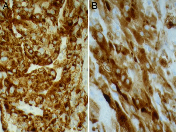
Strong uniform staining of the tumour cells for human melanoma black-45 (A) and similar staining for melanoma antigen recognised by T Cell-1(B). IHC, ×40.
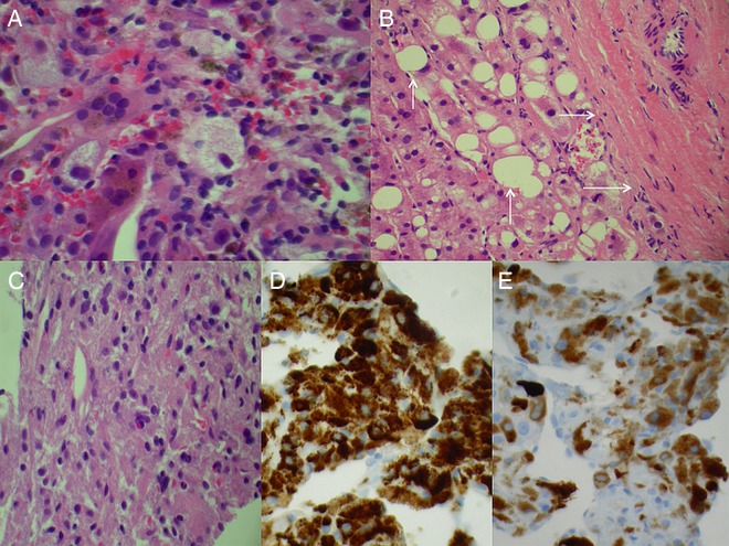
Core needle biopsy of hepatic tumour taken prior to referral showing (A) histiocytes and inflammatory cells suggesting pseudocyst, (B) reactive hepatocytes with steatosis (vertical arrows) adjacent to fibrous tissue (horizontal arrows) possibly representing the tumour capsule, (C) cells resembling degenerative histiocytes or hepatocytes in H&E-stained sections, (D) same cells with strong uniform staining for human melanoma black-45 and (E) same cells staining for melanoma antigen recognised by T Cell-1 on IHC performed retrospectively for both markers. Original magnification ×40.
References
-
- Folpe AL. Neoplasms with perivascular epithelioid cell differentiation (PEComas). In: Flectcher CDM. ed World Health Organization classification of tumours: pathology and genetics of soft tissue and bone. Lyon, France: IARC Press, 2002:221–2
-
- Ishak KG. Mesenchymal tumors of the liver. In: Okuda K, Peters RL. eds Hepatocellular carcinoma. New York: John Wiley & Sons, 1976:247–307
-
- Tsui WM, Colombari R, Portmann BC, et al. Hepatic angiomyolipoma: a clinicopathologic study of 30 cases and delineation of unusual morphologic variants. Am J Surg Pathol 1999;23:34–48 - PubMed
-
- Szekely E, Schaff Z, Madaras L, et al. Trabecular angiomyolipoma mimicking hepatic cell carcinoma. Pathol Oncol Res 2000;6:224–6 - PubMed
Publication types
MeSH terms
LinkOut - more resources
Full Text Sources
Other Literature Sources
Medical