. 2014 Aug;197(4):1137-51.
doi: 10.1534/genetics.114.166421. Epub 2014 Jun 6.

Chromatin organization and remodeling of interstitial telomeric sites during meiosis in the Mongolian gerbil (Meriones unguiculatus)

Affiliations

Chromatin organization and remodeling of interstitial telomeric sites during meiosis in the Mongolian gerbil (Meriones unguiculatus)

Roberto de la Fuente et al. Genetics. 2014 Aug.

Abstract

Telomeric DNA repeats are key features of chromosomes that allow the maintenance of integrity and stability in the telomeres. However, interstitial telomere sites (ITSs) can also be found along the chromosomes, especially near the centromere, where they may appear following chromosomal rearrangements like Robertsonian translocations. There is no defined role for ITSs, but they are linked to DNA damage-prone sites. We were interested in studying the structural organization of ITSs during meiosis, a kind of cell division in which programmed DNA damage events and noticeable chromatin reorganizations occur. Here we describe the presence of highly amplified ITSs in the pericentromeric region of Mongolian gerbil (Meriones unguiculatus) chromosomes. During meiosis, ITSs show a different chromatin conformation than DNA repeats at telomeres, appearing more extended and accumulating heterochromatin markers. Interestingly, ITSs also recruit the telomeric proteins RAP1 and TRF1, but in a stage-dependent manner, appearing mainly at late prophase I stages. We did not find a specific accumulation of DNA repair factors to the ITSs, such as γH2AX or RAD51 at these stages, but we could detect the presence of MLH1, a marker for reciprocal recombination. However, contrary to previous reports, we did not find a specific accumulation of crossovers at ITSs. Intriguingly, some centromeric regions of metacentric chromosomes may bind the nuclear envelope through the association to SUN1 protein, a feature usually performed by telomeres. Therefore, ITSs present a particular and dynamic chromatin configuration in meiosis, which could be involved in maintaining their genetic stability, but they additionally retain some features of distal telomeres, provided by their capability to associate to telomere-binding proteins.

Keywords: Mongolian gerbil; RAP1; TRF1; chromatin; interstitial telomeres; meiosis.


Figures

Figure 1
Figure 1
FISH for telomeric DNA repeats (green) and counterstaining of the chromatin with DAPI (blue) on metaphase chromosomes from bone marrow spreads of M. unguiculatus. (A) Metaphase plate and (B) organized karyotype from this cell. The signal of the probe is notably present on the pericentromeric region of every autosome, including the telocentric ones (asterisks). (C) Telomeric signal in one selected telocentric chromosome. The presence of telomeric sequences is clearly evident on the centromeric region and more intense than in the telomeres. (D) A selected chromosome pair showing differences in the intensity of telomeric signal at the centromere. (E) Detail of the sex chromosomes. The X chromosome exhibits two discrete dots of the probe in the pericentromeric region, even smaller than those on the telomeres. No signal of ITS is detected on the Y chromosome.
Figure 2
Figure 2
Double immunolocalization of centromeric proteins (green) and SYCP3 (red) and FISH for telomeric DNA repeats (blue) on spread spermatocytes. (A) Late zygotene. The telomeric DNA signal is detected over the pericentromeric region of every bivalent, coinciding with the signal of the ACA serum. Note the late-synapsing bivalent (asterisk) enlarged in A′ and A′′. Sex chromosomes (XY) lack interstitial the PNA signal, which is observed only on their telomeres. (B and C) Pachytene. Differences between the signals of the ACA serum and the telomeric repeats are evident at the centromeres. They are indicated by arrows in the enlarged bivalents (asterisk) shown in B′ and B′′ and C′ and C′′. The ITS signal occupies a larger domain than the distal ones (arrowheads). (D) Early diplotene. The same features are observed at the ITSs, while distal signals tend to fragment and partially disorganize.
Figure 3
Figure 3
FISH for telomeric DNA repeats (green) and double immunolocalization of SYCP3 (red) and H3K9Me3 (blue) proteins on spread spermatocytes. (A) Late zygotene. The signal of the anti-H3K9Me3 antibody reveals the presence of this protein in the pericentromeric region of the autosomes and also on two highly heterochromatic bivalents (H and h). Additionally, sex chromosomes show a notable signal of this histone. (B) Early pachytene. H3K9Me3 is clearly visible on the pericentromeric region corresponding to the spread chromatin loops, as occurs with the nontelomeric DNA repeats. A selected bivalent (asterisk) is enlarged in B′–B′′′. (C) Late pachytene. The histone signal is maintained on the autosomes, whereas it is restricted to the centromeric region of the X (arrow). (C′–C′′′) Selected bivalent from C. (D) Early diplotene. The signal of the probe and H3K9Me3 are still detectable on the pericentromeric region of the autosomes.
Figure 4
Figure 4
Double immunolocalization of SYCP3 (red) and RAP1 (blue) proteins combined with FISH for (TTAGGG)n repeats (green) on spread spermatocytes. (A–A′′′) Early pachytene. RAP1 is clearly seen at chromosome telomeres but is not detectable in the pericentromeric region of the bivalents. (B–B′′′) Late pachytene. RAP1 is detected on the telomeric DNA-containing chromatin loops of the pericentromeric region. Bivalents marked with an asterisk in both A and B are enlarged in the right column.
Figure 5
Figure 5
Triple immunolabeling of spread spermatocytes with antibodies against RAP1 (green), SYCP3 (red), and H3K9Me3 (blue). Bivalents indicated with an asterisk are enlarged in the details. (A–A′′′) Early pachytene. RAP1 is visible only on the telomeres of both the autosomal bivalents and sex chromosomes, but is not detected on the pericentromeric region at this stage, as detailed in a selected bivalent (A′–A′′′). (B–B′′′) Late pachytene. Each bivalent exhibits RAP1 on the pericentromeric region at the end of this stage, maintaining the signal of H3K9Me3. (C–C′′′) Early diplotene. No obvious differences are visible from late pachytene, excepting a slightly higher condensation of the chromatin, as shown by the signals of H3K9Me3 and RAP1 proteins. Both proteins maintain their pattern during the beginning of this stage. (D–D′′′) Late diplotene. RAP1 has detached from the pericentromeric region of the autosomes and is not detected in some telomeres. H3K9Me3 has begun to dissociate from a few bivalents.
Figure 6
Figure 6
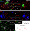
(A–D) Triple immunolabeling of spread spermatocytes with antibodies against centromeric proteins (blue), γH2AX (green), and SYCP3 (red). (A) γH2AX appears greatly concentrated over the sex chromosomes (XY). Labeling is also observed over some autosomes, where γH2AX mostly appears as small foci located over the synaptonemal complex. Two bivalents (asterisks) are enlarged in B–B′′ and C–C′′. While most γH2AX foci lie outside the centromeric regions (B–B′′), in some rare occasions γH2AX signal may overlap with the centromeric signal (C–C′′). (D) Late pachytene. γH2AX appears only on the sex chromosomes while autosomal foci are no longer detected. (E–H) Triple immunolabeling of spread spermatocytes with antibodies against H3K9Me3 (blue), RAD51 (green), and SYCP3 (red). (E) Zygotene. Some of the autosomal bivalents are still undergoing synapsis while others have completed their synapsis. In both cases, abundant RAD51 foci appear scattered along SCs. The pericentromeric regions, labeled with H3K9Me3 (which also labels the sex chromosomes and two autosomal bivalents), are mostly devoid of RAD51 foci (see enlarged detail of asterisk-marked bivalent in F–F′′). On some occasions, RAD51 foci appear close to the pericentromeric area but usually with no overlapping (see detailed bivalent in G–G′′). (H) Pachytene. Most RAD51 foci have disappeared. Some signal is still present in some autosomes and mainly on the sex chromosomes (XY). No overlap of RAD51 and pericentromeric regions was observed. (I–J′′) Triple immunolabeling of spread spermatocytes with antibodies against RAP1 (blue), MLH1 (green), and SYCP3 (red). (I) Pachytene spermatocyte in which the distribution of MLH1 foci, corresponding to the location of chiasmata, can be appreciated. Most MLH1 foci appear located outside the pericentromeric regions. However, in some cases there is overlap with the RAP1 signal (asterisk). The overlapping can be appreciated in the enlarged detail shown (J–J′′). (K) Analysis of chiasmata distribution along bivalent #10. Top left image shows an enlargement of bivalent #10 where the MLH1 focus is visible. A schematic representation of the bivalent is depicted on the top right. Below the image, intervals show the different locations considered for chiasmata. The chart shows the distribution of chiasmata in bivalent #10 analyzed in 115 spermatocytes. The frequency of chiasma occurrence within each interval of the bivalent is represented. Crossing over is rarely found near the proximal region (intervals 1 and 2) while the highest incidence takes place within the middle segment of the bivalent (intervals 4–6).
Figure 7
Figure 7
Triple immunolabeling of spermatocytes with antibodies against centromeric proteins (blue), SUN1 (green), and SYCP3 (red). (A) Pachytene spread spermatocyte in which SUN1 can be clearly seen at the ends of bivalents. Most centromeric regions do not show any SUN1 signal, but one bivalent shows a small signal at the centromere (arrow). (B–B′′) An enlarged bivalent showing a centromeric SUN1 signal (arrow). The synaptonemal complex appears stretched at this region (arrowhead). (C) Pachytene squashed spermatocyte. Five different focal planes were superimposed to give rise to a single image. The three-dimensional organization of the nucleus was preserved, and the attachment point of bivalents at the nuclear periphery can be observed. A small metacentric chromosome, similar to that shown in B–B′′, shows a three-point attachment configuration: two at telomeres (arrowheads) and one at the centromere (arrow), which presents a SUN1 signal. Note the presence of many centromeric signals, corresponding to metacentric chromosomes, located in the nuclear interior. For a complete reconstruction of the nucleus, see File S1.
Figure 8
Figure 8
Schematic representation illustrating the different patterns of chromatin configuration of distally vs. interstitially located telomeric DNA repeats during prophase I. A submetacentric autosomal bivalent from early pachytene (A–C) to late diplotene (J–L) is depicted, in which a synaptonemal complex (double red and yellow lines) and RAP1-TRF1 complexes (purple spheres) are represented. Chromatin loops of each homolog are colored in light and dark gray, respectively, while those loops containing telomeric (TTAGGG)n repeats are depicted in green. Also, chromatin loops containing both telomeric DNA repeats and H3K9Me3 histone are represented in blue. Note that RAP1–TRF1 complexes are present only in the pericentromeric region during late pachytene–early diplotene (D, E, G, and H). By contrast, RAP1–TRF1 complexes are always detected in the telomeric loops as a constitutive factor (C, F, I, and L). It is worth noting the different level of condensation of the chromatin during diplotene (G–L), in which the loops and the protein complexes appear more compacted. The same situation takes place in telocentric chromosomes.

Similar articles

Cited by

References

    1. Abuín M., Martínez P., Sánchez L., 1996. Localization of the repetitive telomeric sequence (TTAGGG)n in four salmonid species. Genome 39: 1035–1038. - PubMed
    1. Adelfalk C., Janschek J., Revenkova E., Blei C., Liebe B., et al. , 2009. Cohesin SMC1beta protects telomeres in meiocytes. J. Cell Biol. 187: 185–199. - PMC - PubMed
    1. Ahmed E. A., van der Vaart A., Barten A., Kal H. B., Chen J., et al. , 2007. Differences in DNA double strand breaks repair in male germ cell types: lessons learned from a differential expression of Mdc1 and 53BP1. DNA Repair (Amst.) 6: 1243–1254. - PubMed
    1. Ahmed E. A., Philippens M. E., Kal H. B., de Rooij D. G., de Boer P., 2010. Genetic probing of homologous recombination and non-homologous end joining during meiotic prophase in irradiated mouse spermatocytes. Mutat. Res. 688: 12–18. - PubMed
    1. Ahmed E. A., Sfeir A., Takai H., Scherthan H., 2013. Ku70 and non-homologous end joining protect testicular cells from DNA damage. J. Cell Sci. 126: 3095–3104. - PMC - PubMed

Publication types