. 2014 Jun 6;2(6):e12036.
doi: 10.14814/phy2.12036. Print 2014 Jun 1.

Hindlimb venous distention evokes a pressor reflex in decerebrated rats

Affiliations

Hindlimb venous distention evokes a pressor reflex in decerebrated rats

Katsuya Yamauchi et al. Physiol Rep. .

Abstract

The distention of small vessels caused by an increase in blood flow to dynamically exercising muscles has been proposed as a stimulus that activates the thin fiber (groups III and IV) afferents evoking the exercise pressor reflex. This theory has been supported by evidence obtained from both humans and animals. In decerebrated unanesthetized rats with either freely perfused femoral arteries or arteries that were ligated 3 days before the experiment, we attempted to provide evidence in support of this theory by measuring arterial pressure, heart rate, and renal sympathetic nerve discharge while retrogradely injecting Ringer's solution in increasing volumes into the femoral vein just as it excited the triceps surae muscles. We found that the pressor response to injection was directly proportional to the volume injected. Retrograde injection of volumes up to and including 1 mL had no significant effect on either heart rate or renal sympathetic nerve activity. Cyclooxygenase blockade with indomethacin attenuated the reflex pressor response to retrograde injection in both groups of rats. In contrast, gadolinium, which blocks mechanogated channels, attenuated the reflex pressor response to retrograde injection in the "ligated rats," but had no effect on the response in "freely perfused" rats. Our findings are consistent with the possibility that distension of small vessels within exercising skeletal muscle can serve as a stimulus to the thin fiber afferents evoking the exercise pressor reflex.

Keywords: Autonomic nervous system; dynamic exercise; peripheral artery disease; thin fiber muscle afferents.


Figures

Figure 1.
Figure 1.
Effects of retrograde injection of five volumes of Ringer's solution (0.1 mL, 0.25 mL, 0.5 mL, 0.75 mL, and 1.0 mL) into the femoral vein on arterial blood pressure and heart rate in eight “sham” (i.e., freely perfused) rats and eight “ligated” rats. Filled bars represent baseline values and open bars represent peak values evoked by injection (mean ± standard errors). Abbreviations: MAP, mean arterial pressure. HR, heart rate. Number signs (#) depict significant difference (P < 0.05) between corresponding baseline and peak values before and after retrograde injection of Ringer's solution into the femoral vein. Asterisks (*) depict significant differences (P < 0.05) between the pressor responses to retrograde injection in “ligated” rats and the pressor responses to retrograde injection in “sham” rats for a given volume.
Figure 2.
Figure 2.
Individual records of the effect of retrograde injection of 1.0 mL of Ringer's solution into the femoral vein on renal sympathetic nerve activity (RSNA), heart rate (HR), and arterial blood pressure (ABP). Horizontal bar depicts the injection period. Note that the pressor response to injection is greater in the “ligated” rat (B) than it is in the “freely perfused” rat (A).
Figure 3.
Figure 3.
Effects of retrograde injection of five volumes of Ringer's solution (0.1 mL, 0.25 mL, 0.5 mL, 0.75 mL, and 1.0 mL) into the femoral vein on renal sympathetic nerve activity (RSNA) in eight “freely perfused” and “eight ligated” rats. Bars indicate changes from baseline values and represent means ± standard errors.
Figure 4.
Figure 4.
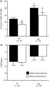
The pressor and heart rate responses to retrograde injection of Ringer's solution (1.0 mL) into the femoral vein before (filled bars) and after femoral vein injection of indomethacin (open bars) in 10 “freely perfused rats” (FP) and in 10 “ligated” (Lig) rats. Number sign (#) represents a significant difference (P < 0.05) between the pressor response before and after indomethacin. Asterisk (*) represents significant difference (P < 0.05) in the pressor response to injection between “freely perfused” and “ligated” rats.
Figure 5.
Figure 5.
The pressor and heart rate responses to retrograde injection of Ringer's solution (1.0 mL) into the femoral vein before (filled bars) and after femoral vein injection of Gadolinium (open bars) in 10 “freely perfused” (FP) rats and in eight “ligated” (Lig) rats. Number sign (#) represents a significant difference (P < 0.05) between the pressor response before and after Gadolinium. Asterisk (*) represents significant difference (P < 0.05) in the pressor response to injection between “freely perfused” and “ligated” rats.
Figure 6.
Figure 6.
The pressor and heart rate responses to retrograde injection of Ringer's solution (1.0 mL) into the femoral vein before (filled bars) and after femoral vein injection of Lidocaine (open bars) in five “freely perfused” (FP) rats and in six “ligated” (Lig) rats. Number sign (#) represents a significant difference (P < 0.05) between the pressor response before and after Lidocaine. Asterisk (*) represents significant difference (P < 0.05) in the pressor response to injection between “freely perfused” and “ligated” rats.
Figure 7.
Figure 7.
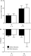
The pressor and heart rate responses to retrograde injection of Ringer's solution (1.0 mL) into the femoral vein before (filled bars) and after jugular vein injection of Lidocaine (open bars) in four “freely perfused” (FP) rats and in four “ligated” (Lig) rats. Note that lidocaine injected into the jugular vein had no effect on the pressor responses to retrograde injection of Ringer's solution in either the FP or the Lig rats.
Figure 8.
Figure 8.
The effects of sectioning either the L2–S3 dorsal roots (A and B) or sectioning the L2–S3 dorsal and ventral roots (C and D) on the pressor responses to retrograde injection of four volumes of Ringer's solution into the femoral vein. Filled bars represent pressor responses before cutting and open bars represent pressor responses after cutting. A and C show data obtained from six “freely perfused” rats and B and D show data obtained from eight (B) and six (D) “ligated” rats. Note that rats having their dorsal and ventral roots sectioned were different than those having only their dorsal roots cut. Number signs (#) represent significant difference (P < 0.05) between the pressor response to retrograde injection before and after cutting the roots.

References

    1. Adreani C. M., Kaufman M. P. 1998. Effect of arterial occlusion on responses of group III and IV afferents to dynamic exercise. J. Appl. Physiol.; 84:1827-1833. - PubMed
    1. Adreani C. M., Hill J. M., Kaufman M. P. 1997. Responses of group III and IV muscle afferents to dynamic exercise. J. Appl. Physiol.; 82:1811-1817. - PubMed
    1. Amann M., Blain G. M., Proctor L. T., Sebranek J. J., Pegelow D. F., Dempsey J. A. 2010. Group III and IV muscle afferents contribute to ventilatory and cardiovascular response to rhythmic exercise in humans. J. Appl. Physiol.; 109:966-976. - PMC - PubMed
    1. Amann M., Runnels S., Morgan D. E., Trinity J. D., Fjeldstad A. S., Wray D. W. 2011. On the contribution of group III and IV muscle afferents to the circulatory response to rhythmic exercise in humans. J. Physiol.; 589:3855-3866. - PMC - PubMed
    1. Andrews C. J., Andrews W. H., Orbach J. 1972. A sympathetic reflex elicited by distension of the mesenteric venous bed. J. Physiol.; 226:119-131. - PMC - PubMed

LinkOut - more resources