. 2014 Jun;26(6):2524-2537.
doi: 10.1105/tpc.114.123349. Epub 2014 Jun 6.

An Uncharacterized Apocarotenoid-Derived Signal Generated in ζ-Carotene Desaturase Mutants Regulates Leaf Development and the Expression of Chloroplast and Nuclear Genes in Arabidopsis

Affiliations

An Uncharacterized Apocarotenoid-Derived Signal Generated in ζ-Carotene Desaturase Mutants Regulates Leaf Development and the Expression of Chloroplast and Nuclear Genes in Arabidopsis

Aida-Odette Avendaño-Vázquez et al. Plant Cell. 2014 Jun.

Abstract

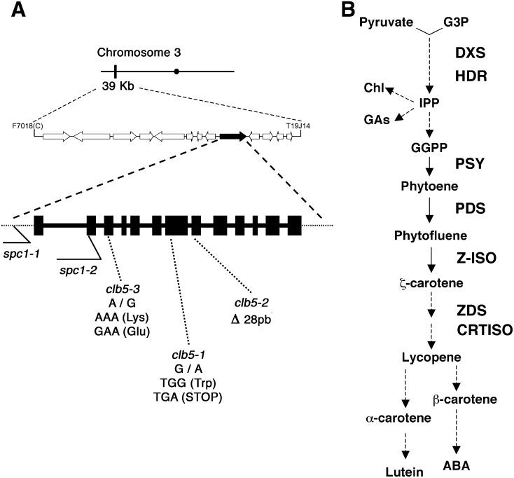
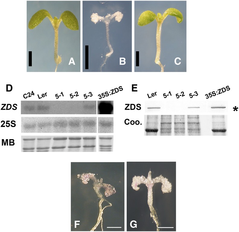
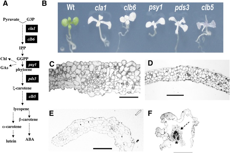
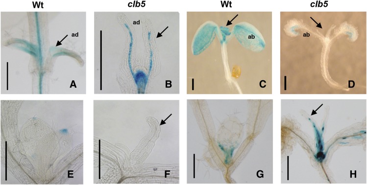
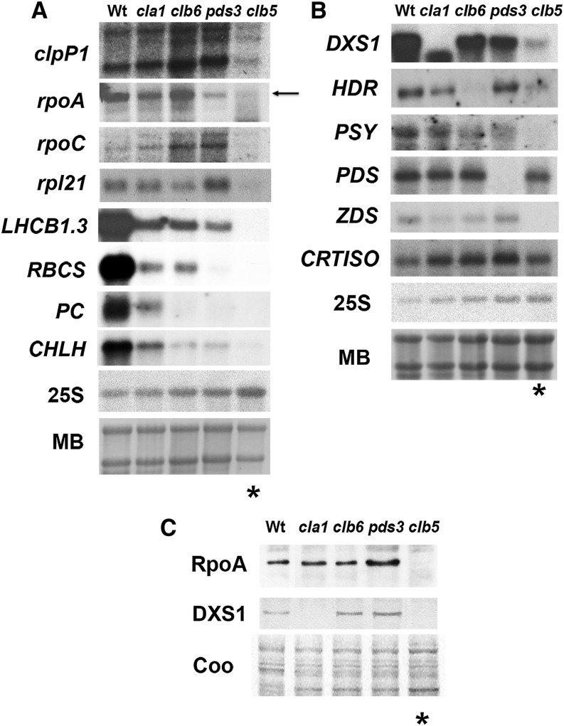
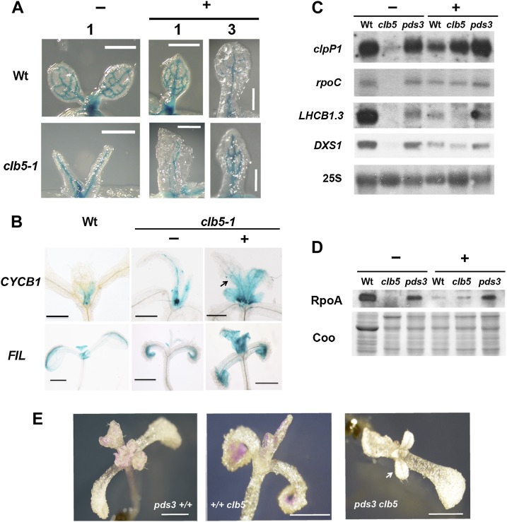
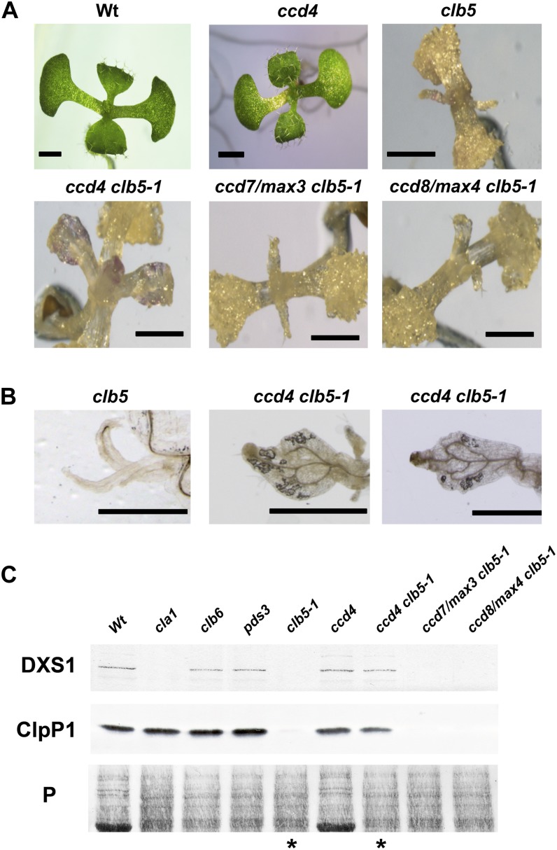
In addition to acting as photoprotective compounds, carotenoids also serve as precursors in the biosynthesis of several phytohormones and proposed regulatory signals. Here, we report a signaling process derived from carotenoids that regulates early chloroplast and leaf development. Biosynthesis of the signal depends on ζ-carotene desaturase activity encoded by the ζ-CAROTENE DESATURASE (ZDS)/CHLOROPLAST BIOGENESIS5 (CLB5) gene in Arabidopsis thaliana. Unlike other carotenoid-deficient plants, zds/clb5 mutant alleles display profound alterations in leaf morphology and cellular differentiation as well as altered expression of many plastid- and nucleus-encoded genes. The leaf developmental phenotypes and gene expression alterations of zds/clb5/spc1/pde181 plants are rescued by inhibitors or mutations of phytoene desaturase, demonstrating that phytofluene and/or ζ-carotene are substrates for an unidentified signaling molecule. Our work further demonstrates that this signal is an apocarotenoid whose synthesis requires the activity of the carotenoid cleavage dioxygenase CCD4.


Figures

Figure 1.
Figure 1.
Mapping of the clb5-1 Mutation. (A) Representation of the chromosome (black bar) and ZDS gene location (black arrow). The structure of the ZDS gene is shown with 13 exons (black boxes) and introns (line). The locations of the mutations for each clb5 and the previously reported spc1 alleles are indicated. (B) Simplified diagram of the carotenoid biosynthetic pathway. Chl, chlorophyll; GAs, gibberellins; GGPP, geranylgeranyl diphosphate; G3P, glyceraldehyde-3-phosphate; HDR, 1-hydroxy-2-methyl-2-(E)-butenyl 4-diphosphate reductase; IPP, isopentenyl pyrophosphate; Z-ISO, 15-cis-ζ-carotene isomerase.
Figure 2.
Figure 2.
Molecular Complementation of clb5 and Characteristics of the clb5 Alleles. (A) to (C) Phenotypes of a representative 8-d-old wild-type Ler seedling (A), a 16-d-old clb5-1 mutant (B), and an 8-d-old T2 transgenic clb5-1 homozygous complemented line (C). Bars = 1 mm. (D) RNA gel blot analysis of ZDS transcript from 8-d-old wild-type (C24 and Ler ecotypes) and complemented mutant (35S:ZDS) plants and from 15-d-old clb5 seedlings. Fifteen micrograms of total RNA was loaded in each lane. Hybridization of the same membrane with the 25S rRNA and the methylene blue (MB)–stained gels are shown as loading controls. The RNA gel blots are representative of two independent biological experiments. (E) Expression of the ZDS protein in the clb5 alleles. ZDS protein was immunodetected in protein extracts from 8-d-old wild-type (Ler) and complemented transgenic (35S:ZDS) plants or from 15-d-old clb5 alleles. Ten micrograms of Ler, 5 μg of 35S:ZDS, and 15 μg of the clb5 mutant total protein extracts were run in each lane. A Coomassie blue (Coo)–stained gel run in parallel is shown as a loading control. The asterisk marks the ZDS protein. The protein blot is representative of three independent biological experiments. (F) and (G) Phenotypes of 10-d-old clb5-1 (F) and clb5-2 (G) alleles. Bars = 0.5 mm. [See online article for color version of this figure.]
Figure 3.
Figure 3.
Morphological Analyses of the clb5 Mutant. (A) Simplified diagram of the carotenoid pathway as described in Figure 1. The black boxes indicate the steps of the pathway altered in mutants used in this study. (B) Morphology of albino mutants that affect carotenoid biosynthesis. Plants were grown in a 16/8-h light/dark period for 8 d for the wild type and 15 d for the mutants. (C) to (F) Light micrographs of transverse sections of wild-type (C), clb6 (D), pds3 (E), and clb5-1 (F) seedling leaves. In (F), vascular tissue is marked with an asterisk and mesophyll tissue is marked with a dashed arrow. Plants were grown on MS medium for 21 d, and the second leaf of a representative seedling for each phenotype was fixed for light microscopic analysis. Bars = 100 μm.
Figure 4.
Figure 4.
Expression Pattern of Leaf Developmental Markers in the clb5-1 Mutant. GUS expression pattern is shown for the gPHB-GUS adaxial marker in 7-d-old wild-type (A) and 10-d-old clb5-1 (B) plants, the pFIL:GUS ventral marker in 7-d-old wild-type (C) and 10-d-old clb5-1 (D) plants, the DR5:GUS synthetic auxin reporter in wild-type (E) and 14-d-old clb5-1 (F) plants, and pCYCB1:D-box-GUS in 6-d-old wild-type (G) and 14-d-old clb5-1 (H) plants. Arrows indicate primary leaves. ab, abaxial; ad, adaxial. Bars = 0.5 mm.
Figure 5.
Figure 5.
Expression of Nucleus- and Chloroplast-Encoded Transcripts and Proteins. (A) Expression of chloroplast marker genes: clpP1, rpoA, rpoC, rpl21, LHCB1.3, RBCS, PC, and CHLH. (B) Nucleus-encoded genes involved in the carotenoid biosynthetic pathway: DXS1, HDR, PSY, PDS, ZDS, and CRTISO probes. Each lane contains 15 μg of total RNA from 8-d-old wild-type and 15-d-old mutant seedlings. The hybridization of the 25S rRNA and methylene blue (MB)–stained gels are shown as loading controls. These data are representative of two independent biological experiments. (C) Analysis of the protein levels of RpoA and nucleus-encoded DXS1. Total protein extracts were isolated from 8-d-old wild-type (Ler) and 15-d-old cla1-1, clb6, pds3, and clb5-1 mutant seedlings. Immunoblotting was performed with specific antibodies against the RpoA and DXS1 proteins. Each lane contains 15 μg of total protein extract. A Coomassie blue–stained gel run in parallel with the same samples is shown as a loading control (Coo). Asterisks mark the clb5 mutant lanes. The protein blots shown are representative from three independent biological replicates.
Figure 6.
Figure 6.
Inhibition of PDS Improves Leaf Development and Gene Expression of the clb5-1 Mutant. (A) Morphology of the first (1) or third (3) leaf in 8-d-old wild-type or 15-d-old clb5 seedlings treated with (+) or without (−) fluridone. The GUS expression pattern corresponds to the pREV:GUS vascular marker. (B) GUS expression pattern of the pCYCB1:D-box-GUS and pFIL:GUS markers of 8-d-old wild-type and 15-d-old clb5-1 seedlings treated with (+) or without (−) fluridone. (C) Transcript levels of clpP1, rpoC, LHCB1.3, and DXS1 genes. Each lane contains 5 μg of total RNA. The 25S rRNA hybridization is shown as a loading control. (D) RpoA protein levels of Ler wild-type, clb5-1, and pds3 mutant seedlings treated with (+) or without (−) fluridone. Each lane contains 10 μg of total protein extract from the same developmental age as in (C). The blot was developed using RpoA antibody. A Coomassie blue–stained gel run in parallel with the same samples is shown as a loading control (Coo). The gels shown are representative from three independent biological replicates. (E) Seedling morphology of 20-d-old single pds3 (pds3+/+) and clb5-1 (+/+clb5) mutants and double pds3 clb5 mutants. Bars = 1 mm.
Figure 7.
Figure 7.
Role of CCDs in the Morphological and Expression Defects of the clb5-1 Mutant. (A) Morphology of seedlings from 8-d-old wild-type and ccd4 mutant seedlings or 16-d-old clb5-1, ccd4 clb5-1, ccd7/max3 clb5-1, and ccd8/max4 clb5-1 mutant seedlings. Bars = 1 mm. (B) Close-ups of the primary leaf of 16-d-old clb5 and two representative ccd4 clb5-1 leaves. Bars = 1 mm. (C) Analysis of the protein levels of the nucleus-encoded DXS1 and the chloroplast-encoded ClpP1. Total protein extracts were isolated from 8-d-old wild-type (Ler) and ccd4 mutant seedlings or from 15-d-old cla1-1, clb6, pds3, clb5-1, ccd4 clb5-1, ccd7/max3 clb5-1, and ccd8/max4 clb5-1 mutant seedlings. Immunoblotting was performed with polyclonal antibodies against the DXS1 and ClpP proteins as described in Methods. Each lane contains 15 μg of total protein extract. The Ponceau-stained membrane is shown as a loading control (P). Asterisks mark the lanes corresponding to clb5-1 and ccd4 clb5-1 mutants. The protein gel shown is representative from three independent biological replicates. [See online article for color version of this figure.]

References

    1. Ahrazem O., Trapero A., Gómez M.D., Rubio-Moraga A., Gómez-Gómez L. (2010). Genomic analysis and gene structure of the plant carotenoid dioxygenase 4 family: A deeper study in Crocus sativus and its allies. Genomics 96: 239–250. - PubMed
    1. Alder A., Jamil M., Marzorati M., Bruno M., Vermathen M., Bigler P., Ghisla S., Bouwmeester H., Beyer P., Al-Babili S. (2012). The path from β-carotene to carlactone, a strigolactone-like plant hormone. Science 335: 1348–1351. - PubMed
    1. Auldridge M.E., Block A., Vogel J.T., Dabney-Smith C., Mila I., Bouzayen M., Magallanes-Lundback M., DellaPenna D., McCarty D.R., Klee H.J. (2006a). Characterization of three members of the Arabidopsis carotenoid cleavage dioxygenase family demonstrates the divergent roles of this multifunctional enzyme family. Plant J. 45: 982–993. - PubMed
    1. Auldridge M.E., McCarty D.R., Klee H.J. (2006b). Plant carotenoid cleavage oxygenases and their apocarotenoid products. Curr. Opin. Plant Biol. 9: 315–321. - PubMed
    1. Barkan A., Goldschmidt-Clermont M. (2000). Participation of nuclear genes in chloroplast gene expression. Biochimie 82: 559–572. - PubMed