. 2014 Sep:101:68-75.
doi: 10.1016/j.pep.2014.05.011. Epub 2014 Jun 5.

Expression, purification and reconstitution of the 4-hydroxybenzoate transporter PcaK from Acinetobacter sp. ADP1

Affiliations

Expression, purification and reconstitution of the 4-hydroxybenzoate transporter PcaK from Acinetobacter sp. ADP1

Christian Pernstich et al. Protein Expr Purif. 2014 Sep.

Abstract

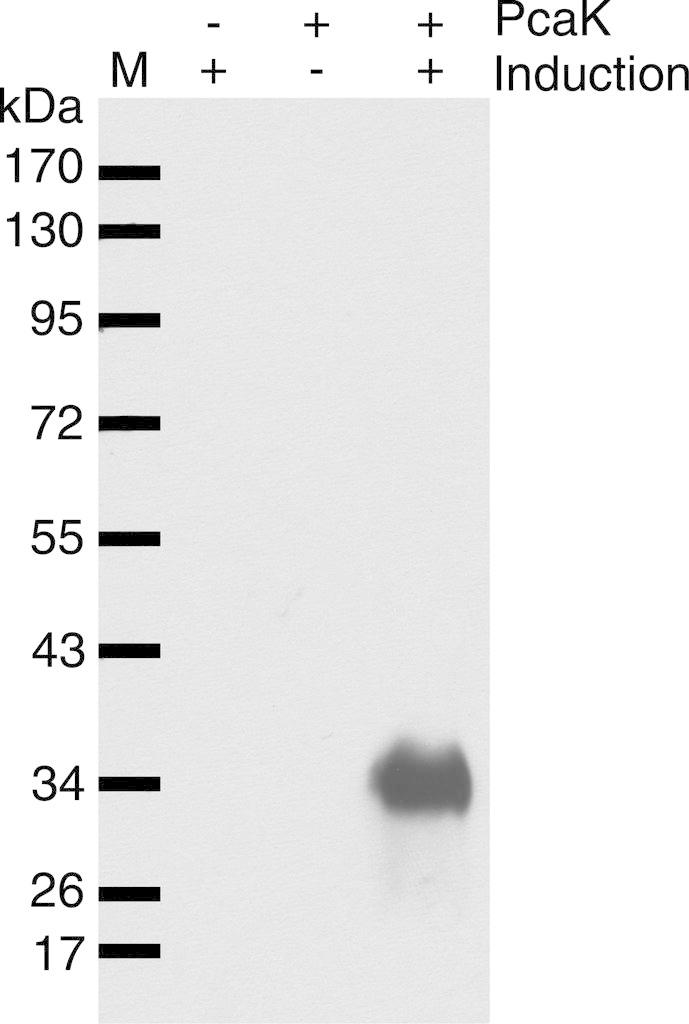
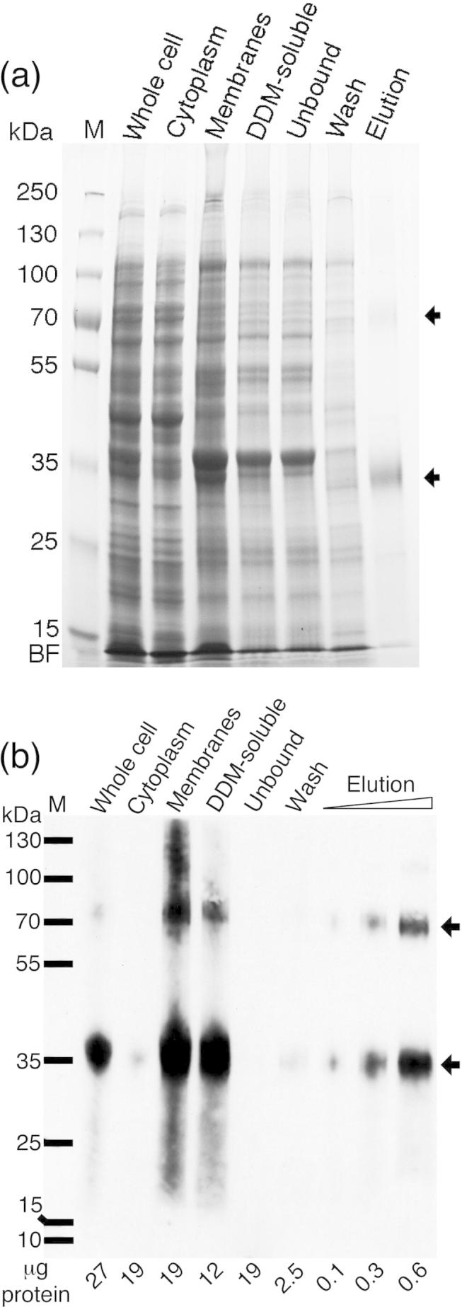
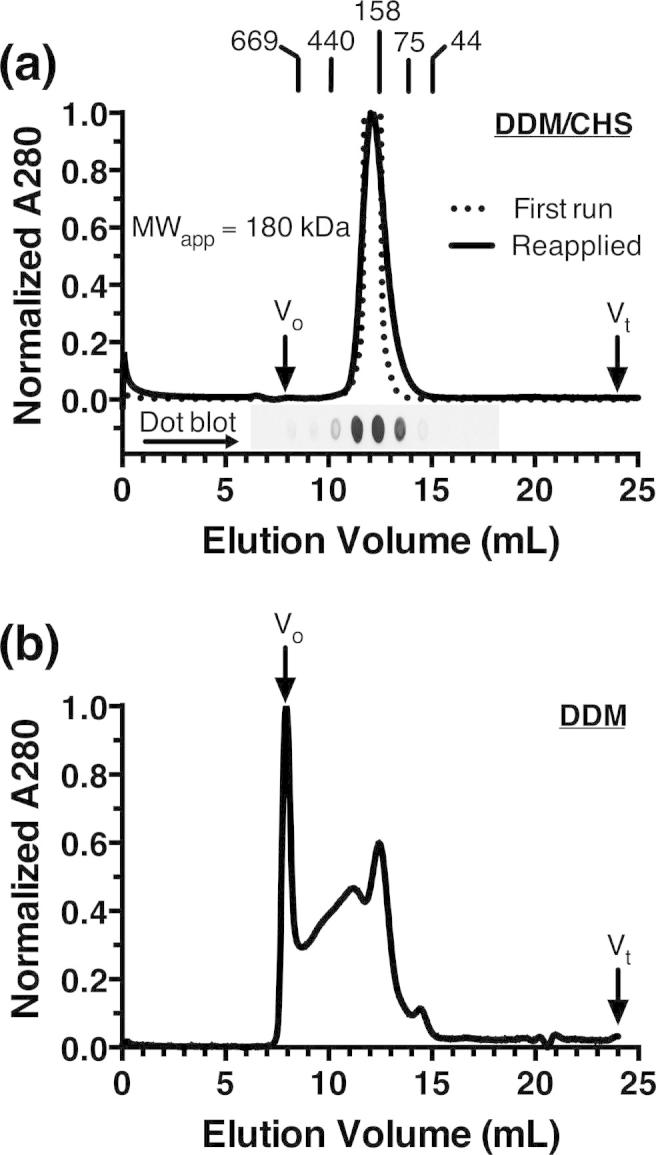
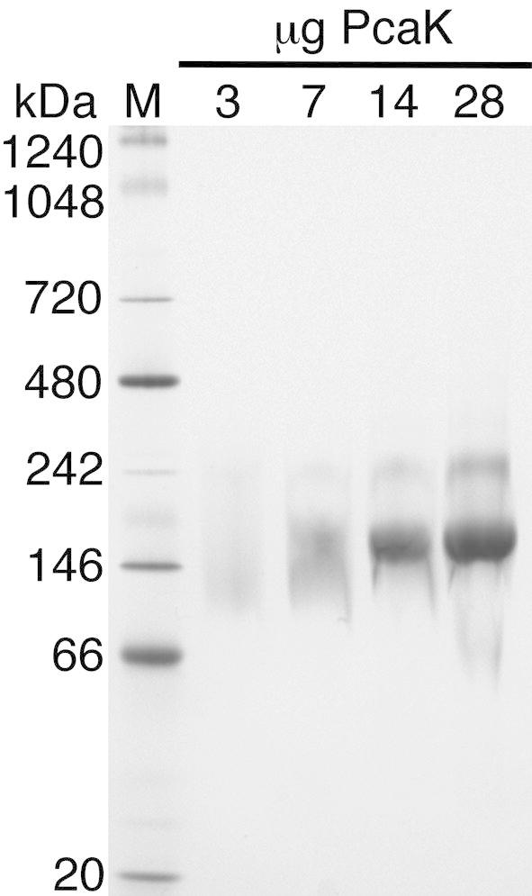
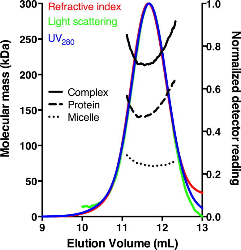
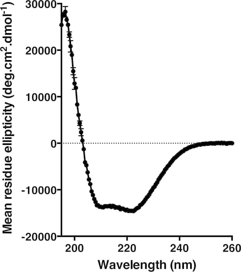
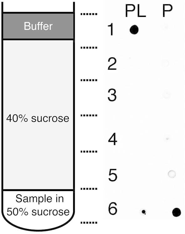
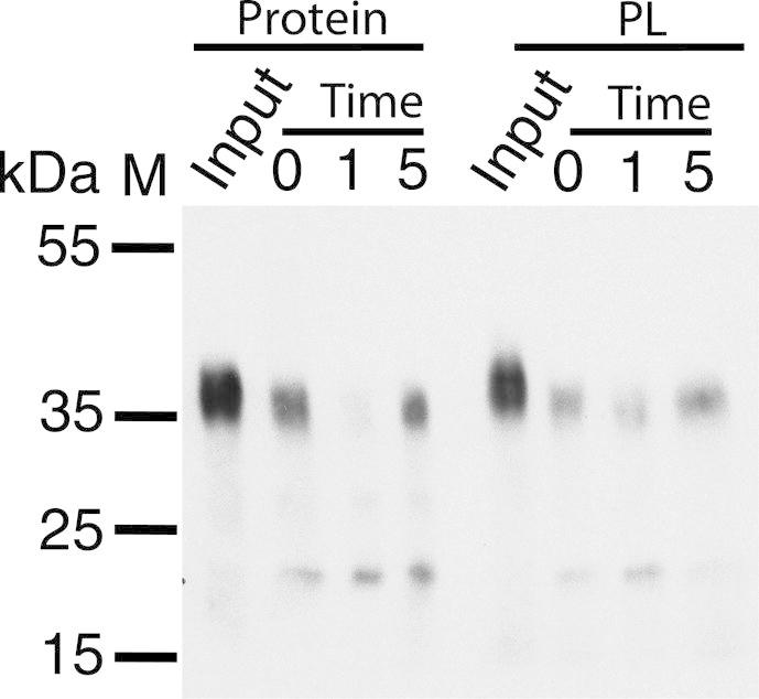
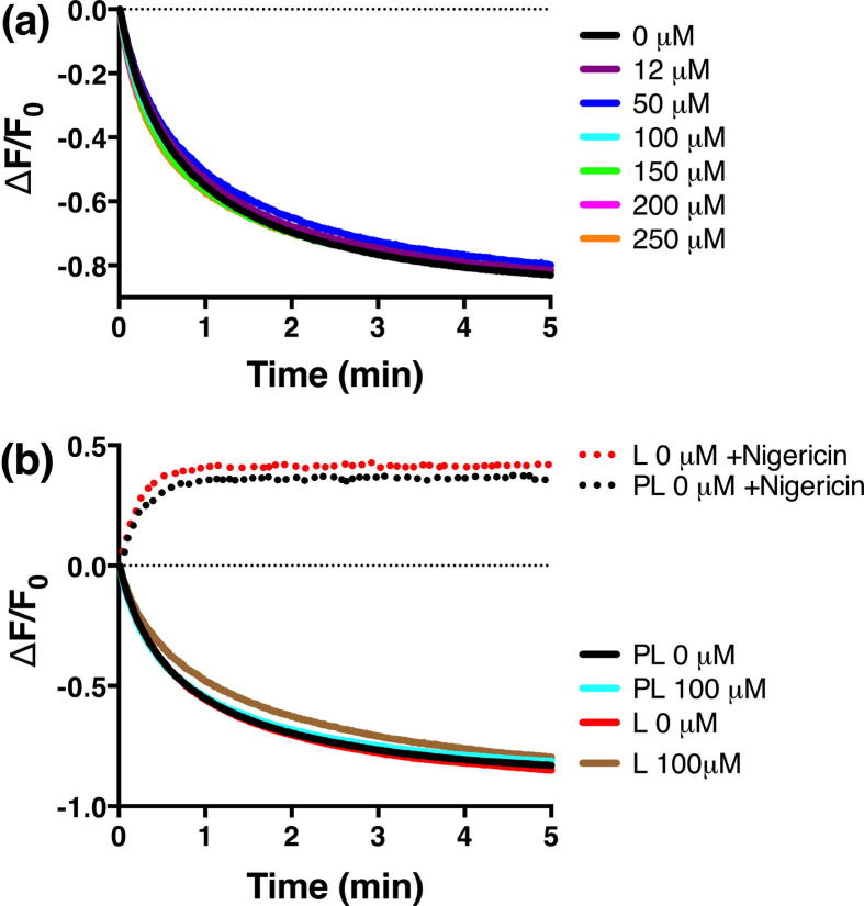
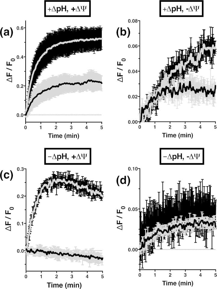
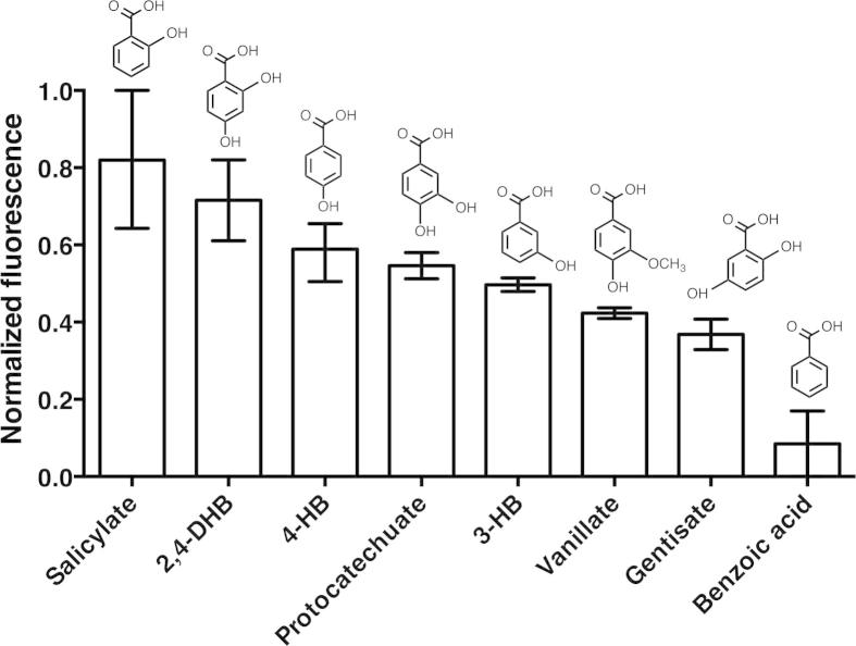
The aromatic acid:H(+) symporter family of integral membrane proteins play an important role in the microbial metabolism of aromatic compounds. Here, we show that the 4-hydroxybenzoate transporter from Acinetobacter sp. ADP1, PcaK, can be successfully overexpressed in Escherichia coli and purified by affinity chromatography. Affinity-purified PcaK is a stable, monodisperse homotrimer in the detergent n-dodecyl-β-d-maltopyranoside supplemented with cholesteryl hemisuccinate. The purified protein has α-helical secondary structure and can be reconstituted to a functional state in synthetic proteoliposomes. Asymmetric substrate transport was observed when proteoliposomes were energized by applying an electrochemical proton gradient (Δμ‾H(+)) or a membrane potential (ΔΨ) but not by ΔpH alone. PcaK was selective in transporting 4-hydroxybenzoate and 3,4-dihydroxybenzoate over closely related compounds, confirming previous reports on substrate specificity. However, PcaK also showed an unexpected preference for transporting 2-hydroxybenzoates. These results provide the basis for further detailed studies of the structure and function of this family of transporters.

Keywords: Enzyme purification; Membrane proteins; Membrane transport; Recombinant protein expression; Reconstitution of membrane transporters.


Figures

Fig. 1
Fig. 1
Small-scale expression analysis of recombinant PcaK in Escherichia coli. SDS–PAGE gels were Western blotted and probed with a V5 antibody directed toward an artificial C-terminal V5 epitope. M, molecular weight marker in kDa as shown. 10 μg total protein was loaded in each lane.
Fig. 2
Fig. 2
Purification of PcaK. (a) Cell fractions and column purification fractions were loaded onto SDS–PAGE gels and stained with Coomassie. (b) Equivalent gel subject to Western blotting with anti-V5. Purified PcaK migrated as two bands corresponding to the apparent molecular weights of PcaK monomer (32 kDa) and dimer (70 kDa). Fractions are: Whole cell, total cell lysate; Cytoplasm, supernatant after ultracentrifugation; Membranes, pellet after ultracentrifugation; DDM-soluble, membranes solubilized in detergent DDM, supernatant after ultracentrifugation; Unbound, IMAC column flow-through; Wash, column wash fraction; Elution, column eluent. M, molecular weight marker in kDa as shown. μg protein, μg total protein loaded per lane. BF, buffer front.
Fig. 3
Fig. 3
Size-exclusion chromatography of purified PcaK. (a) In DDM/CHS mixed micelles a single well-resolved peak is observed at an apparent molecular weight of 180 kDa relative to known protein standards as shown. Immunoblot of column fractions (‘dot blot’) confirms that the peak corresponds to PcaK. (b) In contrast, chromatograms collected under conditions of DDM alone, without CHS added to purification buffers or the size exclusion buffer, show substantial heterogeneity.
Fig. 4
Fig. 4
Blue native PAGE gel of PcaK after size exclusion chromatography. Total weight of loaded protein in each lane is shown. When sufficient protein is loaded a single major band is observed at approximately 180 kDa, in agreement with size-exclusion chromatograms. M, molecular weight marker in kDa.
Fig. 5
Fig. 5
Molecular mass determination from size-exclusion chromatography with in-line multi-angle static light scattering (SEC-MALS). Normalized outputs from refractive index, light scattering and absorbance detectors are overlaid. Black lines show the molecular mass calculations across the peak for the protein-detergent complex (solid, 226 ± 2 kDa), protein component (dashed, 152 ± 2 kDa) and micelle component (dotted, 74 ± 2 kDa).
Fig. 6
Fig. 6
Circular dichroism spectrum of PcaK displaying a classical alpha-helical signature.
Fig. 7
Fig. 7
Sucrose flotation assay demonstrating reconstitution of PcaK. Fractions were removed from a discontinuous sucrose gradient as indicated and the presence of PcaK determined by dot blot with anti-V5. Proteoliposomes (PL) migrate to the buffer interface under ultracentrifugation while unreconstituted protein remains at the bottom of the gradient, confirmed by a protein-only control (P).
Fig. 8
Fig. 8
Susceptibility of the C-terminal V5 tag to proteolysis as a measure of protein orientation. Western blotting shows that Proteinase K immediately digests the C-terminal V5 epitope of PcaK in detergent micelles (Protein) and after reconstitution into proteoliposomes (PL), suggesting that the C-terminal of the reconstituted protein is exposed at the proteoliposome exterior. Time in minutes.
Fig. 9
Fig. 9
Influx assays under applied (ΔμH+) (inside negative and alkaline). (a) Influx assays in proteoliposomes at different concentrations of 4-HB as shown. (b) Comparison of data from proteoliposomes (PL) to control experiments in the absence of protein (L). Traces are the mean of at least two replicates with error bars omitted for clarity.
Fig. 10
Fig. 10
Substrate efflux (inside positive and acidic). Proteoliposomes, grey smoothed line on black error bars; liposome controls, black smoothed line on grey error bars. Experiments were performed in the presence of (a) (ΔμH+), (b) ΔpH only, (c) ΔΨ only or (d) in the absence of any energetic gradient. All traces are mean ± range of at least two replicates after subtracting relevant control experiments absent substrate.
Fig. 11
Fig. 11
Substrate specificity of reconstituted PcaK under outward-directed ΔΨ. Data are mean ± range of at least two independent experiments with different protein and proteoliposome preparations, relative to a control absent substrate.

References

    1. Gibson D.T., Parales R.E. Aromatic hydrocarbon dioxygenases in environmental biotechnology. Curr. Opin. Biotechnol. 2000;11:236–243. - PubMed
    1. Cao B., Nagarajan K., Loh K.-C. Biodegradation of aromatic compounds: current status and opportunities for biomolecular approaches. Appl. Microbiol. Biotechnol. 2009;85:207–228. - PubMed
    1. Fuchs G., Boll M., Heider J. Microbial degradation of aromatic compounds – from one strategy to four. Nat. Rev. Microbiol. 2011;9:803–816. - PubMed
    1. Kirk T.K., Farrell R.L. Enzymatic “combustion”: the microbial degradation of lignin. Annu. Rev. Microbiol. 1987;41:465–505. - PubMed
    1. Bugg T.D.H., Ahmad M., Hardiman E.M., Rahmanpour R. Pathways for degradation of lignin in bacteria and fungi. Nat. Prod. Rep. 2011;28:1883–1896. - PubMed

Publication types

MeSH terms