. 2014 Sep;357(3):645-65.
doi: 10.1007/s00441-014-1906-9. Epub 2014 Jun 8.

The cell-cell junctions of mammalian testes: I. The adhering junctions of the seminiferous epithelium represent special differentiation structures

Affiliations

The cell-cell junctions of mammalian testes: I. The adhering junctions of the seminiferous epithelium represent special differentiation structures

Lisa M Domke et al. Cell Tissue Res. 2014 Sep.

Abstract

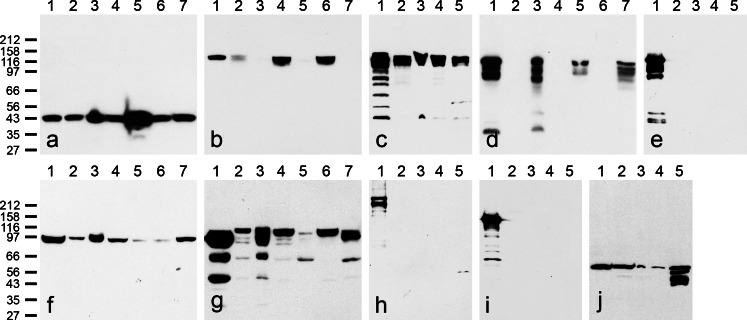
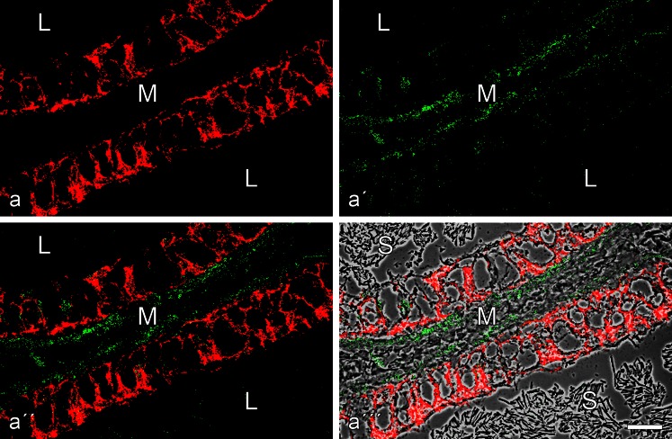
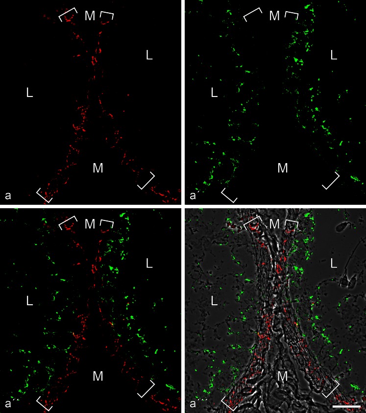
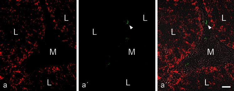
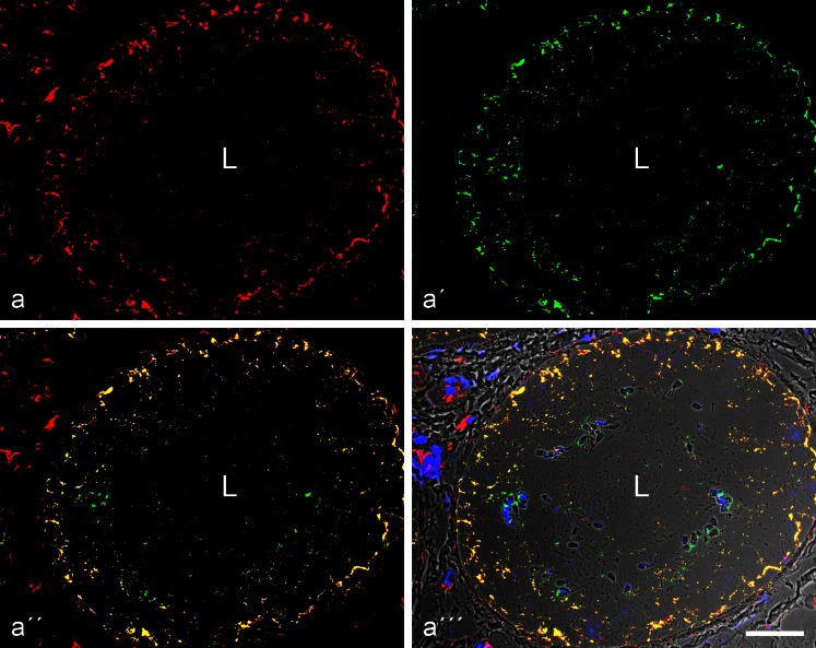
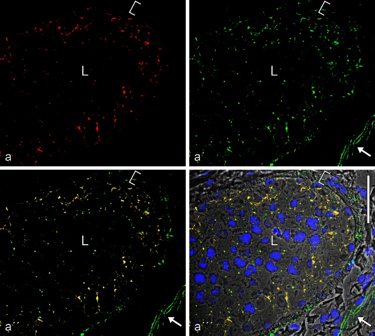
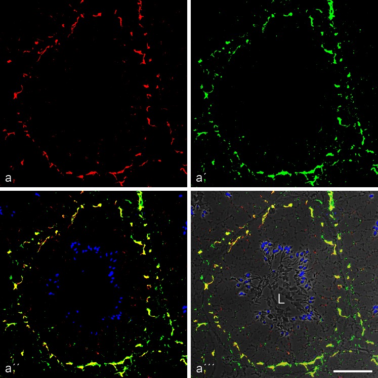
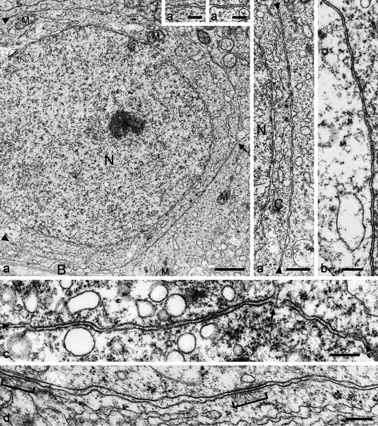
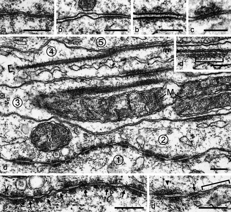
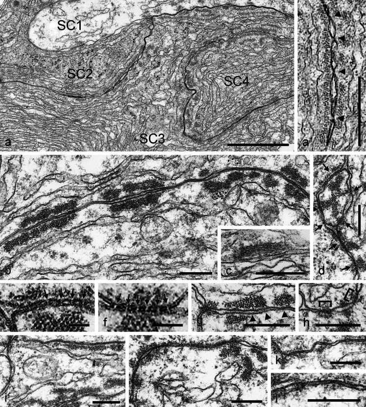
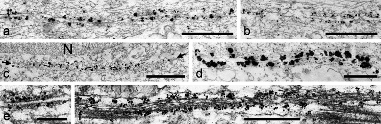
The seminiferous tubules and the excurrent ducts of the mammalian testis are physiologically separated from the mesenchymal tissues and the blood and lymph system by a special structural barrier to paracellular translocations of molecules and particles: the "blood-testis barrier", formed by junctions connecting Sertoli cells with each other and with spermatogonial cells. In combined biochemical as well as light and electron microscopical studies we systematically determine the molecules located in the adhering junctions of adult mammalian (human, bovine, porcine, murine, i.e., rat and mouse) testis. We show that the seminiferous epithelium does not contain desmosomes, or "desmosome-like" junctions, nor any of the desmosome-specific marker molecules and that the adhering junctions of tubules and ductules are fundamentally different. While the ductules contain classical epithelial cell layers with E-cadherin-based adherens junctions (AJs) and typical desmosomes, the Sertoli cells of the tubules lack desmosomes and "desmosome-like" junctions but are connected by morphologically different forms of AJs. These junctions are based on N-cadherin anchored in cytoplasmic plaques, which in some subforms appear thick and dense but in other subforms contain only scarce and loosely arranged plaque structures formed by α- and β-catenin, proteins p120, p0071 and plakoglobin, together with a member of the striatin family and also, in rodents, the proteins ZO-1 and myozap. These N-cadherin-based AJs also include two novel types of junctions: the "areae adhaerentes", i.e., variously-sized, often very large cell-cell contacts and small sieve-plate-like AJs perforated by cytoplasm-to-cytoplasm channels of 5-7 nm internal diameter ("cribelliform junctions"). We emphasize the unique character of this epithelium that totally lacks major epithelial marker molecules and structures such as keratin filaments and desmosomal elements as well as EpCAM- and PERP-containing junctions. We also discuss the nature, development and possible functions of these junctions.


Figures

Fig. 1
Fig. 1
Identification of proteins and glycoproteins of cell-cell adhering junctions in dissected tissue parts of mammalian testis, enriched in seminiferous tubules, or excurrent duct tissues, demonstrated after SDS-PAGE of total protein lysates by immunoblotting with specific antibodies a Antibodies against β-actin identify a single polypeptide band at 43 kDa in cultured human HaCaT keratinocytes (lane 1 in all blots shown, i.e. aj), bovine testis (lane 2) and epididymis (lane 3), boar testis (lane 4) and boar excurrent duct-containing tissue (lane 5), rat testis (lane 6) and rat excurrent duct tissue (lane 7). b Antibodies against N-cadherin recognize this glycoprotein in testicular tissue of bull (lane 2), boar (lane 4) and rat (lane 6) but not to a significant extent in excurrent duct tissues of bovine (lane 3), porcine (lane 5) and murine (rat, lane 7) origin. c As in (b), N-cadherin is detected to comparable intensities in near-equal amounts of testicular tissue of bovine (lane 2), rat (lane 3), mouse (lane 4) and human (lane 5) origin. d In a mutually exclusive way, E-cadherin is recognized in the excurrent duct tissues of bovine (lane 3), porcine (lane 5) and murine (rat, lane 7) origin but not in the corresponding testicular tissues enriched in seminiferous tubules (material in lanes 2, 4 and 6 of bovine, porcine, and murine origin). e E-cadherin is not detected in tissue material from microdissected testicular tissues enriched in seminiferous tubules of bovine (lane 2), rat (lane 3), mouse (lane 4) and human (lane 5) origin. f The major plaque protein of AJs, β-catenin, is detected in all these tissues (lanes 27 as in a, b and d), although in different intensities (note the weak reaction in lanes 5 and 6). g Similarly, the AJ plaque protein p120 is found in all samples, although at a rather low intensity in boar excurrent duct tissues (lane 5; note, however, a major proteolytic degradation product of about 62 kDa in lanes 3, 5 and 7). h In comparison with human HaCaT keratinocytes (lane 1), desmoplakin is not recognized in bovine (lane 2), rat (lane 3), mouse (lane 4,) and human (lane 5) seminiferous tubules of testis. i Correspondingly, desmoglein Dsg-2 (HaCaT cells, lane 1) is not detected in bovine (lane 2), rat (lane 3), mouse (lane 4) and human (lane 5) testicular tissue containing seminiferous tubules. j Control showing the presence of vimentin in all tissues shown in (h–j)
Fig. 2
Fig. 2
Double-label immunofluorescence microscopy of a near-longitudinal cryostat section through frozen rat testis tissue containing excurrent ducts (L lumen; M mesenchymal space with interstitial cells) after reactions with antibodies to E-cadherin (a, red) or N-cadherin (a', green), demonstrating the mutually exclusive localization of both cadherins (a''; a''' with phase contrast background). Note the extensive and intensive E-cadherin reaction along the plasma membranes of the ductal epithelium (a, a'') as well as the weak reaction of N-cadherin in some of the interstitial cells of the mesenchyme (green in a'). Note that the ductal lumen is filled with masses of aggregated spermatozoa (S in a'''). Bar 20 μm
Fig. 3
Fig. 3
This double-label immunofluorescence microscopy shows the specific immunostaining of the AJs connecting excurrent duct epithelial cells of rat testis after reactions with the armadillo plaque proteins p120 (a, red, mouse mAb) and β-catenin (a', green, rabbit antibodies), resulting in colocalization indicated by the yellow merger staining (a'', on a phase contrast background) in the subapical zonula adhaerens as well as in the numerous AJs along the lateral membrane-membrane contacts. By contrast in (b), colocalization of β-catenin with the non-armadillo plaque protein, myozap (red, mouse mAb), indicates that in this case protein myozap also occurs in the subapical zonula but is not detectable in significant amounts in most of the lateral membrane junctions. L lumen; M mesenchymal region; S aggregates of spermatozoa. Bar 20 μm
Fig. 4
Fig. 4
Double-label immunofluorescence microscopy of cryostat cross-sections through tubuli seminiferi of frozen bull testis after reactions with antibodies against E-cadherin (red; a, a'', a''') and N-cadherin (green; a', a'', a'''). In a''', the reactions are shown on a phase contrast background. Note the mutually exclusive immunostaining of cell-cell junctions of the adherens type, N-cadherin-based ones (green) in the Sertoli cells and spermatogonia of the tubuli seminiferi and E-cadherin-containing junctions exclusively in a special layer of myoid cells surrounding the tubuli (demarcated by the parentheses). M mesenchymal region with interstitial cells; L lumen of the seminiferous tubules, with individual spermatids (e.g., on the right-hand side of a'''). Bar 20 μm
Fig. 5
Fig. 5
Double-label immunofluorescence microscopy showing the reactions of antibodies to N-cadherin (a, red, mouse mAb) and desmoplakin (b, green, guinea pig antibodies) on a cross-section through tubuli seminiferi of bull testis (L, tubular lumen; M, mesenchymal space). While the N-cadherin reaction identifies the AJs of the Sertoli and spermatogonial cell layer (a and a'' show the reaction in the three neighbouring tubular structures) there is no desmoplakin reaction (a'; for a visualization “control” this picture has been selected as, accidentally, a very small artifical green particle is seen here, denoted by a white arrowhead). Bar 20 μm
Fig. 6
Fig. 6
Double-label immunofluorescence microscopy of cross-sections through seminiferous tubules of frozen bull testis, showing the near-complete colocalization of β-catenin (a, red, mouse mAb) and N-cadherin (a', green, rabbit antibodies) in the AJs of the Sertoli cell layer of the tubules, demonstrated by the yellow merged colour (a'', a'''; the latter is presented on a phase contrast background and with nuclei stained blue with DAPI). Note also β-catenin-positive structures in several types of interstitial cells. L, lumen. Bar 50 μm
Fig. 7
Fig. 7
Double-label immunofluorescence microscopy of cross-sections through seminiferous tubules of frozen bull testis, showing the near-complete colocalization of N-cadherin (a, red, mouse mAb) and the plaque protein p0071 (a', green, guinea pig antibodies) in the AJs of the Sertoli cells, demonstrated by the yellow merger colour (a'', a'''; the latter is shown on a phase contrast background and with DAPI-staining of nuclei). Note also the intensive green immunostaining of the p0071 reaction in the junctions connecting endothelial cells in blood and lymph vessels (here indicated, e.g., by the arrow in the lower right-hand corner of a'a'''). L, lumen. Bar 50 μm
Fig. 8
Fig. 8
Double-label immunofluorescence microscopy of a cross-section through a seminiferous tubule of frozen mouse testis tissues, showing the very frequent colocalization of the plaque protein of the striatin family (a, red, mouse mAb) and β-catenin (a', green, rabbit antibodies) in the basal parts of Sertoli cells and in spermatogonial cells, demonstrated by the yellow merger colour (a'', a'''; the latter is shown on a phase contrast background and with DAPI-staining of the nuclei). Note also the DAPI-stained elongated spermatid heads typical for rodents. L, lumen of the seminiferous tubules. Bar 50 μm
Fig. 9
Fig. 9
Electron micrographs of ultrathin sections through seminiferous tubules of boar (aa''') and bull (bd) testis, showing a survey picture including large parts of a nucleus (N) and details of the very extended, rather regularly narrow-spaced (membrane-to-membrane interspace 8–18 nm) area adhaerens junctions; B, basal lamina; M, mitochondrion (a'a''' present details at higher magnification; C, cytoplasm). Such extended, narrow-spaced plasma membrane connections of the “minimal plaque material” AJ type are also seen in bovine Sertoli cells (bd) and only occasionally rather thin, loosely and irregularly arranged plaque-like structures are detected (see, e.g., d, parentheses). Note that these MPM-AJ associations are also maintained at sites where the plasma membranes of three cells meet (c, arrow). Bars (a) 1 μm, (a''') 500 nm, (a', a'', bd) 200 nm
Fig. 10
Fig. 10
Electron micrographs of ultrathin sections through testicular tissue of a bull, showing various subtypes and aspects of AJs connecting Sertoli cells in the tubuli seminiferi. a Junctions characterized by a rather narrow distance between the membrane profiles (8–18 nm) loosely associated with some cytoplasmic plaque material that is highly variable in size and configuration. b, b' Overview (b) and partial magnification (b') of a region containing an AJ with a strictly planar arrangement of 6- to 7-nm-thick membranes, an intermembrane space of 8–18 nm, with serially arranged “punctate midline” granules of 2–5 nm diameter (b') and a general but loose cytoplasmic plaque coverage. c This small and rather narrow junction (ca. 5 nm intermembrane distance) is covered asymmetrically with irregularly shaped cytoplasmic plaque material. d Survey micrograph showing five tight-packed Sertoli cell processes and extended cell–cell contact regions (areae adhaerentes) between 5 pairs of cells (numbered 15). Note in these Sertoli cell processes, the dense package of, e.g., mitochondria (M) and the so-called “ectoplasmic specializations”, i.e., cortical paracrystalline actin microfilament bundles that in some regions are parallel to—and rather closely associated with—cell–cell junction plasma membrane regions, often revealing lateral up to 4-nm-thick cross-bridges between the filament paracrystals and the plasma membrane (see, e.g., the bundle in the upper right of cell process numbered 2 and the parenthesis in the insert labeled d'). Note also the extended region with cell–cell junctions of the MPM-AJ type connecting cells numbered 1 and 2 in (d) (with higher partial magnifications in d'' and d'''), showing numerous, closely spaced, dense arrangements of typical AJs with cytoplasmic plaque material separated by tight-adpressed special junctions of 10–30 nm diameter (arrows in d'', d'''). All three major junction types are seen side-by-side in (d'''): a punctum adhaerens (arrowhead), a tightly adpressed membrane junction (arrow) and a region of the MPM-AJ type (parenthesis; cf. Fig. 10). Bars (a, b, d, d'', d''') 200 nm, (b', c, d') 100 nm
Fig. 11
Fig. 11
Electron micrographs of ultrathin sections through bovine testicular tissue, showing details of adherens regions (areae adhaerentes) and cribelliform junctions connecting Sertoli cells of a specific subtype. Sertoli cells of this subtype are characterized by a high packing density of endoplasmic reticulum cisternae in a cytoplasm of marked electron density and with extended regions of cell–cell junctions of the adherens type as well as some rather small cribelliform junctions and frequent junction-associated, paracrystalline actin microfilament bundles (“ectoplasmic specializations”). a Interdigitating processes of Sertoli cells (SC1SC4) are connected by extended plasma membrane regions of “normal” intermembrane distance AJs, interspersed with small tightly adpressed membrane junctions some of which even suggest direct molecular interaction (a', b, d). Distinct narrow channels between the cytoplasms of two Sertoli cells are indeed resolved in some very thin sections and appear as sieve-plate junctions (some positions are denoted by arrows in d and some of them are shown at higher magnification in e and f): cribelliform junctions (areae cribelliformes; e, f). The channel-like cell–cell continuities of these cribelliform junctions (e, f) have an inner “pore” diameter of 6–7 nm and a total length of 6–9 nm. Note that these cell–cell channels are often also characterized by electron-dense, plaque-like structures on one or on both sides of the channel (arrowheads or brackets in d, f, h, j). All in all, a major part of the plasma membrane indicates a junction-like association with adjacent actin filament bundles, which are often cross-bridged to the plasma membrane by short structures (c, g, i, j; see also arrowheads in g). Not infrequently, these parallel and close-spaced membrane-membrane junction-like structures are coated with loose and irregularly shaped cytoplasmic dense materials (jl). Bars (a) 1 μm, (a') 500 nm, (bd, gl) 200 nm, (e, f) 50 nm
Fig. 12
Fig. 12
Immunoelectron microscopy of bull testicular tissue using antibodies against β-catenin (ad), N-cadherin (e) and striatin (f). The silver-enhanced immunogold grains show specific binding of β-catenin in the extended regions of these Sertoli cell contacts with neighboring cells, including very long (4–6 μm) stretches with almost continuous β-catenin labelling (a–d). The reaction antibodies used in the preparation (d) have been enlarged by an especially intensive silver enhancement. All the diverse morphological subforms of Sertoli–Sertoli cell junctions also appear positive for N-cadherin (e.g., e) as well as for the other major adherens plaque proteins such as striatin (f) Bars (c) 1 μm, (d, f) 500 nm, (a, b, e) 200 nm

Comment in

References

    1. Akat K, Bleck CKE, Lee Y-MA, Haselmann-Weiss U, Kartenbeck J. Characterization of a novel type of adherens junction in meningiomas and the derived cell line HBL-52. Cell Tissue Res. 2008;331:401–412. - PubMed
    1. Altorfer J, Fukuda T, Hedinger C. Desmosomes in human seminiferous epithelium. An electron microscopic study. Virchows Arch B. 1974;16:181–194. - PubMed
    1. Arikkath J. N-cadherin: stabilizing synapses. J Cell Biol. 2010;189:397–398. - PMC - PubMed
    1. Baccetti B, Bigliardi E, Talluri MV, Burrini AG. The Sertoli cell in lizards. J Ultrastruct Res. 1983;85:11–23. - PubMed
    1. Barth M, Schumacher H, Kuhn C, Akhyari P, Lichtenberg A, Franke WW. Cordial connections: molecular ensembles and structures of adhering junctions connecting interstitial cells of cardiac valves in situ and in cell culture. Cell Tissue Res. 2009;337:63–77. - PubMed

Publication types

MeSH terms

LinkOut - more resources