. 2014 Jun 4;82(5):1088-100.
doi: 10.1016/j.neuron.2014.04.020.

Microsecond dissection of neurotransmitter release: SNARE-complex assembly dictates speed and Ca²⁺ sensitivity

Affiliations

Microsecond dissection of neurotransmitter release: SNARE-complex assembly dictates speed and Ca²⁺ sensitivity

Claudio Acuna et al. Neuron. .

Abstract

SNARE-complex assembly mediates synaptic vesicle fusion during neurotransmitter release and requires that the target-SNARE protein syntaxin-1 switches from a closed to an open conformation. Although many SNARE proteins are available per vesicle, only one to three SNARE complexes are minimally needed for a fusion reaction. Here, we use high-resolution measurements of synaptic transmission in the calyx-of-Held synapse from mutant mice in which syntaxin-1 is rendered constitutively open and SNARE-complex assembly is enhanced to examine the relation between SNARE-complex assembly and neurotransmitter release. We show that enhancing SNARE-complex assembly dramatically increases the speed of evoked release, potentiates the Ca(2+)-affinity of release, and accelerates fusion-pore expansion during individual vesicle fusion events. Our data indicate that the number of assembled SNARE complexes per vesicle during fusion determines the presynaptic release probability and fusion kinetics and suggest a mechanism whereby proteins (Munc13 or RIM) may control presynaptic plasticity by regulating SNARE-complex assembly.


Figures

Figure 1
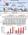
Figure 1. Syntaxin-1Open mutation promotes SNARE-complex assembly
A. Diagram of SNARE/SM-protein complex assembly from synaptobrevin/VAMP (SybVAMP), Syntaxin-1 (Synt1), SNAP-25, and Munc18-1 during synaptic vesicle fusion. Prior to fusion, Munc18-1 is bound to closed syntaxin-1 (Synt1Closed) which does not require the syntaxin-1 N-terminus (Dulubova et al., 1999). When fusion is initiated, syntaxin-1 is converted to an open conformation (Synt1Open), probably by Munc13 (Ma et al., 2011), and partly assembled trans-SNARE complexes are formed (Step A). Munc18-1 remains bound to syntaxin-1 throughout SNARE-complex assembly, but its binding now requires the syntaxin-1 N-terminus (Dulubova et al., 2007; Khvotchev et al., 2007; Zhou et al., 2013). Subsequenty, Ca2+ binds to synaptotagmin (Syt) to trigger completion of SNARE-complex assembly and fusion pore opening (Step B). Thus, two major conformational transitions occur during fusion: the switch of syntaxin-1 from closed to open, and the folding of the syntaxin-1, SNAP-25, and synaptobrevin SNARE motifs into the four-helical bundle of the SNARE complex. B. SNARE-complex assembly measured by co-immunoprecipitation of SNARE-proteins expressed in transfected HEK293 cells is enhanced by opening syntaxin-1 (top, representative immunoblots; bottom, summary graphs of SNARE complexes measured via co-immunoprecipitation with synaptobrevin-2 of syntaxin-1 (left) or SNAP-25 (right)). HEK293 cells were co-transfected with a constant amount of expression vectors encoding synaptobrevin-2 (Syb2), SNAP-25, and Munc18-1, and increasing amounts of expression vectors encoding either Syntaxin-1WT or Syntaxin-1Open. Immunoprecipitates were analyzed by immunoblotting, and quantified using 125I-labeled secondary antibodies (means ± SEM; n=4; *p<0.05, **p<0.01 by Student’s t-test).
Figure 2
Figure 2. Syntaxin-1Open mutation increases and accelerates Ca2+-triggered neurotransmitter release
A. Left, fluorescent image of the calyx-of-Held synapse (green, presynaptic terminal; red, postsynaptic MNTB neuron, visualized after loading of impermeable fluorescent dyes via patch pipettes). Right, schematic representation of the recording configuration. B. Single action-potential evoked EPSCs in Syntaxin-1WT (black) or Syntaxin-1Open (red) synapses. Left panels show representative traces for experiments performed without (top) or with (bottom) 2 mM kynurenic acid (Kyn). Right panels show cumulative distributions and summary graphs of the EPSC amplitudes obtained without (middle) or with Kyn (right) in Syntaxin-1WT (black) or Syntaxin-1Open (red) terminals. C. EPSC properties obtained from the experiment in B in Syntaxin-1WT (black) or Syntaxin-1Open (red) terminals. Summery graphs show various EPSC parameters without (left) or with (right) kynurenic acid in the bath. Data shown are means ± SEM; numbers of synapses analyzed are shown in graphs. Statistical significance was assessed with the Kolmogorov-Smirnov test for cumulative distributions and Student’s t-test for summary graphs (*P < 0.05, **P <0.01, ***P <0.001).
Figure 3
Figure 3. Short-term plasticity and response to extended stimulus trains in syntaxin-1Open synapses
A. Paired-pulse ratio monitored in Syntaxin-1WT and Syntaxin-1Open synapses. Panels show representative traces (left) and summary graphs (right) for inter-stimulus intervals of 5, 10, 20, 50, 100, 1000 ms. B. Representative EPSC traces of the first and last 20 responses triggered by a 10 s, 100 Hz action-potential train. C. Summary plots of absolute (left, in nA) and normalized (right) average responses for the first 25 stimuli obtained with 10 Hz (top), 25 Hz (middle), and 100 Hz (bottom) stimulus trains in Syntaxin-1WTand Syntaxin-1Open synapses. Note the increase in synaptic depression at the beginning of the train and the continued absolute enhancement of release at the end of the train in Syntaxin-1Open synapses. D. Frequency-dependent enhancement of transmitter release in Syntaxin-1Open terminals. EPSC ratios (Syntaxin-1Open/Syntaxin-1WT) for 10, 25 and 100 Hz trains were computed after binning synaptic responses compared to normalized control (dotted line). Data shown are means ± SEM; number of synapses analyzed is given in the graphs. Statistical significance was assessed using Student’s t-test (*P < 0.05, **P <0.01, ***P <0.001).
Figure 4
Figure 4. Syntaxin-1Open mutation decreases the size of the RRP
A. Estimation of RRP size using a prolonged presynaptic depolarization stimulus. Left, representative traces of Ca2+-currents (ICa2+) recorded from presynaptic terminals that were depolarized from −80 to 0 mV for 50 ms to induce maximal opening of Ca2+-channels (left top), and of the postsynaptic EPSC induced by the resulting release of the entire RRP (left bottom). Middle and right, cumulative distribution and summary plot of peak EPSC amplitudes triggered by sustained presynaptic depolarization in Syntaxin-1WT and Syntaxin-1Open synapses. B. Multiple EPSC parameters obtained from the experiments in A, including total charge transfer of the RRP (100 ms time-interval), 20–80% rise-time, latency (delay between the presynaptic Ca2+-current and 10% of postsynaptic EPSC), and fast and slow decay time constants obtained after double exponential fitting of EPSC decays. C. Measurements of RRP size by flash photolysis of caged Ca2+. Left, light-evoked EPSCs recorded from Syntaxin-1WT (black) and Syntaxin-1Open (red) synapses in response to maximal Ca2+-elevations triggered by strong flashes of UV-light delivered to presynaptic terminals loaded with caged Ca2+ and the low affinity Ca2+-indicator Fura 6F (see text and Supplementary Methods for details). Middle, EPSCs triggered by maximal presynaptic Ca2+-uncaging (Ca2+ ≥ 15 µM) were deconvolved with a representative mEPSC waveform, integrated, and then plotted as a function of time. Right, estimation of averaged light-evoked RRP size for control and Syntaxin-1Open mutant synapses at 100 ms time-intervals. Data shown are means ± SEM; number of synapses analyzed is shown in the graphs. Statistical significance was assessed using the Kolmogorov-Smirnov test for cumulative distributions and Student’s t-test for summary graphs (*P < 0.05). All measurements were performed in 2 mM Kyn.
Figure 5
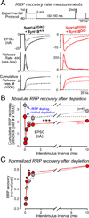
Figure 5. Opening syntaxin-1 affects vesicle replenishment dynamics
A. Experimental protocol and representative traces of RRP recovery experiments in Syntaxin-1WT (left) and Syntaxin-1Open (right) synapses. The RRP was fully depleted by a 50 ms presynaptic depolarizing stimulus, and the RRP recovery was then measured after a defined interstimulus interval (ISI) by application of a second 40 ms presynaptic depolarizing stimulus. In the example shown, the ISI was 200 ms. Postsynaptic EPSCs were recorded at −80 mV holding potentials, converted into release rates by deconvolution (middle), and then integrated over time (bottom). B and C. Summary plots of absolute RRP measured during the recovery experiments (B) and the relative replenishment rates calculated from the absolute RRP (C). Measurements were performed in Syntaxin-1WT (grey) and Syntaxin-1Open (red) synapses. Data shown are means ± SEM; number of synapses analyzed is shown in the graphs. Statistical significance was assessed using ANOVA, following by a Bonferroni test for multiple comparisons (*, p<0.05). All measurements were performed in 2 mM Kyn.
Figure 6
Figure 6. Syntaxin-1Open mutation enhances the Ca2+-sensitivity of neurotransmitter release
A. Representative Ca2+-uncaging experiments in Syntaxin-1WT (left) and Syntaxin-1Open (right) synapses using a weak (top) or strong (bottom) flash of UV-light to produce small or intermediate elevations of Ca2+ which were measured with Fura 4F as Ca2+-indicator dye. Intraterminal [Ca2+] elevations and corresponding EPSCs were simultaneously recorded using dual whole-cell patch-clamp and ratiometric Ca2+-fluorometry measurements from the same presynaptic terminals; EPSCs were then converted into release rates by deconvolution. B. Summary plot of the Ca2+-dependence of peak release rates in Syntaxin-1WT (black) and Syntaxin-1Open (red) synapses. Each data point (circle) represents the measurement results from a single synapse (n= 44 and 42 for Syntaxin-1WT and Syntaxin-1Open synapses, respectively). Solid lines show the fit of the data to the revised dual-Ca2+-sensor model (Sun et al., 2007; Fig. S5A), which in contrast to previous models takes into account the amount of SNARE complexes mediating exocytosis of individual synaptic vesicles. The left-shift in mutant synapses could be readily explained by increasing the Ks 7-fold (see text and Supp. Materials), which in turn decreased the apparent EC50 for Ca2+ in triggering release ~2-fold. Statistical significance was assessed using ANCOVA (***, p<0.001). For a plot of the synaptic delays as a function of the Ca2+-concentration, see Fig. S5B. C. Modeling of local presynaptic Ca2+-signals and postsynaptic EPSCs in control and mutant synapses using the dual-Ca2+-sensor model of release. An idealized representation of the Ca 2+ transient waveform during a single action potential at the calyx-of-Held (top) was simulated as a Gaussian function with half-width of 0.18 ms and followed with a double exponential function with time constants of 2ms (75%) and 0.2ms (25%). The corresponding transmitter release rates (middle) were predicted with the modified dual-Ca2+-sensor model using a Ks=1 for Syntaxin-1WT synapses (left) and a Ks=7 for Syntaxin-1Open synapses (right); all other parameters were kept identical. The experimentally measured EPSCs (dotted lines; obtained in the presence of 2 mM Kyn) are displayed and superimposed with the simulated EPSCs by convolution of the idealized release rate and the measured average mEPSC (bottom).
Figure 7
Figure 7. The Syntaxin-1Open mutation accelerates fusion-pore opening
A. Left, properties of single vesicle fusion events recorded from Syntaxin-1WT (black) and Syntaxin-1Open (red) synapses. Representative traces are shown at low (top) and at high resolution (bottom; averages of 156 and 186 events from a single Syntaxin-1WT and Syntaxin-1Open synapse, respectively). B–D. Cumulative distributions (left) and summary graph (right) of the mEPSC frequency (B), amplitude (C), and rise times (D) in control (black) and Syntaxin-1open (red) synapses E. Schematic in-scale representation of synaptic vesicle exocytosis highlighting the relative sizes of synaptic vesicles, the fusion pore, the width of the synapse, and the synaptic cleft. F and G. Dose-dependent actions of the competitive AMPA-receptor antagonist γ-DGG on mEPSC parameters. Representative traces are shown in F, and summary graphs in G of the mEPSC amplitudes (top), rise times (middle), and decay kinetics (bottom). H and I. Same as in F and G, but using the non-competitive AMPA-receptor antagonist GYKI53655. Data shown are means ± SEM. Number of experiments analyzed is shown in the graphs. Significance was assessed by the Kolmogorov-Smirnov test for cumulative distributions and Student’s t-test for summary graphs (*,p<0.05; **, p<0.01; ***, p<0.001).

References

    1. Arancillo M, et al. Titration of Syntaxin1 in mammalian synapses reveals multiple roles in vesicle docking, priming, and release probability. J Neurosci. 2013;33:16698–16714. - PMC - PubMed
    1. Augustin I, Rosenmund C, Südhof TC, Brose N. Munc13-1 is essential for fusion competence of glutamatergic synaptic vesicles. Nature. 1999;400:457–461. - PubMed
    1. Bezprozvanny I, Scheller RH, Tsien RW. Functional impact of syntaxin on gating of N-type and Q-type calcium channels. Nature. 1995;378:623–626. - PubMed
    1. Bollmann JH, Sakmann B. Control of synaptic strength and timing by the release-site Ca2+ signal. Nat Neurosci. 2005;8(4):426–434. - PubMed
    1. Bollmann JH, Sakmann B, Borst JG. Calcium sensitivity of glutamate release in a calyx-type terminal. Science. 2000;289:953–957. - PubMed

Publication types