. 2014 Jun 9:5:3986.
doi: 10.1038/ncomms4986.

Differential developmental requirement and peripheral regulation for dermal Vγ4 and Vγ6T17 cells in health and inflammation

Affiliations

Differential developmental requirement and peripheral regulation for dermal Vγ4 and Vγ6T17 cells in health and inflammation

Yihua Cai et al. Nat Commun. .

Erratum in

Abstract

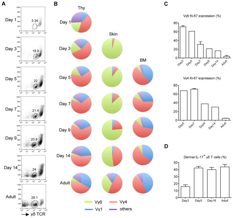
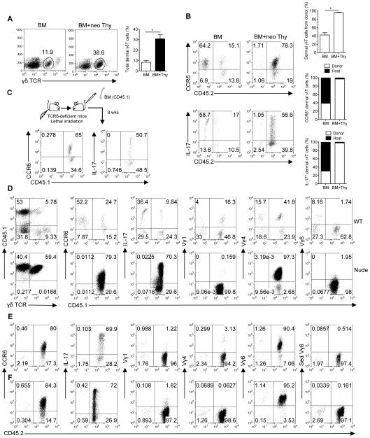
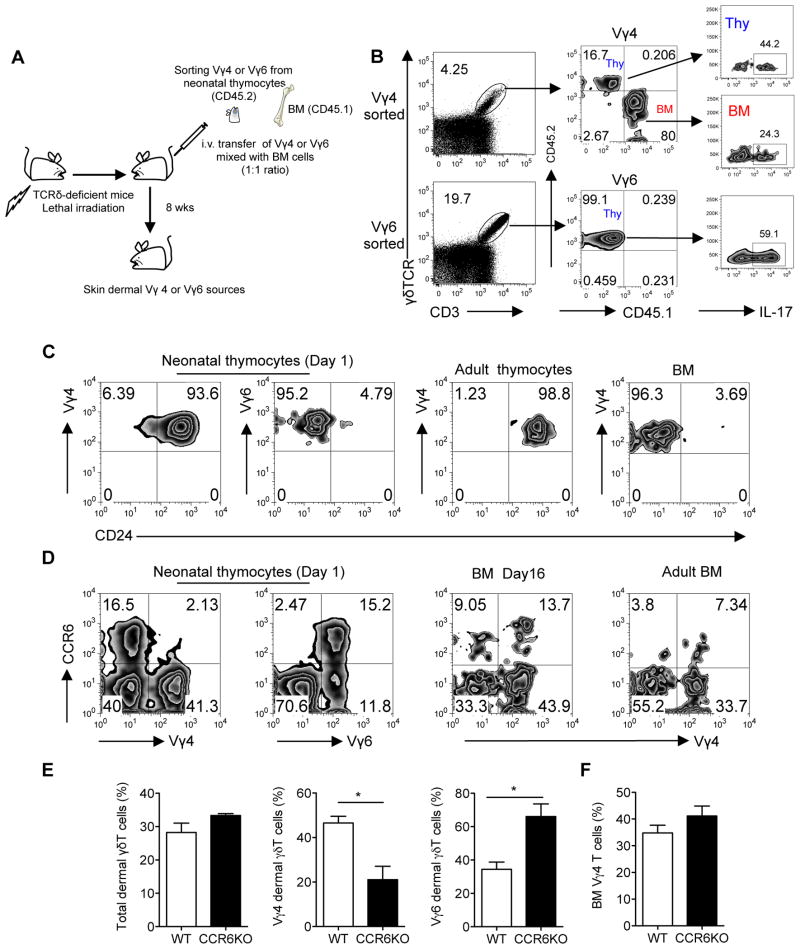
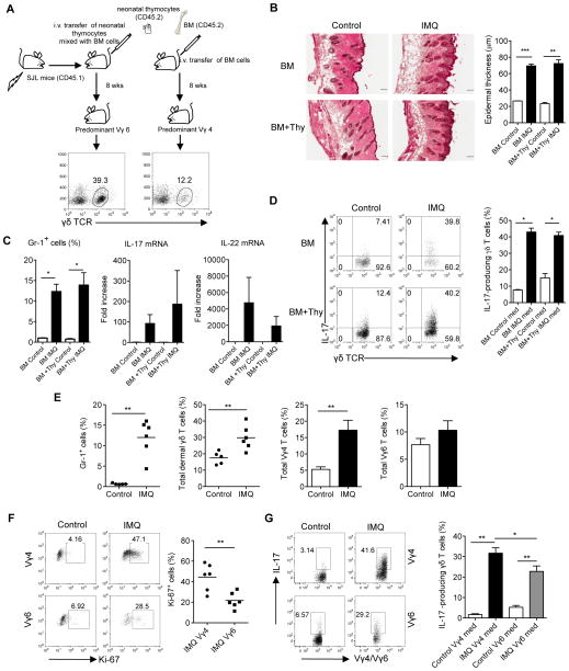
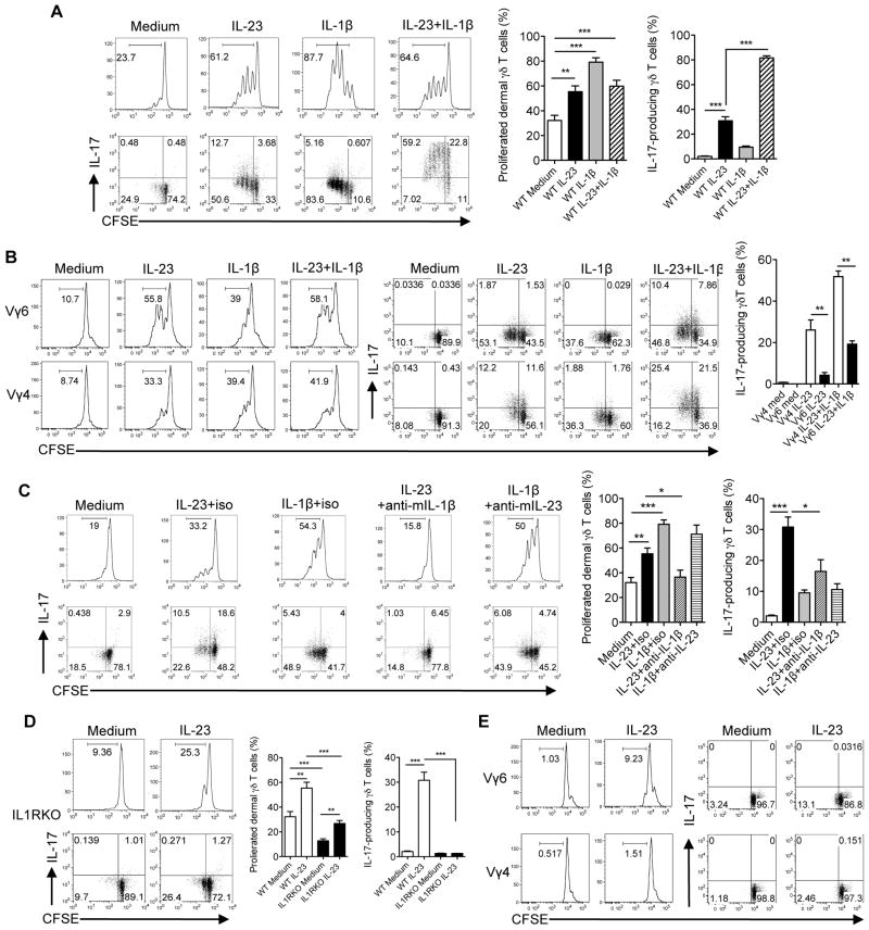
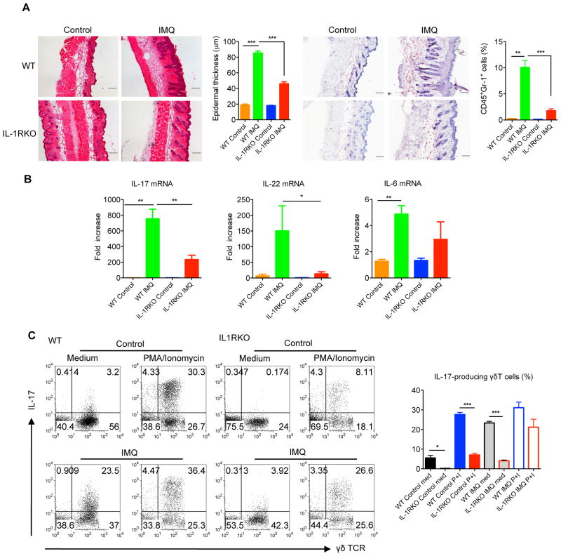
Dermal IL-17-producing γδT cells have a critical role in skin inflammation. However, their development and peripheral regulation have not been fully elucidated. Here we demonstrate that dermal γδT cells develop from the embryonic thymus and undergo homeostatic proliferation after birth with diversified TCR repertoire. Vγ6T cells are bona fide resident, but precursors of dermal Vγ4T cells may require extrathymic environment for imprinting skin-homing properties. Thymic Vγ6T cells are more competitive than Vγ4 for dermal γδT cell reconstitution and TCRδ(-/-) mice reconstituted with Vγ6 develop psoriasis-like inflammation after IMQ-application. Although both IL-23 and IL-1β promote Vγ4 and Vγ6 proliferation, Vγ4 are the main source of IL-17 production that requires IL-1 signalling. Mice with deficiency of IL-1RI signalling have significantly decreased skin inflammation. These studies reveal a differential developmental requirement and peripheral regulation for dermal Vγ6 and Vγ4 γδT cells, implying a new mechanism that may be involved in skin inflammation.


Figures

Figure 1
Figure 1. Development of dermal γδT cells
(A) Whole skin cell suspensions prepared from C57Bl/6 WT mice at different days (n=3–5) after birth were stained with CD3 and γδTCR and the frequency of γδTCRint dermal γδT cells was analyzed by flow cytometry. Flow plots gated on CD3+ cells are representative of three independent experiments with similar results. (B) Thymocytes (Thy), whole skin cells and BM cells from different days after birth were stained with CD3, γδTCR and Vγ (Vγ 1, 4, 6) mAbs. Percentages of Vγ1, Vγ4, and Vγ6 γδT cells were analyzed by flow cytometry and summarized in pie chart. (C) Percentages of Ki-67 expression on dermal Vγ4 and Vγ6 T cells were analyzed by flow cytometry. Cells were gated on CD3intγδTCRint Vγ4+ or Vγ6+ cells. Data are shown as mean ± SEM. (D) Whole skin cells were stimulated with PMA plus ionomycin for 5 hours and the percentage of IL-17-producing γδT cells gated on CD3intγδTCRint cells were analyzed by flow cytometry. Data are shown as mean ± SEM.
Figure 2
Figure 2. Fetal thymus is required for dermal γδT cell development and Vγ6 T cell reconstitution while dermal Vγ4 T cells originate mainly from BM
BM cells or BM cells plus neonatal thymocytes (BM+neo Thy) from C57Bl/6 WT mice (CD45.2) were transplanted into lethally irradiated SJL mice (CD45.1, n=4–5). (A) Eight weeks after reconstitution, percentage of dermal γδ T cells gated on CD3+ cells from recipient mice was analyzed by flow cytometry and total frequency of dermal γδT cells was summarized. Data are shown as mean ± SEM. *p < 0.05 (unpaired Student’s t test). (B) Whole skin cells were stimulated with PMA plus ionomycin for 5 hours. CCR6+ and IL-17+CD3intγδTCRint cells were analyzed by flow cytometry. Flow plots were gated on CD3intγδTCRint cells. Percentages of dermal γδ T cells from donor are shown as mean ± SEM. *p < 0.05 (unpaired Student’s t test). Degree of chimerism of CCR6+ and IL-17-producing dermal γδ T cells are also shown as open bars (donor) and filled bars (host). Data are representative of at least three independent experiments with similar results. (C) BM cells from SJL mice (CD45.1) were transplanted into lethally irradiated TCRδ KO mice (CD45.2). After 8 weeks of reconstitution, CCR6+ and IL-17- producing dermal γδ T cells were analyzed by flow cytometry. Flow plots gated on CD3+γδTCR+ cells are representative of two independent experiments with similar results. (D) BM cells from SJL mice (CD45.1) were transplanted into lethally irradiated C57Bl/6 WT mice or Nude mice (CD45.2). After 8 weeks of reconstitution, percentage of dermal γδ T cells gated on CD3+ cells was detected by flow cytometry. Furthermore, CCR6 expression and Vγ usage (Vγ1, 4, 6) of dermal γδ T cells and intracellular IL-17 by dermal γδ T cells after PMA plus ionomycin stimulation were also analyzed by flow cytometry. Flow plots were gated on CD3intγδTCRint cells. (E, F) Thymocytes from C57Bl/6 WT neonatal (E) or E18 (F) pups plus BM cells from TCRδ KO mice were transplanted into lethally irradiated SJL mice. After 8 weeks of reconstitution, CCR6 expression and Vγ usage (Vγ1, 4, 6) of dermal γδT cells and intracellular IL-17 after PMA plus ionomycin stimulation were analyzed by flow cytometry. Flow plots were gated on CD3intγδTCRint cells.
Figure 3
Figure 3. Thymic Vγ4 T cells require extrathymic environment for imprinting of skin homing properties
(A, B) Sorted Vγ4 or Vγ6 neonatal thymocytes from C57Bl/6 WT mice (CD45.2) mixed with BM cells from SJL mice (CD45.1) were transferred into lethally irradiated TCRδ KO mice (n=3–5). The ratio of Vγ4 from both sources was around 1:1 as well as Vγ6 to Vγ4 (based on Vγ4 frequency in BM). After 8 weeks of reconstitution, total dermal γδT cells (A) and the sources of Vγ4 or Vγ6 T cells as well as intracellular IL-17 stimulated with PMA plus ionomycin (B) were determined by flow cytometry. Flow plots were gated on CD3+ γδTCR+ cells. (C, D) CD24 (C) and CCR6 (D) expression by Vγ4 or Vγ6 cells on neonatal thymocytes, adult thymocytes, BM cells (Day16) and adult BM cells were determined by flow cytometry. Flow plot gated on CD3+ γδTCR+ cells are representative of three independent experiments with similar results. (E, F) Whole skin cells (E) or BM cells (F) from C57Bl/6 WT mice or CCR6KO mice (n=3) were stained with CD3, γδTCR, Vγ4 and Vγ6. Percentages of total dermal γδT cells as well as dermal Vγ4 and Vγ6 cells or BM Vγ4 cells were analyzed by flow cytometry. Data are shown as mean ± SEM and are representative of three independent experiments with similar results. *p < 0.05 (unpaired Student’s t test).
Figure 4
Figure 4. Dermal Vγ6 T cells induce skin inflammation but dermal Vγ4 are preferentially expanded and are the major IL-17 producers
(A) BM cells or BM cells plus neonatal thymocytes (BM+Thy) from C57Bl/6 WT mice (CD45.2) were transplanted into lethally irradiated SJL mice (CD45.1, n=5). After 8 weeks of reconstitution, frequency of dermal γδT cells was detected by flow cytometry. Mice receiving BM cells alone predominantly reconstituted Vγ4 while mice receiving BM plus neonatal thymocytes predominantly reconstituted Vγ6. (B) Reconstituted mice were treated daily for 5 days with IMQ or control cream (Control). Representative H&E-stained sections are shown and epidermal thickness were measured at day 5. Scale bar, 100 μm. Data are shown as mean ± SEM. **p < 0.01, ***p < 0.001 (unpaired Student’s t test). (C) Percentage of CD45+Gr-1+ cells after IMQ treatment was analyzed by flow cytometry. IL-17 and IL-22 mRNA levels were measured by qPCR. Data are shown as mean ± SEM. *p < 0.05 (unpaired Student’s t test). (D) Intracellular IL-17 production by skin dermal γδT cells from IMQ-treated or control mice was determined by flow cytometry (without stimulation). Flow plots were gated on CD3+ cells. Data are shown as mean ± SEM. *p < 0.05 (unpaired Student’s t test). (E) C57Bl/6 WT mice (n=5–6) were treated daily for 5 days with IMQ or control cream (Control). Percentages of CD45+Gr-1+ cells, total dermal γδT cells, dermal Vγ4 and Vγ6 cells were analyzed by flow cytometry. Data are shown as mean ± SEM and are representative of two independent experiments with similar results. **p < 0.01 (unpaired Student’s t test). (F, G) Ki-67 expression (F) and intracellular IL-17 production (G) by dermal Vγ4 and Vγ6 cells were determined by flow cytometry (without stimulation). Flow plots were gated on CD3intγδTCRint cells. Data are shown as mean ± SEM and are representative of two independent experiments with similar results. *p < 0.05, **p < 0.01 (unpaired Student’s t test).
Figure 5
Figure 5. IL-23 and IL-1β regulate dermal Vγ4 and Vγ6 γδ T cell proliferation and IL-17 production
(A, B) Whole skin cell suspensions were labeled with CFSE and then stimulated with IL-23, IL- 1β or IL-23 plus IL-1β for 3 days. CFSE dilution and intracellular IL-17 production by dermal γδT cells (A) or dermal Vγ4 and Vγ6 γδ T cells (B) were determined by flow cytometry. Flow plots gated on CD3int γδTCRint cells are representative of at least three independent experiments with similar results. Data are shown as mean ± SEM (n=10). **p < 0.01, ***p < 0.001 (unpaired Student’s t test). (C) Whole skin cells suspensions were labeled with CFSE and then stimulated with IL-23, IL-1β, IL-23 plus anti-mouse IL-1β, IL-1β plus anti-mouse IL-23 or isotype control mAb in WT mice for 3 days. CFSE dilution and intracellular IL-17 production by dermal γδT cells were determined by flow cytometry. Flow plots gated on CD3int γδTCRint cells are representative of at least three independent experiments with similar results. Summarized data (n=8–9) are shown as mean± SEM. *p<0.05, **p < 0.01, ***p < 0.001 (unpaired Student’s t test). (D, E) Whole skin cells suspensions from IL-1RI KO mice labeled with CFSE were stimulated with IL- 23 for 3 days. CFSE dilution and intracellular IL-17 production by dermal γδ T cells (D) or dermal Vγ4 and Vγ6 γδ T cells (E) were determined by flow cytometry. Flow plots gated on CD3int γδTCRint cells are representative of at least three independent experiments with similar results. Data are shown as mean ± SEM (n=8–9). **p < 0.01, ***p < 0.001 (unpaired Student’s t test).
Figure 6
Figure 6. CCR6 is essential for γδT cell trafficking from periphery to dermis
(A) Primary murine KC were stimulated with IL-1β for 6 hours. CCL2, CCL5, CCL20, CXCL9, and CXCL10 mRNA levels were measured by qPCR. The figure shows fold changes normalized for β-MG mRNA versus medium alone. Data are shown as mean ± SEM. (B) Sorted γδT cells from spleen and lymph nodes from C57Bl/6 WT mice or CCR6KO mice were adoptively transferred into SJL mice (n=4). After 5–7 days, cells from skin, peripheral blood, lymph nodes and spleen were stained with CD3, γδ TCR and CCR6. Frequency of total γδT cells and CCR6+ γδT cells from donor (CD45.2) were determined by flow cytometry. Flow plots were gated on skin CD3intγδTCRint cells. Data are shown as mean ± SEM. **p < 0.01, n.s., not significant (unpaired Student’s t test).
Figure 7
Figure 7. IL-1R signaling is essential for IMQ-induced skin inflammation and acanthosis
(A) C57Bl/6 WT and IL-1RI KO mice (n=6–8) were treated daily for 5 days with IMQ or control cream (Control). Representative H&E-stained sections and frozen sections stained with Gr-1 are shown. Gr-1 positive cells are brown. Skin tissues were also stained with CD45 and Gr-1 assessed by flow cytometry. Epidermal thickness and percentage of CD45+Gr-1+ cells were measured at day 5. Scale bar, 100 μm. Data are shown as mean ± SEM. **p < 0.01, ***P<0.001 (unpaired Student’s t test). (B) IL-17, IL-22 and IL-6 mRNA levels were measured by qPCR. The figure shows fold changes normalized for β-MG mRNA versus control skin from WT mice. Data are shown as mean ± SEM. *p < 0.05, **p< 0.01 (unpaired Student’s t test). (C) Intracellular IL- 17 production by dermal γδT cells with or without PMA plus ionomycin stimulation was determined by flow cytometry. Flow plots were gated on CD3+ cells. Data are shown as mean ± SEM. *p < 0.05, ***p<0.001 (unpaired Student’s t test).

References

    1. Di Meglio P, Perera GK, Nestle FO. The multitasking organ: recent insights into skin immune function. Immunity. 2011;35:857–869. - PubMed
    1. Payer E, Elbe A, Stingl G. Epidermal T lymphocytes--ontogeny, features and function. Springer seminars in immunopathology. 1992;13:315–331. - PubMed
    1. Sharp LL, Jameson JM, Cauvi G, Havran WL. Dendritic epidermal T cells regulate skin homeostasis through local production of insulin-like growth factor 1. Nat Immunol. 2005;6:73–79. - PubMed
    1. Cai Y, et al. Pivotal Role of Dermal IL-17-Producing gammadelta T Cells in Skin Inflammation. Immunity. 2011;35:596–610. - PMC - PubMed
    1. Gray EE, Suzuki K, Cyster JG. Cutting edge: Identification of a motile IL-17-producing gammadelta T cell population in the dermis. J Immunol. 2011;186:6091–6095. - PMC - PubMed

Publication types

LinkOut - more resources