. 2014 Jul;14(7):1523-32.
doi: 10.1111/ajt.12742. Epub 2014 Jun 6.

Enhancing human islet transplantation by localized release of trophic factors from PLG scaffolds

Affiliations

Enhancing human islet transplantation by localized release of trophic factors from PLG scaffolds

K A Hlavaty et al. Am J Transplant. 2014 Jul.

Abstract

Islet transplantation represents a potential cure for type 1 diabetes, yet the clinical approach of intrahepatic delivery is limited by the microenvironment. Microporous scaffolds enable extrahepatic transplantation, and the microenvironment can be designed to enhance islet engraftment and function. We investigated localized trophic factor delivery in a xenogeneic human islet to mouse model of islet transplantation. Double emulsion microspheres containing exendin-4 (Ex4) or insulin-like growth factor-1 (IGF-1) were incorporated into a layered scaffold design consisting of porous outer layers for islet transplantation and a center layer for sustained factor release. Protein encapsulation and release were dependent on both the polymer concentration and the identity of the protein. Proteins retained bioactivity upon release from scaffolds in vitro. A minimal human islet mass transplanted on Ex4-releasing scaffolds demonstrated significant improvement and prolongation of graft function relative to blank scaffolds carrying no protein, and the release profile significantly impacted the duration over which the graft functioned. Ex4-releasing scaffolds enabled better glycemic control in animals subjected to an intraperitoneal glucose tolerance test. Scaffolds releasing IGF-1 lowered blood glucose levels, yet the reduction was insufficient to achieve euglycemia. Ex4-delivering scaffolds provide an extrahepatic transplantation site for modulating the islet microenvironment to enhance islet function posttransplant.

Keywords: Bioengineering; islet xenotransplantation; regenerative medicine; type 1 diabetes mellitus.


Conflict of interest statement

Disclosure

The authors of this manuscript have conflicts of interest to disclose as described by the American Journal of Transplantation. L.D.S. consults on islet transplantation for Pioneer Biosolutions and has equity in the company.

Figures

Figure 1
Figure 1. Layered PLG scaffold
(A) Cross-sectional view of a layered scaffold (5mm diameter × 3 mm height) demonstrating the outer layers molded around a smaller solid inner layer that does not span the width of the scaffold. (B) Scanning electron micrograph of a layered scaffold cross-sectional view. Outer layers were formed with single emulsion microspheres and NaCl particles to create 250–425 µm pores. Protein-delivering double emulsion microspheres form the center layer. (C) Magnified view showing the porous outer layer for islet seeding and center layer containing protein-releasing microspheres. PLG, poly(lactide-co-glycolide).
Figure 2
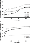
Figure 2. Protein release from PLG scaffolds
Layered scaffolds were fabricated with center layers of microspheres loaded with either (A) Ex4 or (B) IGF-1. The release rate was monitored using radiolabeled proteins (n = 3 per condition). Ex4, exendin-4; IGF-1, insulin-like growth factor-1; PLG, poly(lactide-co-glycolide).
Figure 3
Figure 3. Ex4 and IGF-1 bioactivity following release
(A) Rat insulinoma (RINm5f) β cells were treated for 1 day with conditioned medium previously incubated with Ex4 loaded scaffolds for 8 days. Insulin release from the cells was quantified by ELISA. Controls included medium incubated with scaffolds without Ex4 or medium alone. Error bars represent SEM and *p<0.05. Differences between the Ex4 scaffold and Ex4 conditions or the blank scaffold and control conditions were not significant. (B) INS-1 cells were treated for 1 day with conditioned medium previously incubated with IGF-1 loaded scaffolds for 8 days. P-ERK was evaluated by Western blot analysis. Control conditions included cells prior to treatment (time Oh) or cells treated with control medium from incubation with scaffolds without encapsulated IGF-1, which are denoted as controls A and B, respectively. Ex4, exendin-4; IGF-1, insulin-like growth factor-1.
Figure 4
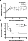
Figure 4. Graft function for human islets transplanted on 6% IGF-1 loaded scaffolds
Blood glucose was measured over time from mice transplanted with 1500–2000 islet equivalents on (A) 6% 50:50 scaffolds containing IGF-1 (n = 8) and blank scaffolds (n = 8), *p<0.05. (B) A Kaplan–Meier plot represents animals with functioning grafts, where NS indicates “not significant.” Graft failure is defined as three consecutive glucose measurements greater than 250 mg/dL. IGF-1, insulin-like growth factor-1.
Figure 5
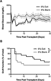
Figure 5. Graft function for human islets transplanted on 6% Ex4 loaded scaffolds
Blood glucose was measured over time from mice transplanted with 1500–2000 islet equivalents on (A) 6% 50:50 scaffolds containing Ex4 (n = 19) and blank scaffolds (n = 17). (B) A Kaplan–Meier plot demonstrating graft function for the 6% 50:50 Ex4 scaffolds and blank scaffolds. Graft failure is defined as three consecutive glucose measurements greaterthan 250 mg/dL, *p<0.05. Ex4, exendin-4.
Figure 6
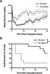
Figure 6. Graft function for human islets transplanted on 3% Ex4 loaded scaffolds
Blood glucose was measured over time from mice transplanted with 1500–1800 islet equivalents on (A) 3% 50:50 scaffolds containing Ex4(n = 10) and blank scaffolds (n = 10). (B) A Kaplan-Meier plot demonstrating graft function for the 3% 50:50 Ex4 scaffolds and blank scaffolds. Graft failure is defined as three consecutive glucose measurements greaterthan 250 mg/dL, **p<0.01. Ex4, exendin-4.
Figure 7
Figure 7. Intraperitoneal glucose tolerance test for human islets transplanted on 3% Ex4 scaffolds
Blood glucose measurements (A) and area under the curve (AUC) (***p< 0.001) (B) for naïve mice (n = 6) or mice receiving Ex4 scaffolds (n = 4) or blank scaffolds (n = 5) following challenge with an intraperitoneal dextrose injection 11 days posttransplant. Ex4, exendin-4.
Figure 8
Figure 8. Human islets transplanted on 3% Ex4 scaffolds have a greater islet area and demonstrate more proliferation
Quantification of islet area in 3% Ex4 scaffolds (A) shows a greater area than in blank scaffolds (B), **p<0.01 (n>40 islets) (C). Islets transplanted on 3% Ex4 scaffolds (D) show more proliferating nuclei (white arrows) within islets compared to blank scaffolds (E). Within insulin-positive areas, the ratio of proliferating cells (Ki67+) to nuclei is greater in 3% Ex4 scaffolds than in blank scaffolds (F), ***p < 0.001 (n >35 islets).

Similar articles

Cited by

References

    1. Shapiro AM, Lakey JR, Ryan EA, et al. Islet transplantation in seven patients with type 1 diabetes mellitus using a glucocorticoid-free immunosuppressive regimen. N Engl J Med. 2000;343:230–238. - PubMed
    1. Shapiro AM, Ricordi C, Hering BJ, et al. International trial of the Edmonton protocol for islet transplantation. N Engl J Med. 2006;355:1318–1330. - PubMed
    1. Pambianco G, Costacou T, Ellis D, Becker DJ, Klein R, Orchard TJ. The 30-year natural history of type 1 diabetes complications: The Pittsburgh Epidemiology of Diabetes Complications Study experience. Diabetes. 2006;55:1463–1469. - PubMed
    1. Barton FB, Rickels MR, Alejandro R, et al. Improvement in outcomes of clinical islet transplantation: 1999–2010. Diabetes Care. 2012;35:1436–1445. - PMC - PubMed
    1. Barshes NR, Wyllie S, Goss JA. Inflammation-mediated dysfunction and apoptosis in pancreatic islet transplantation: Implications for intrahepatic grafts. J Leukoc Biol. 2005;77:587–597. - PubMed

Publication types