. 2014 Dec;74(12):1210-25.
doi: 10.1002/dneu.22199. Epub 2014 Jun 18.

Loss of Tau results in defects in photoreceptor development and progressive neuronal degeneration in Drosophila

Affiliations

Loss of Tau results in defects in photoreceptor development and progressive neuronal degeneration in Drosophila

Bonnie J Bolkan et al. Dev Neurobiol. 2014 Dec.

Abstract

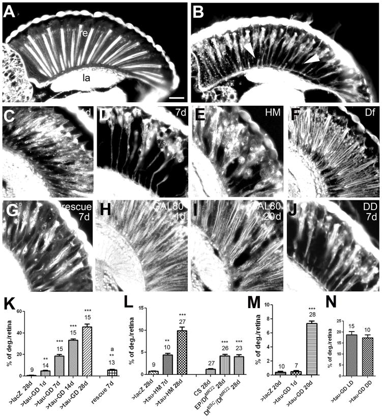
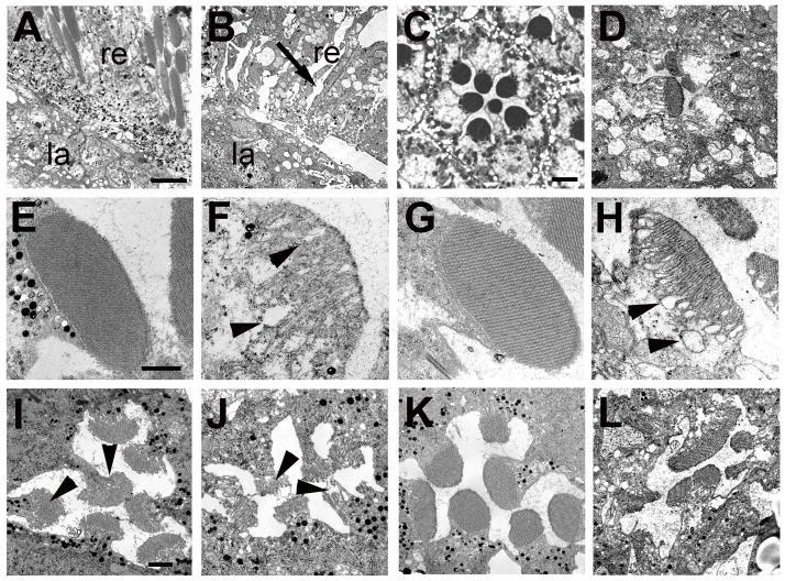
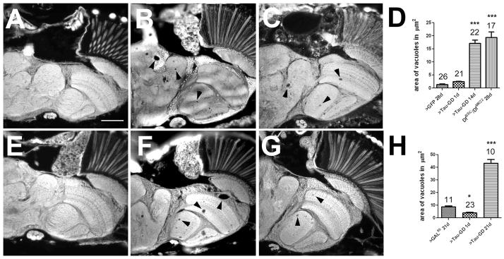
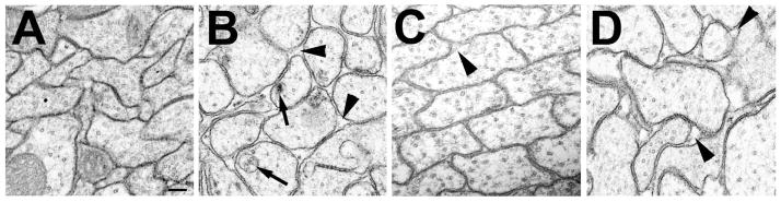
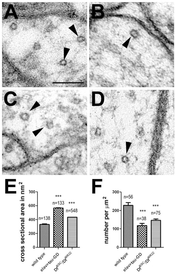
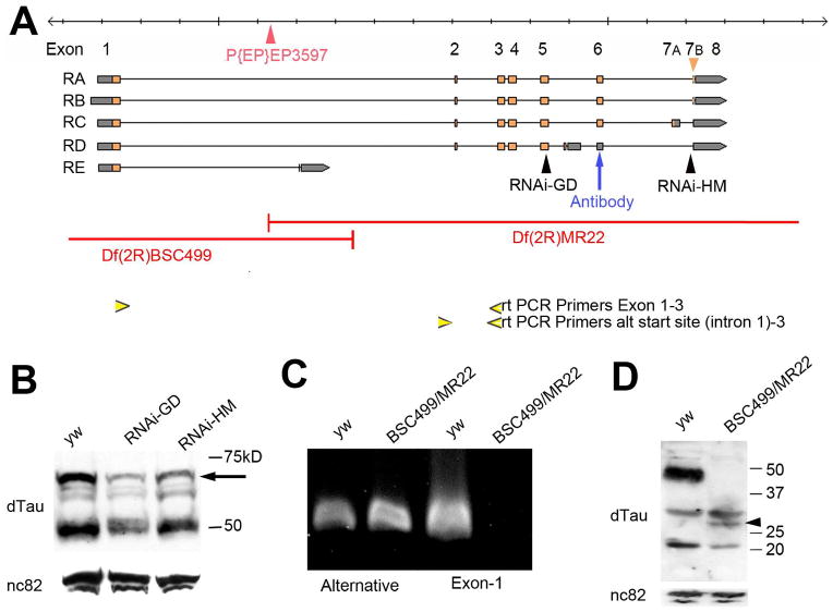
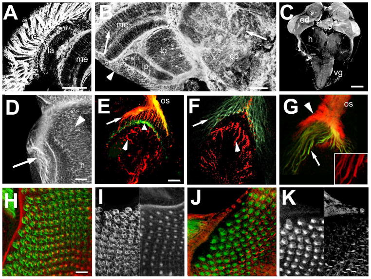
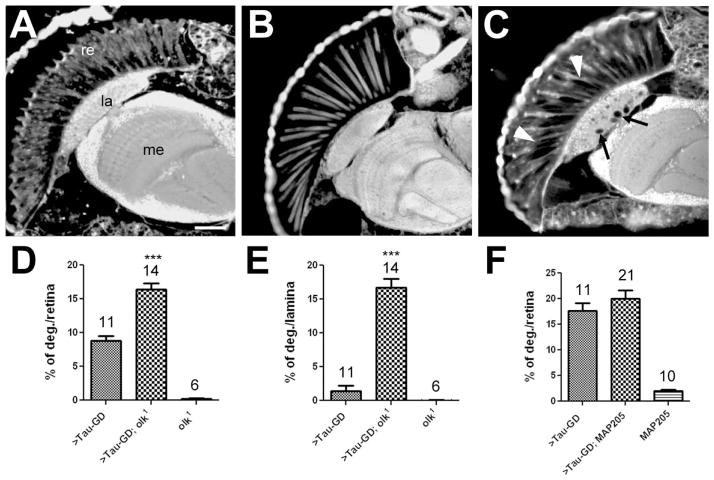
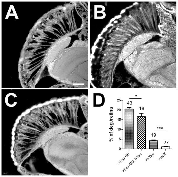
Accumulations of Tau, a microtubule-associated protein (MAP), into neurofibrillary tangles is a hallmark of Alzheimer's disease and other tauopathies. However, the mechanisms leading to this pathology are still unclear: the aggregates themselves could be toxic or the sequestration of Tau into tangles might prevent Tau from fulfilling its normal functions, thereby inducing a loss of function defect. Surprisingly, the consequences of losing normal Tau expression in vivo are still not well understood, in part due to the fact that Tau knockout mice show only subtle phenotypes, presumably due to the fact that mammals express several MAPs with partially overlapping functions. In contrast, flies express fewer MAP, with Tau being the only member of the Tau/MAP2/MAP4 family. Therefore, we used Drosophila to address the physiological consequences caused by the loss of Tau. Reducing the levels of fly Tau (dTau) ubiquitously resulted in developmental lethality, whereas deleting Tau specifically in neurons or the eye caused progressive neurodegeneration. Similarly, chromosomal mutations affecting dTau also caused progressive degeneration in both the eye and brain. Although photoreceptor cells initially developed normally in dTau knockdown animals, they subsequently degenerated during late pupal stages whereas weaker dTau alleles caused an age-dependent defect in rhabdomere structure. Expression of wild type human Tau partially rescued the neurodegenerative phenotype caused by the loss of endogenous dTau, suggesting that the functions of Tau proteins are functionally conserved from flies to humans.

Keywords: Drosophila; eye development; neurodegeneration; tau.


Conflict of interest statement

The authors declare no conflict of interest.

Figures

Figure 1
Figure 1
Decreasing dTau levels in the eye causes progressive retinal degeneration. A: A four-week-old GMR-Gal4/UAS-LacZ control fly shows an intact retina (re) and underlying lamina (la). B: In contrast, vacuoles (arrowheads) can be detected in the retina of a 7d old GMR-GAL4; tauGD25023 fly. C: This phenotype is already detectable at 1-day-old but increases with age (7d in D). E: Retinal degeneration also occurs in a 4-week-old GMR-GAL4; tau HM05101 fly and a tauDf(3R)MR22/Df(3R)BSC499 fly aged for four weeks (F). G: Co-expression of UAS-dTau with tauGD25023 reduces retinal degeneration when comparing to the age-matched control in (D). H: A GMR-GAL4; tauGD25023; tubP-GAL80ts fly raised at the permissive temperature does not show retinal degeneration when 1d old. I: However, aged for 20d at the restrictive temperature, these flies again show retinal degeneration. J: Aging GMR-GAL4; tauGD25023 flies in constant darkness did not prevent the retinal degeneration observed when 7d old. Quantification of the retinal degeneration observed in GMR-GAL4; tauGD25023 (K), GMR-GAL4; tau HM05101 and tauDf(3R)MR22/Df(3R)BSC499 (L), and in GMR-GAL4; tauGD25023; tubP-GAL80ts (M). N: Quantification of the retinal degeneration in flies raised in a light:dark cycle or in constant darkness. The number of analyzed retinae and the SEMs are indicated for each bar. ** p<0.01, *** p<0.001. Scale bar in A=20μm.
Figure 2
Figure 2
Transmission electron microscopy reveals degeneration of rhabdomeres. A: A GMR-GAL4/UAS-LacZ fly at 36 hours post eclosion shows an intact retina (re) and lamina (la). B: In a 36hr old GMR-GAL4; tauGD25023 fly the retina is disorganized and vacuoles have formed in the retina (re, arrow). C: An intact ommatidia in a 36h old GMR-GAL4/UAS-LacZ control with seven photoreceptors present. D: In contrast, a 36h old GMR-GAL4; tauGD25023 fly shows a disrupted ommatidial structure with only a few abnormal looking photoreceptors present. Whereas control flies have highly structured rhabdomeres at this age (E), the rhabdomeres of the photoreceptors that are present in GMR-GAL4; tauGD25023 have an abnormally loose structure with loops forming that are mostly found in proximity to the cell cytoplasm (F, arrowheads). G: In contrast, the rhabdomeres are intact in a 1d old tauDf(3R)MR22/Df(3R)BSC499 fly but become more loose when aged for 7d (H). I: P11 control pupa showing the developing seven rhabdomeres (arrowheads). J: In an age-matched GMR-GAL4; tauGD25023 fly, the rhabdomeres already look abnormal although all photoreceptors are present at this stage. K: Pharate adult control fly. L: A pharate adult GMR-GAL4; tauGD25023 fly has lost several photoreceptors and the remaining photoreceptors are abnormally shaped and have irregular rhabdomeres. Scale bars in A=6μm, in C=2μm, in E=1μm, and in I=1μm.
Figure 3
Figure 3
Loss of dTau causes degeneration in the brain. A: No degeneration is detectable in a 1d old tauGD25023; elav-GAL4 fly. However after 14d several vacuoles have formed in the brain (arrowheads, B). C: Vacuoles can also be found in a 28d old tauDf(3R)MR22/Df(3R)BSC499 fly. D: Quantification of the area of vacuoles. E: tauGD25023; elav-GAL4/tubP-GAL80ts flies do not reveal vacuolization at 1d but vacuoles have formed after being aged for 21d at the restrictive temperature (F). G: A elav-GAL4/tubP-GAL80ts aged for 21d at the restrictive temperature also shows a few small vacuoles. H: Quantification of the vacuoles when raised at the restrictive temperature. D, H: The number of analyzed flies and the SEMs are indicated for each bar. * p<0.05, *** p<0.001. Scale bar in A=50μm.
Figure 4
Figure 4
Decreasing the levels of dTau affects axonal morphology. A: Transmission electron microscopy reveals tightly packed axons in a 1d old wild type fly. B: In a 1d old the tauGD25023; elav-GAL4 fly the axons look more rounded and extracellular gaps are visible (arrowheads). In addition, intracellular inclusions and vacuoles can be detected (arrows). C: In tauDf(3R)MR22/Df(3R)BSC499, the axons are tighter packed and only a few extracellular gaps are visible (arrowhead) at 1d of age, however, the number and size of these gaps increases with age as seen in a 16d old fly (D). Scale bar in A=200μm.
Figure 5
Figure 5
Decreasing the levels of dTau affects microtubule morphology and density. A: Microtubules (arrowheads) in a wild type fly. B: Less and larger microtubules are found in a tauGD25023;elav-GAL4. C: A 1d old tauDf(3R)MR22/Df(3R)BSC499 fly also has larger microtubules and this in confirmed in a 14d old fly (D). E: Quantification of the microtubule mean cross sectional area in nm2. The number of measured individual microtubules and the SEMs are indicated. F: Number of microtubules found per μm2 axon area. The number of analyzed axons (from at least three animals and at least 6 images) and the SEMs are indicated. All flies were 1d old. *** p<0.001. Scale bar in A=100nm.
Figure 6
Figure 6
The dTau locus encodes several alternative transcripts and protein isoforms. A; Map of the dTau locus (modified from flybase.org). The five annotated transcripts are shown and the areas deleted in Df(3R)BSC499 and tauDf(3R)MR22 are indicated by red lines. The small 7B exon only present in the RA and RB transcript is pointed out by an orange arrowhead. Also indicated are the locations of the tauEP3597 insertion (red arrowhead), the regions targeted by the RNAi construct (black arrowheads), and the locations of the antibody epitope (blue arrow). The yellow arrowheads represent the positions of the primers used for the PCR reactions. B: Western blot from head extracts using anti-dTau shows multiple bands corresponding to various dTau isoforms. Whereas induction of tauGD8682 via GMR-GAL4 decreased the levels of all protein isoforms, induction tauHM05101 reduced the levels of the largest isoform (arrow) but did not affect smaller isoforms. C: RT-PCRs reveal products in both y w control flies and tauDf(3R)MR22/Df(3R)BSC499 flies when using primes corresponding to the alternative start site and exon 3 (lane 1 and 2). However, when using primers corresponding to exon 1 and exon 3, a band can only be detected in y w controls (lane 3 and 4). D: A Western blot shows a strong 50kD band and several smaller bands in head extracts from y w controls (lane 1). In head extracts from tauDf(3R)MR22/Df(3R)BSC499 flies the 50kD band is absent whereas the smaller bands are still present and a new band is detectable. B, D: Loading controls using nc-82 (anti-Bruchpilot) are shown below.
Figure 7
Figure 7
dTau is expressed in the eye and brain. A, B: Vibratome sections stained with anti-dTau show that dTau is strongly expressed in the adult eye (A, confocal stack of 25 0.5μm optical sections) but it can also be found in the brain (B, confocal stack of 20 images). dTau is detectable in cell bodies localized in the cortex (arrowhead) and in neuronal fibers in the neuropils (arrows). C; In the larval brain, strongest expression is found in the eye discs (ed) but lower levels of expression are detectable in the proximal half of the hemispheres (h) and in the ventral ganglia (vg). D: Weak expression can also be detected in the outer optic anlagen (arrowhead, the arrow points to the photoreceptor axons). E: In wild type, dTau (red) co-localizes with chaoptin (24B10, green) in photoreceptor axons projections (arrow) but does not appear to be present in their terminals (broad arrowhead). It can also be seen in fibers in the developing lamina (arrowhead). F: In GMR-GAL4; tauGD25023, dTau is still present in the fibers in the lamina (arrowhead) but is not detectable in photoreceptor axons (arrow). G: In contrast, in tauDf(3R)MR22/Df(3R)BSC499 dTau is still present in photoreceptor axons, though at much lower levels than in wild type (arrows and inset). In addition, there is a strong staining in the optic stalk (os) that does not co-localize with 24B10 (arrowhead). H: dTau is also detectable in the eye disc (stack of 15 optical images of 0.5μm), co-localizing with 24B10. However, the dTau appears restricted to axons (I, right panel), whereas 24B10 also outlines the photoreceptor cell bodies (I, left panel). J, K: In eye discs from tauDf(3R)MR22/Df(3R)BSC499 flies, the pattern again looks different with a less distinct and punctuate dTau staining (K, right panel). Scale bar in A=50μm, in B=25μm, in C=100μm, in D=20μm, in E=25μm, and in H=20μm
Figure 8
Figure 8
A mutation in MAP1B/Futsch enhances the dTau phenotype. In contrast to a 3d old GMR-GAL4; tauGD25023 fly (A), a 3d old futscholk1 mutant does not show retinal degeneration (B). C: A 3d old futscholk1; GMR-GAL4; tauGD25023 fly shows a more severe retinal degeneration (arrowheads) and also vacuole formation in the lamina (arrows). Quantification of the degeneration in the retina (D) and lamina (E). F: Co-expressing MAP205 with GMR-GAL4; tauGD25023 had no effect on the retinal degeneration when analyzed at 7d. The number of analyzed retinae and the SEMs are indicated for each bar. *** p<0.001. Scale bar in A=25μm.
Figure 9
Figure 9
Expression of human Tau suppresses the retinal degeneration caused by the loss of dTau. A: 7d old GMR-GAL4; tauGD25023 fly. B: An age-matched GMR-GAL4; UAS-hTau23 fly also shows some vacuoles in the retina. C: Co-expressing hTau23 with GMR-GAL4; tauGD25023 suppresses the retinal degeneration at 7d. D: Quantifying the degeneration in the retina shows a significant decrease when hTau23 is co-expressed with GMR-GAL4; tauGD25023 even though hTau23 expression alone results in a significant retinal degeneration. The number of analyzed retinae and the SEMs are indicated. * p<0.05, *** p<0.001. Scale bar in A=25μm.

Similar articles

Cited by

References

    1. Barten DM, Fanara P, Andorfer C, Hoque N, Wong PY, Husted KH, Cadelina GW, Decarr LB, Yang L, Liu V, Fessler C, Protassio J, Riff T, Turner H, Janus CG, Sankaranarayanan S, Polson C, Meredith JE, Gray G, Hanna A, Olson RE, Kim SH, Vite GD, Lee FY, Albright CF. Hyperdynamic microtubules, cognitive deficits, and pathology are improved in tau transgenic mice with low doses of the microtubule-stabilizing agent BMS-241027. J Neurosci. 2012;32:7137–7145. - PMC - PubMed
    1. Bettencourt da Cruz A, Schwarzel M, Schulze S, Niyyati M, Heisenberg M, Kretzschmar D. Disruption of the MAP1B-related protein FUTSCH leads to changes in the neuronal cytoskeleton, axonal transport defects, and progressive neurodegeneration in Drosophila. Mol Biol Cell. 2005;16:2433–2442. - PMC - PubMed
    1. Carmine-Simmen K, Proctor T, Tschape J, Poeck B, Triphan T, Strauss R, Kretzschmar D. Neurotoxic effects induced by the Drosophila amyloid-beta peptide suggest a conserved toxic function. Neurobiol Dis. 2009;33:274–281. - PMC - PubMed
    1. Cash AD, Aliev G, Siedlak SL, Nunomura A, Fujioka H, Zhu X, Raina AK, Vinters HV, Tabaton M, Johnson AB, Paula-Barbosa M, Avila J, Jones PK, Castellani RJ, Smith MA, Perry G. Microtubule reduction in Alzheimer’s disease and aging is independent of tau filament formation. Am J Pathol. 2003;162:1623–1627. - PMC - PubMed
    1. Chatterjee S, Sang TK, Lawless GM, Jackson GR. Dissociation of tau toxicity and phosphorylation: role of GSK-3beta, MARK and Cdk5 in a Drosophila model. Hum Mol Genet. 2009;18:164–177. - PMC - PubMed

Publication types

MeSH terms

LinkOut - more resources