Review
. 2015 Jan;36(1):3-23.
doi: 10.1038/aps.2014.18. Epub 2014 Jun 9.

Androgen receptor: structure, role in prostate cancer and drug discovery

Affiliations
Review

Androgen receptor: structure, role in prostate cancer and drug discovery

M H Eileen Tan et al. Acta Pharmacol Sin. 2015 Jan.

Abstract

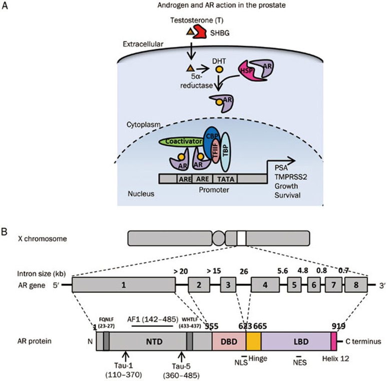
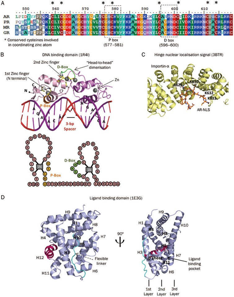
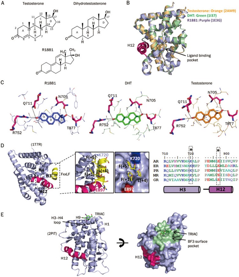
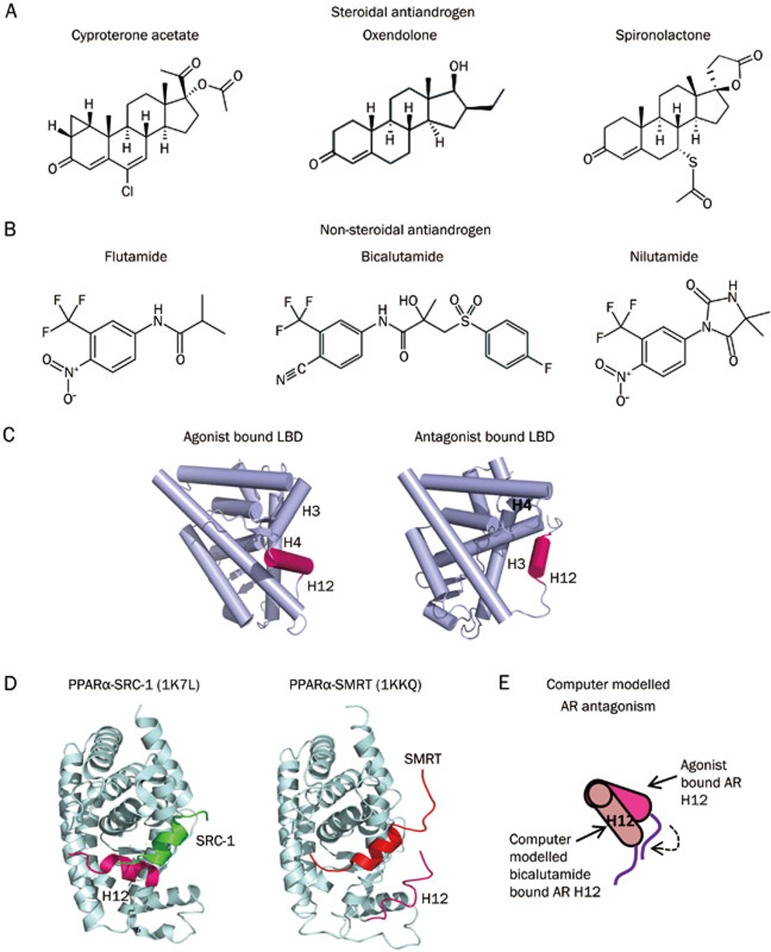
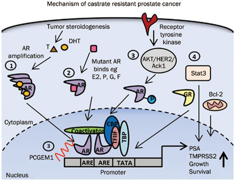
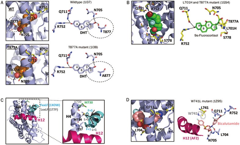
Androgens and androgen receptors (AR) play a pivotal role in expression of the male phenotype. Several diseases, such as androgen insensitivity syndrome (AIS) and prostate cancer, are associated with alterations in AR functions. Indeed, androgen blockade by drugs that prevent the production of androgens and/or block the action of the AR inhibits prostate cancer growth. However, resistance to these drugs often occurs after 2-3 years as the patients develop castration-resistant prostate cancer (CRPC). In CRPC, a functional AR remains a key regulator. Early studies focused on the functional domains of the AR and its crucial role in the pathology. The elucidation of the structures of the AR DNA binding domain (DBD) and ligand binding domain (LBD) provides a new framework for understanding the functions of this receptor and leads to the development of rational drug design for the treatment of prostate cancer. An overview of androgen receptor structure and activity, its actions in prostate cancer, and how structural information and high-throughput screening have been or can be used for drug discovery are provided herein.


Figures

Figure 1
Figure 1
Androgen and AR action. Genome organization of the human androgen receptor gene and the functional domain structure of the androgen receptor protein. (A) Androgen and AR signaling in prostate cells. After testicular synthesis, testosterone is transported to target tissues such as the prostate and becomes converted to dihydrotestosterone (DHT) by 5-α-reductase. DHT binds to the ligand-binding pocket and promotes the dissociation of heat-shock proteins (HSPs) from the AR. The AR then translocates into the nucleus, dimerizes and binds to the androgen response element (ARE) in the promoter region of target genes such as prostate-specific antigen (PSA) and TMPRSS2. At the promoter, the AR is able to recruit members of the basal transcription machinery [such as TATA-box-binding protein (TBP) and transcription factor IIF (TFIIF)] in addition to other coregulators such as members of the p160 family of coactivators and cAMP-response element-binding protein (CREB)-binding protein (CBP). SHBG: serum sex hormone-binding globulin. (B) The androgen receptor gene has been mapped to the long arm of the X-chromosome (locus: Xq11-q12). It contains eight exons interrupted by introns of varying lengths (0.7–2.6 kb) and codes for a protein of 919 amino acids consisting of several functional domains (N-terminal domain (NTD), DNA binding domain (DBD) and ligand binding domain (LBD); amino acid residue numbers are indicated above the AR protein domain map). Exon 1 codes for the NTD, exons 2 and 3 encode the DBD, and exons 4 to 8 encode both the hinge and LBD.
Figure 2
Figure 2
Structures of AR functional domains. (A) Sequence alignment of the DNA binding domain of the androgen receptor (AR), progesterone receptor (PR), mineralocorticoid receptor (MR), and glucocorticoid receptor (GR) performed using ClustalW. *indicates the conserved cysteines involved in coordinating the zinc atom. (B) Top, crystal structure of the AR DBD (pink) (PDB: 1R4I) complexed with its hormone response element (red/purple). The DBD contains two zinc fingers (grey). Each zinc ion is coordinated by four cysteines (yellow). One zinc finger is involved in direct DNA binding mediated by the P-box (orange), which recognizes the specific hormone response element half-site 5′-AGAACA-3′. The other zinc finger is involved in a “head-to-head” receptor dimerization through the D-box (green). Bottom, cartoon representation of the AR DBD. (C) Crystal structure of an AR nuclear localization signal (NLS) peptide (amino acid 621-635) (orange) complexed with importin-α (yellow) (PDB: 3BTR). Residues from the major NLS site 629-RKLKKL-634 contribute to importin-α binding. (D) Crystal structure of the AR ligand binding domain (purple) (LBD) (PDB: 1E3G). The LBD consists of 11 α-helices and two small, two-stranded β-sheets arranged in a typical three-layer antiparallel helical sandwich fold. The long flexible linker between helices 1 and 3 is colored blue.
Figure 3
Figure 3
Structural basis of AR agonism. (A) Chemical structures of testosterone, dihydrotestosterone and R1881. (B) Structural overlay of the AR LBD complexed with testosterone (PDB: 2AM9, orange), dihydrotestosterone (PDB: 1I37, green), and R1881 (PDB: 1E3G, purple). (C) Comparison of the binding of R1881, testosterone, and dihydrotestosterone in the AR ligand-binding pocket. AR LBD residues within a distance of 4 Å are shown. Key residues (N705, Q711, R752, and T877) that form hydrogen bonds with ligands are labeled and shown in stick presentation. Hydrogen bonds are indicated by dotted lines. (D) Structure of the AR LBD (purple) in complex with an FxxLF motif-containing peptide (yellow) (Left panel). The middle panel shows the interface between the AR LBD and the FxxLF motif. Hydrogen bonds are indicated by dotted lines with key residues labeled. A surface view of the motif-binding hydrophobic pocket is shown. A sequence alignment of helices H3 and H12 of the androgen receptor (AR), estrogen receptor (ER), progesterone receptor (PR), mineralocorticoid receptor (MR) and glucocorticoid receptor (GR) was performed using ClustalW. (E) Structure of the AR LBD complexed with 3,3′,5-triiodothyroacetic acid (TRIAC) (PDB: 2PIT). Left: cartoon representation; right: surface view of the binding function (BF3) surface pocket (green).
Figure 4
Figure 4
Structural model of AR antagonism. (A) Chemical structure of the steroidal antiandrogens cyproterone acetate, oxendolone and spironolactone. (B) Chemical structure of the non-steroidal antiandrogens flutamide, bicalutamide and nilutamide. (C) Nuclear receptor H12 helices can adopt different conformations. In an agonist state, the H12 of DHT-bound AR (PDB: 1I37 or 2AMA) is held near H3, H4, and H11, which form a groove for coactivator binding. In an antagonist state, H12 rotates clockwise toward H3 and blocks the coactivator binding site. (D) Structure of the PPARα LBD complexed with SRC-1 coactivator peptide (H12 in agonist conformation) and with the SMRT corepressor peptide (H12 in antagonist conformation). (E) Computer model of antagonist-bound AR shows the predicted displacement of H12.
Figure 5
Figure 5
Androgen and AR action in castration-resistant prostate cancer. Mechanism of castration-resistant prostate cancer. Several mechanisms promote the progression of castration-resistant prostate cancer: (1) AR overexpression coupled with continued tumor steroidogenesis. (2) Promiscuous binding and activation of mutant AR by alternative ligands, such as estrogen (E2), progesterone (P), glucocorticoids (C) and flutamide (F). (3) Ligand-independent mechanisms of AR activation via crosstalk with Akt, HER2, and Ack1 kinases that phosphorylate the AR and via long non-coding RNAs (eg, PCGEM1) that bind to the AR to stimulate transcription of AR target genes. (4) AR-independent pathways, in which cancer cell survival and growth are directed by Stat3 signaling or by upregulation of anti-apoptotic Bcl-2. Glucocorticoid receptor (GR) was found to activate a similar set of AR target genes necessary for survival of cancer cells.
Figure 6
Figure 6
Chemical structure of enzalutamide.
Figure 7
Figure 7
Structural understanding of disease/drug resistance-related androgen receptor mutations. (A) Structural comparison of wild-type (PDB: 1I37) and mutant T877A (PDB: 1I38) AR LBDs in complex with dihydrotestosterone. Key residues involved in hydrogen bonding have been highlighted, with hydrogen bonds indicated by black dotted lines. (B) Structure of the AR LBD double mutant L701H/T877A complexed with 9α-fluorocortisol (PDB: 1GS4). (C) Structural overlay of androgen receptor complexed with FxxLF (PDB: 1XOW) and LxxLL (PDB: 1T7F) motif-containing peptides. Residues V730, M734, and I737 are involved in forming hydrophobic contacts with coactivator peptides. Residue V730 was mutated to M730 to demonstrate an enhanced binding of LxxLL peptides. (D) Structure of AR LBD W741L complexed with bicalutamide (PDB: 1Z95). Residues L704, N705, Q711, and R752 form hydrogen bonds with bicalutamide (indicated by dotted lines). Also shown is the wild-type W741 residue (white) to illustrate a possible steric clash between tryptophan and the B-ring of bicalutamide.

Similar articles

Cited by

References

    1. Mangelsdorf DJ, Thummel C, Beato M, Herrlich P, Schutz G, Umesono K, et al. The nuclear receptor superfamily: the second decade. Cell 1995; 83: 835–9. - PMC - PubMed
    1. Tsai MJ, O'Malley BW. Molecular mechanisms of action of steroid/thyroid receptor superfamily members. Annu Rev Biochem 1994; 63: 451–86. - PubMed
    1. Nuclear Receptors Nomenclature Committee. A unified nomenclature system for the nuclear receptor superfamily. Cell 1999; 97: 161–3. - PubMed
    1. Rosner W, Hryb DJ, Khan MS, Nakhla AM, Romas NA. Sex hormone-binding globulin: anatomy and physiology of a new regulatory system. J Steroid Biochem Mol Biol 1991; 40: 813–20. - PubMed
    1. Baker ME. Albumin, steroid hormones and the origin of vertebrates. J Endocrinol 2002; 175: 121–7. - PubMed

Publication types