. 2014 Sep;234(1):120-33.
doi: 10.1002/path.4388. Epub 2014 Jul 25.

Direct acute tubular damage contributes to Shigatoxin-mediated kidney failure

Affiliations
Free PMC article

Direct acute tubular damage contributes to Shigatoxin-mediated kidney failure

Stefan Porubsky et al. J Pathol. 2014 Sep.
Free PMC article

Abstract

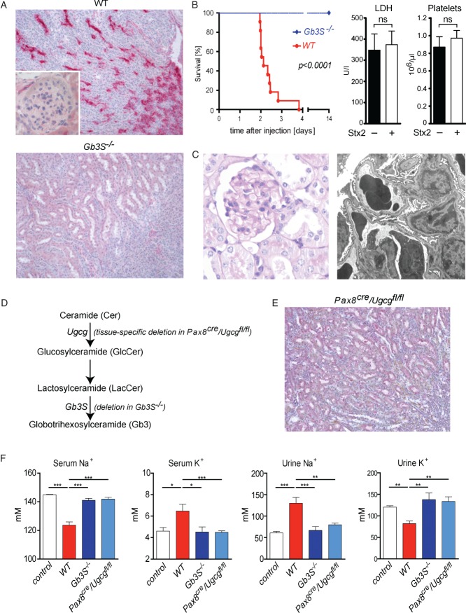
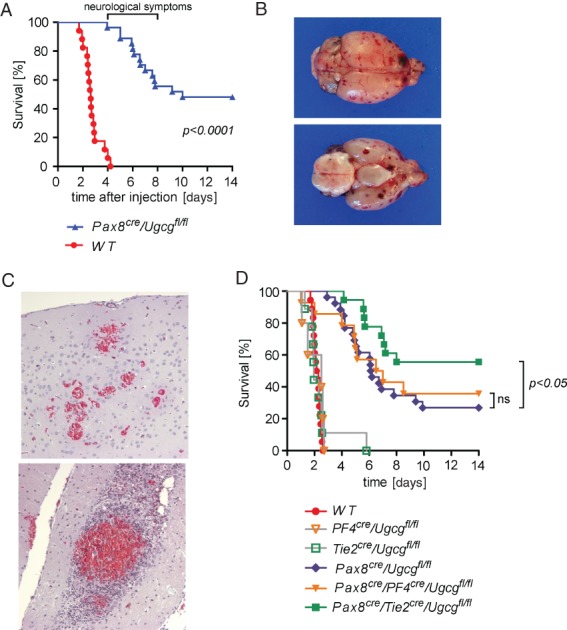
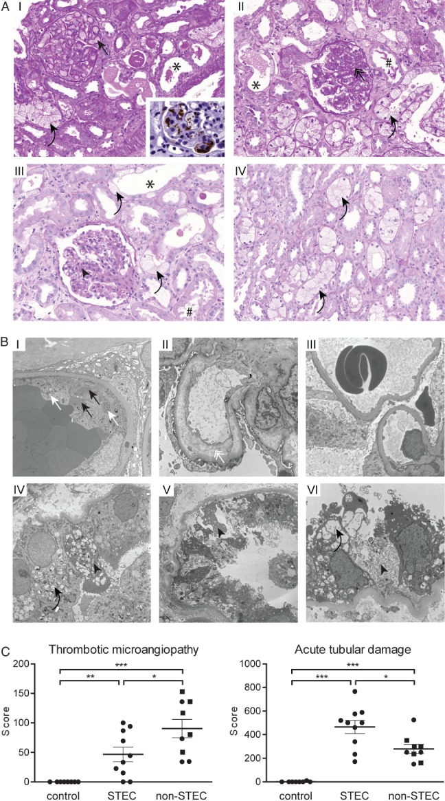
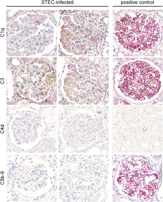
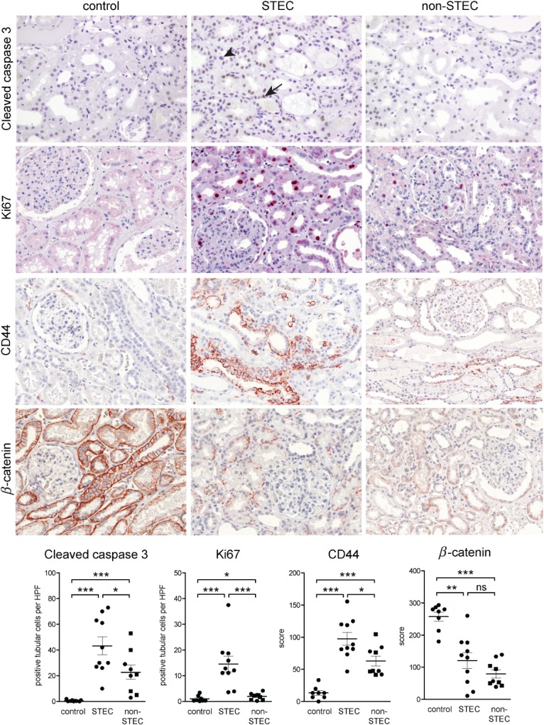
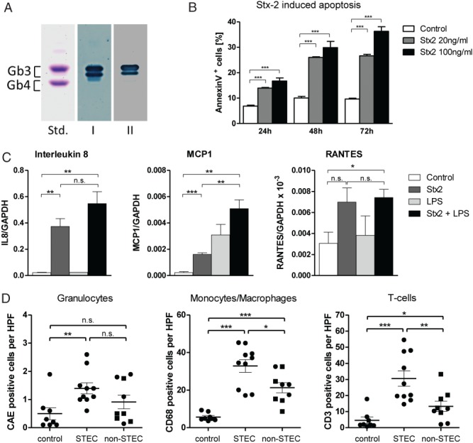
The pathogenesis and therapy of Shigatoxin 2 (Stx2)-mediated kidney failure remain controversial. Our aim was to test whether, during an infection with Stx2-producing E. coli (STEC), Stx2 exerts direct effects on renal tubular epithelium and thereby possibly contributes to acute renal failure. Mice represent a suitable model because they, like humans, express the Stx2-receptor Gb3 in the tubular epithelium but, in contrast to humans, not in glomerular endothelia, and are thus free of glomerular thrombotic microangiopathy (TMA). In wild-type mice, Stx2 caused acute tubular dysfunction with consequent electrolyte disturbance, which was most likely the cause of death. Tubule-specific depletion of Gb3 protected the mice from acute renal failure. In vitro, Stx2 induced secretion of proinflammatory cytokines and apoptosis in human tubular epithelial cells, thus implicating a direct effect of Stx2 on the tubular epithelium. To correlate these results to human disease, kidney biopsies and outcome were analysed in patients with Stx2-associated kidney failure (n = 11, aged 22-44 years). The majority of kidney biopsies showed different stages of an ongoing TMA; however, no glomerular complement activation could be demonstrated. All biopsies, including those without TMA, showed severe acute tubular damage. Due to these findings, patients were treated with supportive therapy without complement-inhibiting antibodies (eculizumab) or immunoadsorption. Despite the severity of the initial disease [creatinine 6.34 (1.31-17.60) mg/dl, lactate dehydrogenase 1944 (753-2792) U/l, platelets 33 (19-124)/nl and haemoglobin 6.2 (5.2-7.8) g/dl; median (range)], all patients were discharged after 33 (range 19-43) days with no neurological symptoms and no dialysis requirement [creatinine 1.39 (range 0.84-2.86) mg/dl]. The creatinine decreased further to 0.90 (range 0.66-1.27) mg/dl after 24 months. Based on these data, one may surmise that acute tubular damage represents a separate pathophysiological mechanism, importantly contributing to Stx2-mediated acute kidney failure. Specifically in young adults, an excellent outcome can be achieved by supportive therapy only.

Keywords: Shigatoxin; Shigatoxin-producing Escherichia coli (STEC); acute renal failure; acute tubular damage; electron microscopy; globoside (Gb3, CD77); thrombotic microangiopathy.


Figures

Figure 1
Figure 1
Stx2 exerts direct toxic effects towards tubular epithelial cells. (A) Indirect immunohistochemistry using primary anti-Gb3 antibodies in combination with alkaline phosphatase-conjugated secondary antibodies visualized Gb3 expression in collecting ducts but not in glomerular or renal endothelial cells of WT mice. In Gb3S-deficient (Gb3S−/−) mice, Gb3 expression was completely abolished; magnification = ×200. (B) WT and Gb3S−/− mice were injected with 0.2 µg Stx2 i.p. While all WT mice died 2–4 days after the injection, all Gb3S−/− mice survived and showed no abnormalities. In moribund Stx2-injected WT mice, serum lactate dehydrogenase (LDH) and platelet counts were measured 36 h after the injection of Stx2. No increase in LDH or decrease of platelet count could be observed, as compared to PBS-injected control WT mice (n = 5/group). (C) Consistent with the absence of Gb3 in the murine glomerulus, TMA could not be detected in the glomeruli of moribund mice. Upon light and transmission electron microscopy, the glomerular morphology was unremarkable; in particular, no signs of endothelial damage or TMA could be detected by electron microscopy: (left panel) PAS staining, magnification = ×400; (right panel) electron micrograph, lead citrate/uranyl acetate staining, magnification = ×6300. (D) Scheme of the synthesis of globotrihexosylceramide (Gb3), starting with ceramide (Cer). Successive additions of one glucose (Glc) and two galactose moieties result in the formation of Gb3. The last step is catalysed by Gb3 synthase (Gb3S) and is defective in Gb3S−/− mice. Tissue-specific depletion of Gb3 was achieved by deletion of the UDP-glucose:ceramide glucosyltransferase (Ugcg) gene, which encodes for the enzyme catalysing the synthesis of glucosylceramide (GlcCer) two steps upstream of Gb3S. (E) Immunohistochemistry for Gb3 performed as in (A), revealed no tubular staining in the kidneys of Pax8cre/Ugcgfl/fl mice as contrasted to WT (cf A); magnification = ×200. (F) 36 h after the Stx2-injection, serum and urine analysis showed profound and significant hyponatraemia and hyperkalaemia mirrored by inverse changes in urine sodium and potassium in WT mice. In contrast, global (Gb3S−/−) as well as tubule-specific depletion of Gb3 (Pax8cre/Ugcgfl/fl) were fully protective. PBS-injected mice served as controls (n = 5/group); bars in (B, F) show mean ± SEM; statistical differences were tested by two-tailed Student's t-test; ns, non-significant; *p < 0.05; **p < 0.01; ***p < 0.001
Figure 2
Figure 2
Mice lacking the Stx2-receptor Gb3 in tubular epithelium developed cerebral purpura after Stx2-injection. (A) Although Pax8cre/Ugcgfl/fl mice were protected from kidney failure and electrolyte dysregulation (shown in Figure 1F), severe and partially fatal neurological symptoms occurred 4–8 days after the Stx2 injection. (B, C) Autopsy revealed diffuse cerebral purpura (B) with bleeding and cerebral TMA; Goldner's trichrome staining, visualizing red blood cells in the haemorrhage foci; magnification = ×200. (D) Depletion of Gb3 in endothelial cells in addition to the tubular cells (Pax8cre/Tie2cre/Ugcgfl/fl, green line) conferred a partial protection from cerebral purpura and improved survival significantly as compared to pure tubular depletion of Gb3 (Pax8cre/Ugcgfl/fl, blue line). In contrast, depletion of Gb3 in platelets in addition to tubular cells (Pax8cre/PF4cre/Ugcgfl/fl, yellow line) did not prove to be protective. As expected, mice with Gb3 deficiency solely in platelets or endothelial cells (PF4cre/Ugcgfl/fl and Tie2cre/Ugcgfl/fl, respectively) died during the early phase, contemporarily with the Stx2-exposed WT mice.
Figure 3
Figure 3
In addition to TMA, STEC-infection caused a profound tubular injury in man. (A) Light microscopy: a part of the biopsies showed glomeruli with congested capillaries filled with red bold cells (I, black arrow) and platelet-containing thrombotic material, visualized as a brown signal in CD61 immunohistochemistry (I, inlay). Other cases predominantly showed glomeruli with segmental sclerosis and wrinkling and collapse of capillaries (II, black double arrow), indicating a preceding but not active TMA. Glomeruli of other biopsies had almost normal architecture, with only a slight mesangial matrix increase (III, black arrowhead). A constant finding in all biopsies was a pronounced acute tubular damage, manifested as epithelial flattening (*) and/or presence of intraluminal cell detritus (#) and/or pronounced vacuolization (I–IV, black curved arrows); PAS staining, magnification = ×250. (B) Transmission electron microscopy of glomeruli (I–III) and tubular epithelium (IV–VI). Active TMA was documented by the presence of thrombotic material composed of platelets (I, white arrows) and fibrin strands (I, black arrows) in glomerular capillaries. Endothelial damage was manifested as absence of endothelial fenestrae and widening of the subendothelial space (II, white double arrow). In two patients the glomeruli showed normal architecture (III). Ultrastructural correlates of the pronounced tubular damage manifested as vacuolization (IV and VI, black curved arrows) and epithelial necrosis (IV–VI, black arrowheads); electron micrographs, lead citrate/uranyl acetate staining; magnification = (glomeruli) × 5000; (tubules) × 6300. (C) The extent of the glomerular TMA and acute tubular damage were compared between normal kidney tissue (tumour-free parts of tumour nephrectomies, control, n = 8), kidney biopsies from STEC-infected patients (STEC, n = 10) and randomly chosen biopsies from patients with STEC-unrelated TMA (non-STEC, n = 9; individuals > 45 years are depicted as squares). Glomerular TMA was semi-quantified on a PAS-stained slide by scoring the extent of TMA in each glomerulus. Acute tubular damage was assessed by building a score reflecting the following three criteria: brush border loss in proximal tubules; epithelial cell flattening; and vacuolization. Although the extent of TMA was less, the tubular damage was significantly higher in the STEC group than in the non-STEC group; data are shown as mean ± SEM; statistical differences were tested by two-tailed Mann–Whitney test; *p < 0.05; **p < 0.01; ***p < 0.001
Figure 4
Figure 4
Immunohistochemistry for complement factors in glomeruli of patients infected with STEC. In the glomeruli of patients with STEC-elicited TMA, the presence of complement factors C1q, C3, C4 and C5b-9 (membrane attack complex) was investigated by immunohistochemistry (alkaline phosphatase/anti-alkaline phosphatase; APAAP method). For C1q, C3 and C5b-9, membranous glomerulopathy served as a positive control. For C4d, an AB0-incompatible kidney transplant showing staining in the peritubular capillaries was used as a positive control. In contrast to the strong staining in the positive control, only faint focal segmental staining for C1q and C3 was detected in STEC-associated TMA. For C4d and C5b-9, virtually no staining was observed in STEC-infected patients; magnification = ×400
Figure 5
Figure 5
Immunohistochemical findings of the STEC-associated acute tubular damage. Expression of cleaved caspase 3, Ki67, β-catenin and CD44 were studied by immunohistochemistry in STEC-infected patients (n = 10) and compared to normal kidney tissue from tumour nephrectomies (control, n = 8) and to kidney biopsies with TMA due to a STEC-unrelated aetiology (non-STEC, n = 9). The acute tubular damage was associated with a significant increase in apoptosis (visualized by cleaved caspase 3), reactive proliferation (visualized by Ki67) and elevated expression of CD44 in both STEC and non-STEC groups. However, for all three parameters, the increase was significantly higher in the STEC than in the non-STEC group. In the STEC group, apoptosis (arrow) and mitosis (arrowhead) could be seen in tubular epithelial cells; β-catenin was significantly down-regulated in renal tubular epithelium of both STEC and non-STEC patients, to a similar extent; magnification = ×100. In the case of immunohistochemistry for cleaved caspase 3 and Ki67, the number of positive tubular cells was counted and expressed as average cell number/high power field (HPF; ×40 objective, ×10 ocular). For evaluation of the immunohistochemistry for β-catenin and CD44, semiquantitative scoring of the staining intensity of tubular cells in each HPF of the biopsy was performed; in the non-STEC group, individuals > 45 years are depicted as squares. Statistical differences were tested by two-tailed Mann–Whitney test; ns, non-significant; *p < 0.05; **p < 0.01; ***p < 0.001
Figure 6
Figure 6
Stx2 effects on human tubular epithelial cells and on interstitial inflammation. (A) Thin-layer chromatography immuno-overlay demonstrated the presence of the Stx2 receptor Gb3 on the human renal tubular epithelial cell line HK-2. Glycosphingolipid aliquots from the HK-2 cells were applied for Gb3 detection, using polyclonal anti-Gb3 antibody (lane I) and Stx2 (lane II). An equimolar mixture of Gb3 and Gb4 (10 µg), prepared from human erythrocytes, served as standard (Std); the two bands of Gb3 are due to different chain lengths of fatty acids in the ceramide portion of Gb3. (B) Apoptosis under the influence of Stx2 was investigated in HK-2 cells; these were exposed for 24, 48 and 72 h to cell culture medium alone (control) or Stx2 at 20 or 100 ng/ml. Apoptosis was measured by flow cytometry as the proportion of Annexin V-positive cells. Stx2 led to a time- and dose-dependent increase of apoptosis in HK-2 cells (n = 4). (C) Expression of selected cytokines in HK-2 cells in response to Stx2 and LPS was investigated; to this end, cells were exposed to Stx2 (100 ng/ml) and/or LPS (1 µg/ml) for 6 h and the expression of IL8, MCP1, RANTES and GAPDH was measured by quantitative RT–PCR. Whereas Stx2 increased the expression of all three cytokines, no further elevation was observed for IL8 and RANTES when the cells were also treated with LPS (n = 3). (D) The presence of granulocytes [positive for chloracetate esterase (CAE)], monocytes/macrophages (CD68-positive) and T cells (CD3-positive) was quantified in kidney biopsies of STEC-infected patients (STEC, n = 10) and compared to normal kidneys (non-tumourous parts of tumour nephrectomies, control, n = 8) and biopsies from patients with STEC-unrelated TMA (non-STEC, n = 9). Whereas the granulocytic infiltrate was elevated in both the STEC and non-STEC groups to a similar extent, a significantly higher increase was observed for monocytes/macrophages and T cells in the STEC than in the non-STEC group; in the non-STEC group, individuals > 45 years are depicted as squares. In (B, C) statistical differences were tested by two-tailed Student's t-test; in (D) two-tailed Mann–Whitney test was used: data are shown as mean ± SEM; n.s., non-significant; *p < 0.05; **p < 0.01; ***p < 0.001

References

    1. Riley LW, Remis RS, Helgerson SD, et al. Hemorrhagic colitis associated with a rare Escherichia coli serotype. N Engl J Med. 1983;308:681–685. - PubMed
    1. Karmali MA, Petric M, Lim C, et al. The association between idiopathic hemolytic uremic syndrome and infection by verotoxin-producing Escherichia coli. J Infect Dis. 1985;151:775–782. - PubMed
    1. MMWR. Update: multistate outbreak of Escherichia coli O157:H7 infections from hamburgers – western United States, 1992–1993. MMWR Morb Mortal Wkly Rep. 42:258–263. - PubMed
    1. Michino H, Araki K, Minami S, et al. Massive outbreak of Escherichia coli O157:H7 infection in schoolchildren in Sakai City, Japan, associated with consumption of white radish sprouts. Am J Epidemiol. 1999;150:787–796. - PubMed
    1. Thorpe CM. Shiga toxin-producing Escherichia coli infection. Clin Infect Dis. 2004;38:1298–1303. - PubMed

Publication types

MeSH terms