. 2014 Jul 8;22(7):961-73.
doi: 10.1016/j.str.2014.04.013. Epub 2014 Jun 5.

Influence of domain interactions on conformational mobility of the progesterone receptor detected by hydrogen/deuterium exchange mass spectrometry

Affiliations

Influence of domain interactions on conformational mobility of the progesterone receptor detected by hydrogen/deuterium exchange mass spectrometry

Devrishi Goswami et al. Structure. .

Abstract

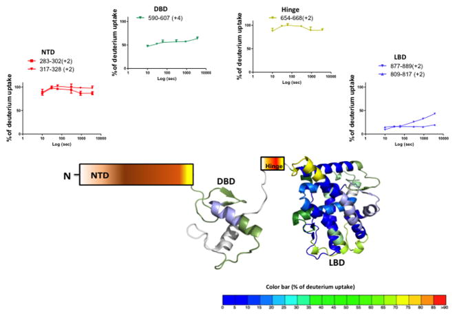
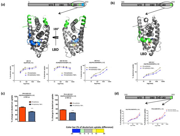
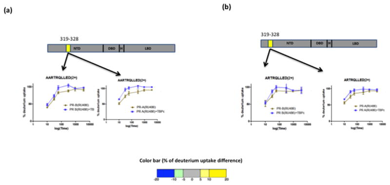
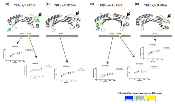
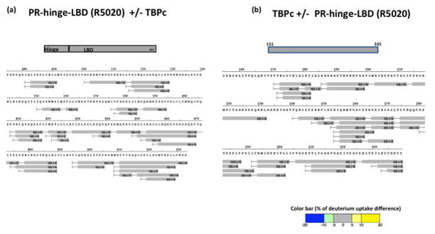
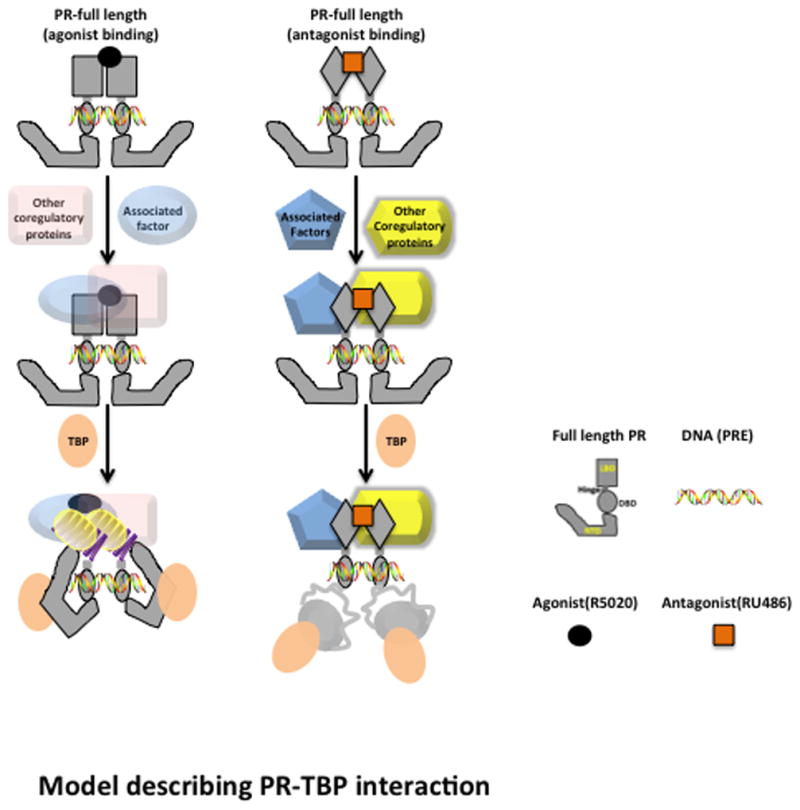
Structural and functional details of the N-terminal activation function 1 (AF1) of most nuclear receptors are poorly understood due to the highly dynamic intrinsically disordered nature of this domain. A hydrogen/deuterium exchange (HDX) mass-spectrometry-based investigation of TATA box-binding protein (TBP) interaction with various domains of progesterone receptor (PR) demonstrate that agonist-bound PR interaction with TBP via AF1 impacts the mobility of the C-terminal AF2. Results from HDX and other biophysical studies involving agonist- and antagonist-bound full-length PR and isolated PR domains reveal the molecular mechanism underlying synergistic transcriptional activation mediated by AF1 and AF2, dominance of PR-B isoform over PR-A, and the necessity of AF2 for full AF1-mediated transcriptional activity. These results provide a comprehensive picture elaborating the underlying mechanism of PR-TBP interactions as a model for studying nuclear receptor (NR)-transcription factor functional interactions.


Figures

Figure 1
Figure 1. Solution state conformational flexibility of full length PR-B
Agonist (R5020) bound full length PR-B was analyzed by hydrogen deuterium exchange studies and the average % of deuterium incorporation across six different time points (0,10,30,60,300,900 and 3600 sec) is overlaid onto the crystal structure of PR LBD (PDB ID: 1A28) and PR DBD (PDB ID: 2C7A). The color is according to the color code at the bottom of the figure. The hinge region and N terminal domain are represented by schematics due to lack of atomic structure. Representative peptide deuterium build up curves from each domain are indicated at top.
Figure 2
Figure 2. Agonist (R5020) bound full length PR when interacted with TBPc shows stabilization at the C terminal ligand binding domain (LBD)
(a) Agonist (R5020) bound PR-B isoform when interacted with TBPc, shows protection from exchange at several regions in LBD as depicted by schematic representations. The average % of deuterium uptake values across six different time points is overlaid (color coded) onto LBD atomic structure (PDB ID: 1A28). The color is according to the color bar at the bottom of the figure. The representative deuterium build-up curves of protected regions of the LBD, including the helix 12 is shown also. (b) Schematic representation along with atomic structure (LBD) overlay and deuterium build-up curve of agonist bound full length PR-A and TBPc interaction. (c) Comparison of deuterium uptake of AF2 regions in agonist bound PR-B and PR-A interaction with TBPc after 900 sec exposure to heavy water. Asterisks indicate significant differences as calculated by the processing software (Pascal et al., 2009). (d) HDX footprint of truncated agonist bound PR-B (233-933) - TBP interaction and corresponding deuterium build-up curve.
Figure 3
Figure 3. Antagonist (RU486) bound full length PR interaction with TBPc
Antagonist bound full length PR-B (a) and PR-A (b) when interacted with TBPc showed slight destabilization at the N terminal domain. Corresponding build-up curves are shown with the schematic representation.
Figure 4
Figure 4. Stabilization of TBPc structure upon interaction with isolated N terminal domain (NTD) and agonist bound full length PR
TBP shows protection from exchange when interacted with isolated NTD of PR-B (a), NTD of PR-A (b), agonist bound full length PR-B (c) and agonist bound full length PR-A (d). Regions of protection are represented by schematics and the average % of deuterium uptake values across six different time points is overlaid (color coded) onto the TBPc crystal structure (PDB ID: 1TGH) according to the color bar at the bottom of the figure. The deuterium build-up curves are shown below the atomic models.
Figure 5
Figure 5. Ligand binding domain (LBD) of PR shows no significant interaction with TBPc by itself
Differential HDX data of agonist bound PR hinge-LBD region with TBPc (a) and vice versa (b) showed no significant interactions. The color coding is according to the color bar at the bottom of the figure.
Figure 6
Figure 6. Folding NTD in the context of full length PR-A in the presence of TBPC as detected by protection against partial proteolysis
Purified PR-A, PR-NTD, PR-LBD, TBPC or an equal molar mixture of PR-A:TBPC, PR-A NTD:TBPC, and PR-LBD:TBPC proteins were subjected to limited proteolysis with trypsin and samples were analyzed by Coomassie blue-stained SDS-PAGE or by immunoblotting. a) Coomassie-stained SDS-PAGE. b) Immunoblotting of limited proteolytic products derived from either full length PR-A or PR-A NTD with antibodies (1294 and 636) specific to distinct epitopes in the NTD or the C-terminal LBD (10A9), c) Immunoblotting with the C-terminal MAb (10A9) of limited proteolytic products generated from the isolated PR-LBD at different time points indicated (0 to 20 min).
Figure 7
Figure 7. A model of TBP-PR NTD interaction to allosterically mediate structural changes in LBD/AF2
Both hormonal agonist and antagonist promote dimerization and binding of PR to progesterone response element (PRE) target DNA. Once DNA-bound, receptor dimers assemble a multi-protein complex of coregulators which differ for an agonist vs. antagonist due at least in part to distinct conformations in the LBD/AF2. In the presence of agonist, TBP binding to the NTD results in conformational rearrangements in the NTD and in the LBD/AF2 through allosteric regulation. These structural rearrangements facilitate the binding and assembly of other co-activators at either LBD/AF2 or AF1 in the NTD. In the presence of antagonist, TBP binding with NTD resulted in a slight destabilization of NTD structure and the interdomain communication with LBD/AF2 was lost.

References

    1. Anderson E, Clarke RB. Steroid receptors and cell cycle in normal mammary epithelium. J Mammary Gland Biol Neoplasia. 2004;9:3–13. - PubMed
    1. Bain DL, Franden MA, McManaman JL, Takimoto GS, Horwitz KB. The N-terminal region of human progesterone B-receptors: biophysical and biochemical comparison to A-receptors. J Biol Chem. 2001;276:23825–23831. - PubMed
    1. Berry M, Metzger D, Chambon P. Role of the two activating domains of the oestrogen receptor in the cell-type and promoter-context dependent agonistic activity of the anti-oestrogen 4-hydroxytamoxifen. EMBO J. 1990;9:2811–2818. - PMC - PubMed
    1. Billas I, Moras D. Allosteric controls of nuclear receptor function in the regulation of transcription. J Mol Biol. 2013;425:2317–2329. - PubMed
    1. Bledsoe RK, Montana VG, Stanley TB, Delves CJ, Apolito CJ, McKee DD, Consler TG, Parks DJ, Stewart EL, Willson TM, et al. Crystal structure of the glucocorticoid receptor ligand binding domain reveals a novel mode of receptor dimerization and coactivator recognition. Cell. 2002;110:93–105. - PubMed

Publication types

MeSH terms