. 2014 Jun 19;40(6):989-1001.
doi: 10.1016/j.immuni.2014.04.019. Epub 2014 Jun 5.

Activation of the aryl hydrocarbon receptor dampens the severity of inflammatory skin conditions

Affiliations

Activation of the aryl hydrocarbon receptor dampens the severity of inflammatory skin conditions

Paola Di Meglio et al. Immunity. .

Abstract

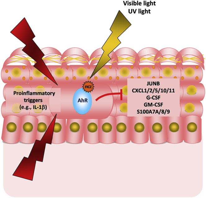
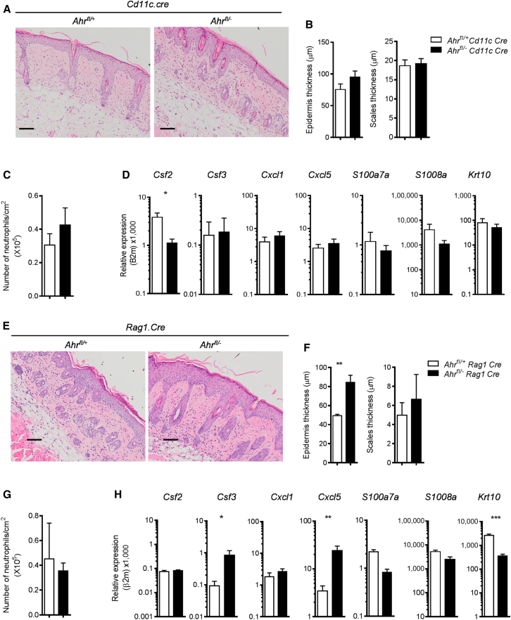
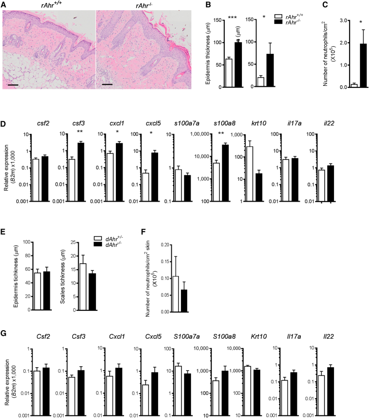
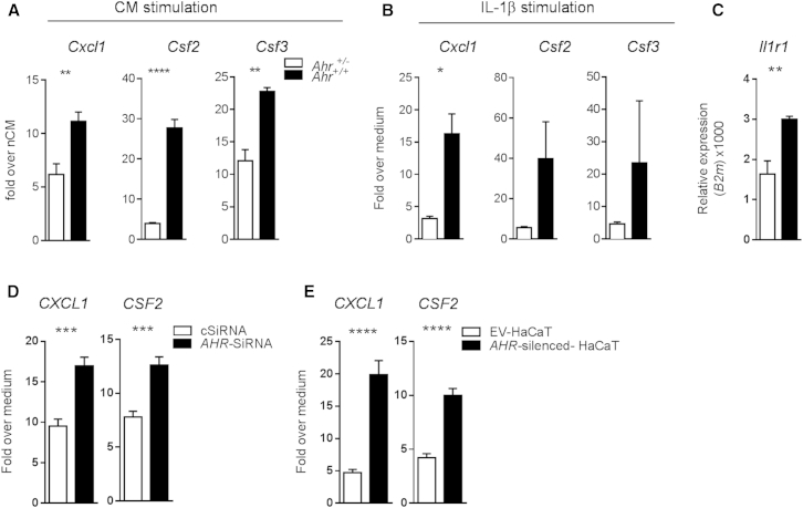
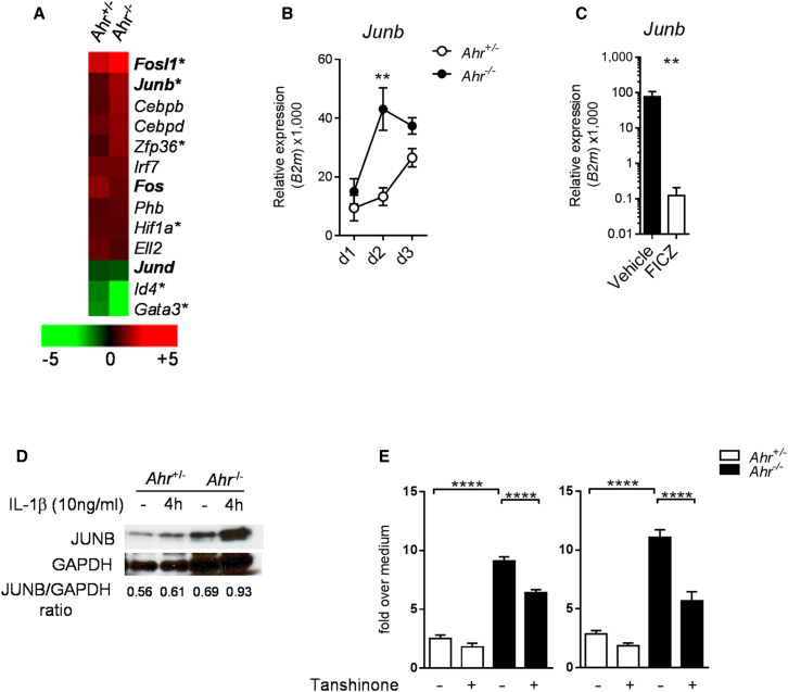
Environmental stimuli are known to contribute to psoriasis pathogenesis and that of other autoimmune diseases, but the mechanisms are largely unknown. Here we show that the aryl hydrocarbon receptor (AhR), a transcription factor that senses environmental stimuli, modulates pathology in psoriasis. AhR-activating ligands reduced inflammation in the lesional skin of psoriasis patients, whereas AhR antagonists increased inflammation. Similarly, AhR signaling via the endogenous ligand FICZ reduced the inflammatory response in the imiquimod-induced model of skin inflammation and AhR-deficient mice exhibited a substantial exacerbation of the disease, compared to AhR-sufficient controls. Nonhematopoietic cells, in particular keratinocytes, were responsible for this hyperinflammatory response, which involved upregulation of AP-1 family members of transcription factors. Thus, our data suggest a critical role for AhR in the regulation of inflammatory responses and open the possibility for novel therapeutic strategies in chronic inflammatory disorders.


Figures

None
Graphical abstract
Figure 1
Figure 1
AhR Ligation in Human Skin Biopsies Modulates Psoriasis-Relevant Genes Lesional (L) and nonlesional (NL) skin biopsies from eight psoriasis patients were quartered: one quarter of each was used as baseline and the remaining three quarters were cultured with either vehicle control, the AhR agonist FICZ, or the AhR antagonist CH-223191 for 16 hr. Whole-skin biopsies from five healthy donors (H) were processed in the same way. All samples were subjected to RNA sequencing. (A) Venn diagram showing genes significantly regulated by either FICZ or CH-223191 in at least one out of the three tissue types analyzed (L, NL, H). (B) Heat map of known AhR-target genes modulated by FICZ and CH-223191. Color indicates mean fold change, with green representing decreased and red increased gene expression. (C) Heat map of genes belonging to the “psoriasis transcriptome” (upregulated genes shown in bold) and modulated by FICZ or CH-223191. Genes are sorted by decreasing fold change for agonist effect on L skin. (D) qPCR validation for six top modulated genes downregulated in L skin by agonist-induced AhR activation. Box and whiskers denoting minimum and maximum values are shown. Wilcoxon signed rank test (for CMPK2) or paired t test (all other genes) was performed. (E) qPCR validation of six top modulated genes upregulated in NL skin by antagonist-induced AhR inhibition. Box and whiskers denoting minimum and maximum values are shown. Wilcoxon signed rank test was performed.
Figure 2
Figure 2
Exacerbated Skin Inflammation in Ahr-Deficient Mice (A) Representative images of H&E staining of skin sections from untreated (baseline) and IMQ-treated Ahr+/− (open circles) and Ahr−/− (filled circles) mice at day 5 (scale bars represent 100 μm). (B) Quantification of epidermal (left) and scale thickness (right) of Ahr+/− (open circles) and Ahr−/− (filled circles) at different time points after initiation of IMQ treatment. (C and D) mRNA expression of proinflammatory mediators and keratinocyte differentiation marker in whole skin from Ahr+/− (open circles) and Ahr−/− (filled circles) mice at day 5. (E) Number of CD4 and γδ T cells expressing IL-17 (left) and IL-22 (right) per cm2 skin of IMQ-treated Ahr+/− (white bars) and Ahr−/− (black bars) mice obtained by intracellular cytokine staining. (F) Number of neutrophils (×105) per cm2 of skin. Plots show mean ± SEM, n = 3–5 mice per group or mean and values of individual mice, n = 8 mice per group. Results from one representative experiment of two independent experiments are shown. p < 0.05, ∗∗p < 0.01, ∗∗∗p < 0.001, and ∗∗∗∗p < 0.0001.
Figure 3
Figure 3
Skin AhR Activation by FICZ Ameliorates IMQ-Induced Psoriasis-like Skin Inflammation (A) Cyp1a1 expression in skin of IMQ-treated C57BL/6 mice receiving vehicle (black bars) or FICZ (white bars) i.p. for 6 days. (B) H&E staining of skin sections (day 6) from IMQ-treated C57BL/6 mice receiving vehicle or FICZ. (C) Quantification of epidermal (left) and scale thickness (right) at day 6 of vehicle-treated (black bars) or FICZ-treated (white bars) mice. (D) mRNA expression of psoriasis-relevant genes in whole skin from IMQ-treated C57BL/6 mice receiving vehicle (black bars) or FICZ (white bars). Plots show mean ± SEM; n = 5 mice per group. Results from one representative experiment of three independent experiments are shown. p < 0.05, ∗∗p < 0.01.
Figure 4
Figure 4
AhR Deficiency in Dendritic Cells or in T and B Cells Does Not Recapitulate the Phenotype of Ahr−/− Mice (A) Representative images of H&E staining of skin sections from imiquimod-treated Ahrfl/+ or Ahrfl/−Cd11c.Cre mice at day 5 (scale bars represent 100 μm). (B) Quantification of epidermal (left) and scale thickness (right) at day 5. (C) Number of neutrophils per cm2 of skin as determined by FACS analysis of Ly6G+ cells. (D) mRNA expression of psoriasis-relevant genes in whole skin from Ahrfl/+ (white bars) or Ahrfl/−Cd11c.Cre mice (black bars) at day 5. (E) Representative images of H&E staining of skin sections from imiquimod-treated Ahrfl/+ or Ahrfl/−Rag1.Cre mice (scale bars represent 100 μm). (F) Quantification of epidermal (left) and scale thickness (right) at day 5. (G) Number of neutrophils per cm2 of skin as determined by FACS analysis of Ly6G+ cells. (H) mRNA expression of psoriasis -relevant genes in whole skin from Ahrfl/+ (white bars) or Ahrfl/−Rag1.Cre mice (black bars) at day 5. Plots show mean + SEM; n = 4–6 mice per group. Results from one representative experiment of two independent experiments per mouse strain are shown. p < 0.05, ∗∗p < 0.01, and ∗∗∗p < 0.001.
Figure 5
Figure 5
Lack of AhR in Nonhematopoietic Cells Recapitulates Exacerbated Skin Inflammation of Ahr−/− Mice (A) Representative images of H&E staining of skin sections from IMQ-treated Ahr+/+Ahr+/+Rag1−/− (rAhR+/+) or Ahr+/+Ahr−/−Rag1−/− (rAhR−/−) BM chimeras (scale bars represent 100 μm). (B) Quantification of epidermal (left) and scale thickness (right) of rAhR+/+ (white bars) or rAhR−/− (black bars) chimeras at day 5. (C) Number of neutrophils per cm2 of skin. (D) mRNA expression of psoriasis-relevant genes in whole skin from rAhR+/+ (white bars) or rAhR−/− (black bars) chimeras at day 5. (E) Quantification of epidermal (left) and scale thickness (right) of Ahr+/−→ Ahr+/+Rag1−/− (dAhR+/−, white bars) or Ahr−/−→ Ahr+/+Rag1−/− (dAhR−/−, black bars) chimeras at day 5. (F) Number of neutrophils per cm2 of skin. (G) mRNA expression of psoriasis-relevant genes in whole skin from dAhR+/− (white bars) or dAhR−/− (black bars) chimeras at day 5. Plots show mean + SEM; n = 5–7 mice per group. Results from one representative experiment of two independent experiments per each set of chimeras are shown. p < 0.05, ∗∗p < 0.01, and ∗∗∗p < 0.001.
Figure 6
Figure 6
Ahr-Deficient Murine and Human Keratinocytes Show Exacerbated Response to Inflammatory Stimuli (A) mRNA expression of proinflammatory mediators in Ahr+/− (white bars) and Ahr/− (black bars) murine keratinocytes stimulated for 24 hr with conditioned medium nCM and iCM. Data are expressed as fold change over stimulation with iCM over nCM medium. (B) mRNA expression of proinflammatory mediators in Ahr+/− (white bars) and Ahr−/− (black bars) murine keratinocytes stimulated for 24 hr with recombinant IL-1β (10 ng/ml). Data expressed as fold change over medium control. (C) Il1r1 mRNA expression in unstimulated murine primary keratinocytes from Ahr+/− (white bars) and Ahr/− (black bars) mice. (D) mRNA expression of proinflammatory mediators in human primary keratinocytes, transiently transfected for 48 hr with a nontargeting control SiRNA (cSiRNA, white bars) or in which AhR was transiently silenced (AhR-SiRNA, black bars), and stimulated for further 24 hr with human recombinant IL-1β (10 ng/ml). (E) mRNA expression of proinflammatory mediators in human keratinocytes HaCaT cell lines, stable transfected with an empty vector (EV-HaCaT, white bars), or in which AhR had been stable silenced (AhR-silenced HaCaT, black bars), and stimulated for 24 hr with human recombinant IL-1β. Data expressed as fold change over medium control. Results from one representative experiment of two or three independent experiments are shown. Plots show mean ± SEM; n = 3–6 wells per group. p < 0.05, ∗∗p < 0.01, and ∗∗∗∗p < 0.0001.
Figure 7
Figure 7
Ahr-Deficient Keratinocytes Display Increased Levels of JunB (A) Heat map visualization of IMQ-regulated, psoriasis-annotated transcription factors in whole skin of Ahr+/− and Ahr−/− mice on day 2 of IMQ treatment. Green color indicates decreased and red color increased gene expression, expressed as mean fold change in IMQ-treated as compared to corresponding untreated mice. AP-1 family transcription factors are marked in bold. Asterisks indicate genes differentially expressed in Ahr/− versus Ahr+/− skin at day 2 of IMQ treatment. (B) Time course of Junb mRNA expression in whole skin from Ahr+/− (open circles) and Ahr−/− (filled circles) mice. (C) Junb mRNA expression in whole skin from IMQ-treated C57BL/6 mice receiving vehicle (black bars) or FICZ (white bars) at day 6. (D) Immunoblot showing JunB protein levels in Ahr+/− (white bars) and Ahr−/− (black bars) murine keratinocytes stimulated for 4 hr with recombinant IL-1β (10 ng/ml). Values denotes JunB/GAPDH densitometry ratio. (E) mRNA expression of Csf2 and Csf3 in murine keratinocytes stimulated for 24 hr with recombinant IL-1β (10 ng/ml) with or without Tanshinone (1 μM). Data expressed as fold change over medium control. Plots show mean ± SEM; n = 3–6 wells or mice per group. Results from one representative experiment of two independent experiments are shown. ∗∗p < 0.01, ∗∗∗p < 0.001, and ∗∗∗∗p < 0.0001.

Comment in

References

    1. Austin L.M., Ozawa M., Kikuchi T., Walters I.B., Krueger J.G. The majority of epidermal T cells in Psoriasis vulgaris lesions can produce type 1 cytokines, interferon-gamma, interleukin-2, and tumor necrosis factor-alpha, defining TC1 (cytotoxic T lymphocyte) and TH1 effector populations: a type 1 differentiation bias is also measured in circulating blood T cells in psoriatic patients. J. Invest. Dermatol. 1999;113:752–759. - PubMed
    1. Bata-Csorgo Z., Szell M. The psoriatic keratinocytes. Expert Rev. Dermatol. 2012;7:473–481.
    1. Bjeldanes L.F., Kim J.Y., Grose K.R., Bartholomew J.C., Bradfield C.A. Aromatic hydrocarbon responsiveness-receptor agonists generated from indole-3-carbinol in vitro and in vivo: comparisons with 2,3,7,8-tetrachlorodibenzo-p-dioxin. Proc. Natl. Acad. Sci. USA. 1991;88:9543–9547. - PMC - PubMed
    1. Bock K.W., Köhle C. Ah receptor: dioxin-mediated toxic responses as hints to deregulated physiologic functions. Biochem. Pharmacol. 2006;72:393–404. - PubMed
    1. Bowcock A.M., Shannon W., Du F., Duncan J., Cao K., Aftergut K., Catier J., Fernandez-Vina M.A., Menter A. Insights into psoriasis and other inflammatory diseases from large-scale gene expression studies. Hum. Mol. Genet. 2001;10:1793–1805. - PubMed

MeSH terms

Associated data