. 2014 Aug 22:274:341-56.
doi: 10.1016/j.neuroscience.2014.05.056. Epub 2014 Jun 5.

Relationships between androgens, serotonin gene expression and innervation in male macaques

Affiliations

Relationships between androgens, serotonin gene expression and innervation in male macaques

C L Bethea et al. Neuroscience. .

Abstract

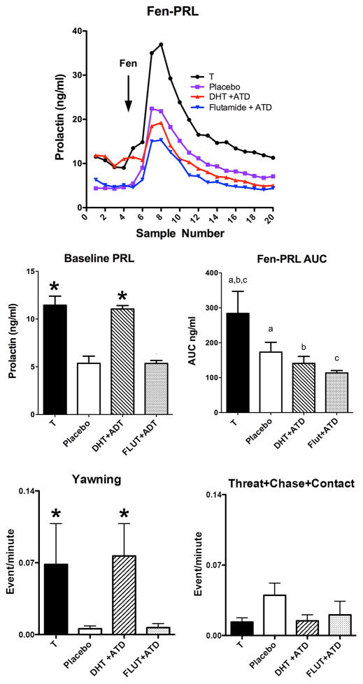
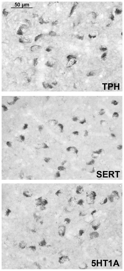
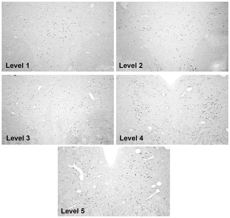
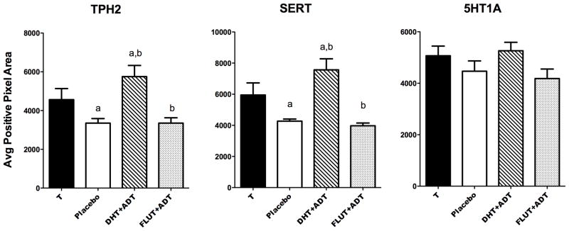
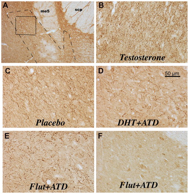
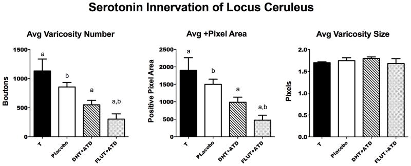
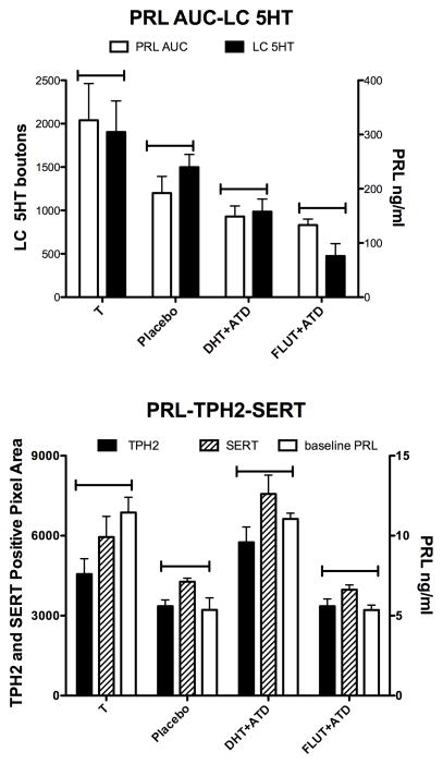
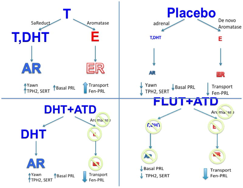
Androgen administration to castrated individuals was purported to decrease activity in the serotonin system. However, we found that androgen administration to castrated male macaques increased fenfluramine-induced serotonin release as reflected by increased prolactin secretion. In this study, we sought to define the effects of androgens and aromatase inhibition on serotonin-related gene expression in the dorsal raphe, as well as serotonergic innervation of the LC. Male Japanese macaques (Macaca fuscata) were castrated for 5-7 months and then treated for 3 months with (1) placebo, (2) testosterone (T), (3) dihydrotestosterone (DHT; non-aromatizable androgen) and ATD (steroidal aromatase inhibitor), or (4) Flutamide (FLUT; androgen antagonist) and ATD (n=5/group). This study reports the expression of serotonin-related genes: tryptophan hydroxylase 2 (TPH2), serotonin reuptake transporter (SERT) and the serotonin 1A autoreceptor (5HT1A) using digoxigenin-ISH and image analysis. To examine the production of serotonin and the serotonergic innervation of a target area underlying arousal and vigilance, we measured the serotonin axon density entering the LC with ICC and image analysis. TPH2 and SERT expression were significantly elevated in T- and DHT + ATD-treated groups over placebo- and FLUT + ATD-treated groups in the dorsal raphe (p < 0.007). There was no difference in 5HT1A expression between the groups. There was a significant decrease in the pixel area of serotonin axons and in the number of varicosities in the LC across the treatment groups with T > placebo > DHT + ATD = FLUT + ATD treatments. Comparatively, T- and DHT + ATD-treated groups had elevated TPH2 and SERT gene expression, but the DHT + ATD group had markedly suppressed serotonin axon density relative to the T-treated group. Further comparison with previously published data indicated that TPH2 and SERT expression reflected yawning and basal prolactin secretion. The serotonin axon density in the LC agreed with the area under the fenfluramine-stimulated prolactin curve, providing a morphological basis for the pharmacological results. This suggested that androgen activity increased TPH2 and SERT gene expression but, aromatase activity, and neural production of estradiol (E), may subserve axonal serotonin and determination of the compartment acted upon by fenfluramine. In summary, androgens stimulated serotonin-related gene expression, but aromatase inhibition dissociated gene expression from the serotonin innervation of the LC terminal field and fenfluramine-stimulated prolactin secretion.

Keywords: 5HT1A; SERT; TPH2; androgen; aromatase; serotonin.


Figures

Figure 1
Figure 1
TOP. Line graph of the PRL response to fenfluramine injection in castrated male monkeys in the 4 groups treated with T, placebo, DHT+ATD and FLUT+ATD that were used in this study. Middle. Histograms illustrating the area under the fenfluramine-stimulated prolactin secretory curve and baseline prolactin across the 4 treatment groups used in this study (out of 6 groups examined for behavior and prolactin secretion in a previous publication (Bethea et al., 2013b). The data have been extracted and reprinted for ease of comparison to data collected in this study. Further statistical analysis was conducted on the 4 included groups. The area under the prolactin curve (fen-PRL AUC) showed a significant difference across the groups (F [3,16] = 4.44; p = 0.019). There was also a significant difference across the groups in baseline prolactin, obtained at 10 min intervals for 1 hour prior to fenfluramine challenge (F [3,20] = 27.40; p < 0.0001). Bottom. Histograms illustrating different components of behavior during the treatment periods. Androgen treatment increased yawning and there was no effect of aromatase inhibition. There was little overt aggressive behavior during the treatment period and no differences between the groups. * different from placebo and FLUT+ATD with with Neuman-Keuls posthoc pairwise comparison (p < 0.05). a,b,c AUC - groups with the same letter were different with Neuman-Keuls posthoc pairwise comparison (p < 0.05).
Figure 2
Figure 2
Photomicrographs of typical digoxigenin ISH signals in the male monkey raphe for TPH2, SERT and 5HT1A. Pictures were obtained from DHT+ATD-treated animals on a Zeiss brightfield microscope with a Microbrightfield Neurolucida camera and Stereoinvestigator software. The DHT+ATD group had robust signals for each gene.
Figure 3
Figure 3
TPH2 digoxigenin ISH staining is shown to illustrate the morphology of the raphe at the 5 levels that were subjected to analysis. Levels 1–3 were obtained from a DHT+ATD-treated animal and levels 4–5 were obtained from a T-treated animal because these groups exhibited the most robust signals. These images were obtained on the Marianas Stereology Workstation with Slidebook 5.0 and represent the typical areas that were analyzed.
Figure 4
Figure 4
Histograms illustrating gene expression for TPH2, SERT and 5HT1A in castrated male macaques treated with T, placebo, DHT+ATD and FLUT+ATD. There was a difference between the groups in TPH2 expression (F [3,14] =6.2; ANOVA p= 0.0067) and in SERT expression (F [3,14] = 8.6; ANOVA p=0.0018). There was no difference post-hoc between T- and DHT+ATD-treated groups. There was no difference between the groups in 5HT1A autoreceptor expression after 3 months of treatment. Groups with the same letter are different by Newman-Keuls posthoc pairwise comparison (p< 0.05).
Figure 5
Figure 5
Photomicrographs of the serotonergic innervation of the locus ceruleus. Panel A indicates the location of the LC noradrenergic neurons (dashed line) and the area examined for serotonin axons and varicosities (solid line). The landmarks in this area include the mesencephalic 5 tract (me5) and the superior cerebellar peduncles (scp). This photograph is representative of level 3 in our analysis and corresponds to level 3 in a previous publication of tyrosine hydroxylasae immunostaining in the locus ceruleus (Bethea et al., 2012a). Panels B, C and D illustrate representative serotonin immunostaining of axons and varicosities over and around the noradrenergic neurons of the locus ceruleus. The serotonin axons are coursing around large unstained neurons (blank areas) in panels D and E. It is difficult to see the neurons under the dense serotonin axons in panels B and C. Panel F illustrates the LC from one animal in which serotonin axons were nearly absent. The serotonin axons are extremely dense in the T-treated male and decrease somewhat in the placebo-treated male. Administration of ATD markedly decreases serotonin axon density in the presence (DHT) or absence (FLUT) of androgens.
Figure 6
Figure 6
Histograms illustrating 3 important parameters of the serotonin innervation of the LC in the 4 treatment groups. There was a significant difference across the groups in the average varicosity number (F [3,13] = 8.675; p = 0.002) and in the average positive pixel area of axons and varicosities (F [3,13] = 8.255; p = 0.0025). Groups with the same letter were different in a posthoc pairwise comparison (Newman Keuls p< 0.05). The average varicosity size did not change. Rather, there were fewer of them across the groups.
Figure 7
Figure 7
Comparisons of endpoints obtained in this study. Top. Both fenfluramine-stimulated PRL secretion (PRL AUC) and the density of serotonin pixels in the LC (LC 5HT) declined with decreased E in the brain due to castration or aromatase block. There was a trend toward correlation between the AUC of fenfluramine-stimulated prolactin secretion and the serotonergic innervation of the LC (F [1,4] = 10.71; r2 = 0.821; p = 0.08). Bottom. Basal PRL, TPH2 gene expression and SERT gene expression all showed stimulation with androgen treatment, either with T or DHT. Aromatase block had no effect on basal PRL, TPH2 expression or SERT expression. Regression analysis of prolactin secretion and TPH2 or SERT gene expression exhibited a positive trend (F [1,4]; r2=0.8; p~0.1). Regression analysis of TPH2 and yawning (F[1,2]=15.17; r2=0.88; p=0.06) and between SERT and yawning (F[1,2]=16.91; r2=0.88; p=0.054) also showed a positive trend (data not shown on graph).
Figure 8
Figure 8
This figure contains a graphic representation of the different treatments, the expected effects of the treatments and the outcomes. The androgens had a similar effect on yawning, basal prolactin secretion, TPH2 gene expression and SERT gene expression. However, divergence was observed in fenfluramine-induced prolactin secretion and serotonin innervation of the LC with aromatase inhibition.

References

    1. Abel KM, Cleare AJ. Peripheral hormonal responses to D-fenfluramine as a probe of central serotonergic function in humans. Psychopharmacology. 1999;142:68–72. - PubMed
    1. Aidara D, Robyn C. The effect of testosterone on the secretion of prolactin. Journal de pharmacologie. 1984;15:309–318. - PubMed
    1. Anderson JR. Non-human primates: a comparative developmental perspective on yawning. Front Neurol Neurosci. 2010;28:63–76. - PubMed
    1. Aston-Jones G, Akaoka H, Charlety P, Chouvet G. Serotonin selectively attenuates glutamate-evoked activation of noradrenergic locus coeruleus neurons. J Neurosci. 1991a;11:760–769. - PMC - PubMed
    1. Aston-Jones G, Chiang C, Alexinsky T. Discharge of noradrenergic locus coeruleus neurons in behaving rats and monkeys suggests a role in vigilance. Prog Brain Res. 1991b;88:501–520. - PubMed

MeSH terms