. 2014 Jul;184(7):1930-9.
doi: 10.1016/j.ajpath.2014.03.011. Epub 2014 Jun 5.

A novel mouse model of endometriosis mimics human phenotype and reveals insights into the inflammatory contribution of shed endometrium

Affiliations

A novel mouse model of endometriosis mimics human phenotype and reveals insights into the inflammatory contribution of shed endometrium

Erin Greaves et al. Am J Pathol. 2014 Jul.

Abstract

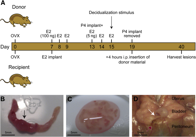
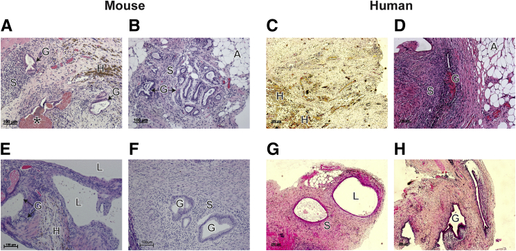
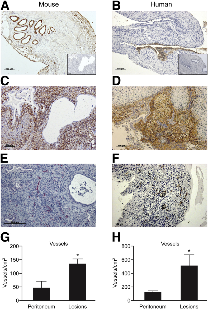
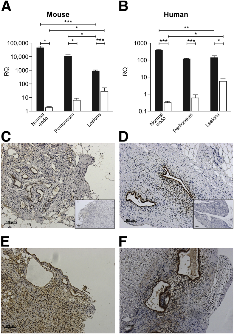
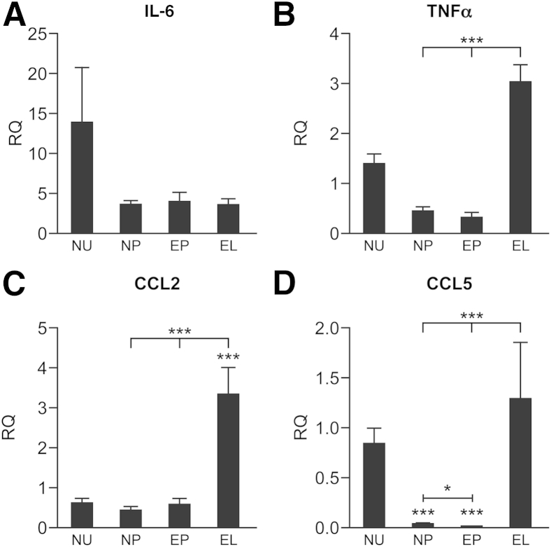
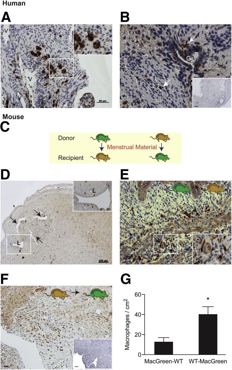
Endometriosis is an estrogen-dependent inflammatory disorder characterized by the presence of endometrial tissue outside the uterine cavity. Patients experience chronic pelvic pain and infertility, with the most likely origin of the tissue deposits (lesions) being endometrial fragments shed at menses. Menstruation is an inflammatory process associated with a dramatic increase in inflammatory mediators and tissue-resident immune cells. In the present study, we developed and validated a mouse model of endometriosis using syngeneic menstrual endometrial tissue introduced into the peritoneum of immunocompetent mice. We demonstrate the establishment of endometriotic lesions that exhibit similarities to those recovered from patients undergoing laparoscopy. Specifically, in both cases, lesions had epithelial (cytokeratin(+)) and stromal (vimentin/CD10(+)) cell compartments with a well-developed vasculature (CD31(+) endothelial cells). Expression of estrogen receptor β was increased in lesions compared with the peritoneum or eutopic endometrium. By performing experiments using mice with green fluorescent protein-labeled macrophages (MacGreen) in reciprocal transfers with wild-type mice, we obtained evidence that macrophages present in the peritoneum and in menses endometrium can contribute to the inflammatory microenvironment of the lesions. In summary, we developed a mouse model of endometriosis that exhibits similarities to human peritoneal lesions with respect to estrogen receptor expression, inflammation, and macrophage infiltration, providing an opportunity for further studies and the possible identification of novel therapies for this perplexing disorder.


Figures

Figure 1
Figure 1
A novel mouse model of endometriosis. A: Schematic showing the timeline of procedures performed on donor and recipient mice. Four hours after P4 withdrawal, mouse menstrual material was collected from donor uteri. The decidualized uterine horn was selected (arrow) and opened longitudinally (B), and the endometrium was removed from the myometrial layer, cut using a scalpel, and resuspended in PBS (C). The tissue was injected into the peritoneum of recipient mice previously primed with an estradiol-17β SILASTIC pellet, and the lesions were allowed to develop for 3 weeks. D: Lesions recovered from recipient mice. The white arrow indicates a neutral lesion associated with the bladder mesentery; red arrow, a peritoneal brown/black lesion.
Figure 2
Figure 2
Mouse lesions mimic human histological phenotypes. H&E stain of mouse lesions recovered from the anterior parietal peritoneum (A), the mesentery associated with the intestines (B), the parietal peritoneum–associated fluid-filled cyst (E), and the posterior parietal peritoneum (F). C, D, G, and H: Human lesions recovered from the peritoneal lining. A, adipose; G, glands; H, hemosiderin; L, cyst lumen; S, stroma. Asterisk in A indicates evidence of hemorrhage.
Figure 3
Figure 3
Mouse and human lesions possess glandular, stromal, and vascular components. In lesions recovered from mice (A, C, and E) or women (B, D, and F), IHC analysis identified cytokeratin-positive epithelial cells (brown positive) (A and B) and stromal fibroblasts (C, vimentin; D, CD10, brown positive in both cases). Endothelial cells were immunopositive for CD31 in mice (E, PermaRed) and humans (F, diaminobenzidine). Insets in A and B show negative controls with omission of primary antibody. G and H: When blood vessels were quantified in peritoneum and lesions (n = 5 mice; n = 5 humans for each sample type), a significantly higher density was detected in the lesions. Data are expressed as means ± SD. P < 0.05 by Student's t-test.
Figure 4
Figure 4
ER expression is comparable in mouse and human lesions. A: Quantitative RT-PCR analysis of ERα (black bars) and ERβ (white bars) in the mouse uterus (n = 6), peritoneum (n = 6), and endometriotic (Endo) lesions (n = 6). B: Quantitative RT-PCR analysis of ERα and ERβ in human endometrium (Endo) (n = 18), peritoneum (n = 19), and peritoneal lesions (n = 19). Quantitative RT-PCR data were analyzed using a one-way analysis of variance and the Newman-Keuls postcomparison test. C: Diaminobenzidine (DAB) IHC analysis using an anti-ERα antibody on mouse lesions revealed moderate immunoreactivity in glandular epithelium. D: DAB IHC analysis using an anti-ERα antibody on human lesions also revealed moderate immunoreactivity in epithelium and stromal areas. Insets in C and D show negative controls with omission of primary antibody. E: DAB IHC analysis using an anti-ERβ antibody on mouse lesions reveals prominent ERβ immunoreactivity in glandular epithelium and stromal regions. F: DAB IHC analysis using an anti-ERβ antibody on human lesions also reveals strong immunoreactivity in glandular and stromal regions. IHC analysis of human lesions was performed on samples dissected during the proliferative (estrogen-dominated) stage of the menstrual cycle. Data are expressed as means ± SD. P < 0.05, ∗∗P < 0.01, and ∗∗∗P < 0.001. RQ, relative quantification.
Figure 5
Figure 5
Analysis of expression of cytokines in mouse tissues revealed altered expression in lesions. Quantitative RT-PCR analysis of inflammatory cytokines in the uterus (NU) and peritoneum (NP) of healthy cycling mice and the peritoneum (EP) and lesions (EL) of mice with endometriosis: IL-6 (A), TNFα (B), CCL2/macrophage chemotactic protein 1 (C), and CCL5/RANTES (D). Quantitative RT-PCR data were analyzed using a one-way analysis of variance and a Newman-Keuls post-comparison test. Data are expressed as means ± SD. P < 0.05, ∗∗∗P < 0.001.
Figure 6
Figure 6
Macrophages from the shed endometrium contribute to endometriotic lesions. A and B: In human peritoneal lesions, immunostaining for CD68 identified macrophages in the stroma, some of which were in close proximity to glandular structures in human lesions. The inset in A shows an enlarged image of stromal macrophages; in B, the inset shows negative control with omission of primary antibody. White arrows indicate macrophages (B). C: Schematic indicating reciprocal transfers of menstrual material between MacGreen mice (green) and WT mice (brown). D: Anti-GFP IHC analysis on MacGreen donor uteri 4 hours after P4 withdrawal revealed macrophages present in the decidua and associated with the uterine lumen (inset). Black arrows indicate an area of the decidua beginning to shed from the basal layer of the endometrium. E: GFP+ macrophages were detected using an anti-GFP antibody in lesions isolated from MacGreen (donor) to WT (recipient) mice, demonstrating the presence of macrophages from the shed endometrium in lesions. Black arrows indicate macrophages. F: GFP+ macrophages were also detected in lesions isolated from WT (donor) to MacGreen (recipient) mice, demonstrating infiltration of host macrophages into lesions. The inset shows negative control with omission of primary antibody. G: Graph showing the respective contribution of donor/recipient to the macrophage composition of mouse endometriotic lesions. Statistical analysis was performed using a Student's t-test. Data are expressed as means ± SD. n = 6 (MacGreen-WT); n = 3 (WT-MacGreen). P < 0.05. G, glands; V, vessels. Scale bars: 50 μmol/L (A, A, inset, and E); 20 μmol/L (B); 100 μmol/L (B, inset); 200 μmol/L (D).

References

    1. Giudice L.C., Kao L.C. Endometriosis. Lancet. 2004;364:1789–1799. - PubMed
    1. Simoens S., Dunselman G., Dirksen C., Hummelshoj L., Bokor A., Brandes I., Brodszky V., Canis M., Colombo G.L., DeLeire T., Falcone T., Graham B., Halis G., Horne A., Kanj O., Kjer J.J., Kristensen J., Lebovic D., Mueller M., Vigano P., Wullschleger M., D'Hooghe T. The burden of endometriosis: costs and quality of life of women with endometriosis and treated in referral centres. Hum Reprod. 2012;27:1292–1299. - PubMed
    1. Young V.J., Brown J.K., Saunders P.T., Horne A.W. The role of the peritoneum in the pathogenesis of endometriosis. Hum Reprod Update. 2013;19:558–569. - PubMed
    1. Trukhacheva E., Lin Z., Reierstad S., Cheng Y.H., Milad M., Bulun S.E. Estrogen receptor (ER) β regulates ERα expression in stromal cells derived from ovarian endometriosis. J Clin Endocrinol Metab. 2009;94:615–622. - PMC - PubMed
    1. Emera D., Romero R., Wagner G. The evolution of menstruation: a new model for genetic assimilation: explaining molecular origins of maternal responses to fetal invasiveness. Bioessays. 2012;34:26–35. - PMC - PubMed

Publication types

Substances