Review
. 2014 Jul:16:66-82.
doi: 10.1016/j.arr.2014.05.003. Epub 2014 Jun 5.

Aging and longevity in the simplest animals and the quest for immortality

Affiliations
Review

Aging and longevity in the simplest animals and the quest for immortality

Ronald S Petralia et al. Ageing Res Rev. 2014 Jul.

Abstract

Here we review the examples of great longevity and potential immortality in the earliest animal types and contrast and compare these to humans and other higher animals. We start by discussing aging in single-celled organisms such as yeast and ciliates, and the idea of the immortal cell clone. Then we describe how these cell clones could become organized into colonies of different cell types that lead to multicellular animal life. We survey aging and longevity in all of the basal metazoan groups including ctenophores (comb jellies), sponges, placozoans, cnidarians (hydras, jellyfish, corals and sea anemones) and myxozoans. Then we move to the simplest bilaterian animals (with a head, three body cell layers, and bilateral symmetry), the two phyla of flatworms. A key determinant of longevity and immortality in most of these simple animals is the large numbers of pluripotent stem cells that underlie the remarkable abilities of these animals to regenerate and rejuvenate themselves. Finally, we discuss briefly the evolution of the higher bilaterians and how longevity was reduced and immortality lost due to attainment of greater body complexity and cell cycle strategies that protect these complex organisms from developing tumors. We also briefly consider how the evolution of multiple aging-related mechanisms/pathways hinders our ability to understand and modify the aging process in higher organisms.

Keywords: Bilateria; Cnidaria; Hydra; Metazoa; Neoblast; Planaria.


Figures

Figure 1
Figure 1
Mechanisms of aging. The diagram illustrates the nine hallmarks of aging as described by López-Otin et al (2013); several of these mechanisms are described in section 2 of this review. Aging animals often display noticeable changes in external appearance, body shape and posture. The center shows a stereotypical silhouette of an aging person with balding head and stooped posture due to various degenerative diseases. Some aging flatworms can show comparable changes such as a surface head groove (asterisk) and a body deformed by cysts (c); note how the digestive tract or gut (g) is pushed to the side. The 2 black dots represent the eyes. The drawing is an original based on micrographs of Macrostomum lignano (Mouton et al., 2009; see section 7).
Figure 2
Figure 2
Misfolding of proteins can lead to degenerative changes associated with aging. This diagram illustrates energy changes associated with protein folding, and shows how misfolding can favor protein aggregation involved in some diseases of aging. “Protein folding allows an amino acid chain to become a functional molecule. Multiple factors in this dynamic process can result in misfolding. A mutation can stabilize an intermediate in the folding process, causing this misfolded state to be favored over the properly folded protein. Furthermore, for many proteins, the functionally folded form is not the lowest energy state of the protein. Many proteins can form stable amyloid fibers or amorphous aggregates that can accumulate and disrupt cellular processes. Given the importance of proper folding to protein function, it is not surprising that each of these misfolding events can be associated with disease.” The figure and text are reprinted from part of a figure in the open-access review of Valastyan and Lindquist (2014).
Figure 3
Figure 3
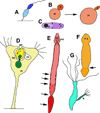
Asymmetric asexual reproduction and senescence. All drawings are original and based on drawings and micrographs in the cited references. A–E. These are examples of asymmetric cell/body division where the “mother” shows signs of senescence (see text for details).A. The sessile, stalked bacterium, Caulobacter crescentus, produces motile swarmer cell progeny (Ackerman et al., 2003). B. Budding yeast (Saccharomyses cerevisiae) mother cells (m) get bigger as they age and produce more ellipsoidal daughters (d; Lee et al, 2012). C. In contrast, fission yeast, such as Schizosaccharomyces pombe, divide symmetrically under normal conditions. But under stress, there is asymmetric accumulation of protein aggregates (p) leading to aging of the cell that retains most of these aggregates. D. Stalked mother cells (m) of some sessile suctorian ciliates, such as Acineta tuberosa (Bardele, 1970) and Tokophrya infusionum (Rudzinska, 1961; Millecchia and Rudzinska, 1970; Karakashian et al., 1984), produce an offspring in a brood pouch (bp) that is born through a birth pore (po). The brood pouch is associated with the contractile vacuole/excretory system (cv; the smaller contractile vacuole of the offspring is shown also, but not labeled). Also note how a portion of the mother’s macronucleus (nu) pinches off into the offspring. E. The flatworm, Stenostomum incaudatum (Sonneborn, 1930; Nuttycombe and Waters, 1938; Newmark and Sánchez Alvarado, 2002), produces a caudal chain of developing offspring. Arrows show locations rostral to the heads of the forming juveniles (note the pinching in of the digestive tract), where they will separate, eventually, from the mother. mo, mouth. F,G. In contrast to the previous examples, asymmetric asexual reproduction in some planaria by fission (F; arrow; Saló, 2006)and hydras via budding (G; b; Bosch, 2009) may not lead to increased senescence of the mother. In these cases, rapid cell proliferation continuously replaces all cells of the body in a short time period, thus continuously renewing the “mother.”
Figure 4
Figure 4
Evolution of choanoflagellate colony to basal metazoans with choanocytes and archeocytes (as found in modern sponges).Single-celled choanoflagellates (A) evolved into colonial forms made of clusters or spheres of cells (B), as still living today. Note the characteristic single flagellum (f) surrounded by a collar (c) made of microvilli. The ancestral metazoan may have evolved from a similar colony. Initially, cell proliferation may have become restricted to a subset of the cells. As the metazoan evolved, cells became specialized for a variety of functions (i.e., division of labor for bodily functions; C). Continued replacement of these various cell types became the task of the ancestral somatic stem cells; in the case of sponges, these were probably choanocytes - cells that retained the basic morphology of the ancestral choanoflagellates (asterisks). As these metazoans evolved into more complex forms with both surface and interior cell types of various kinds (e.g., skeleton-forming cells such as sponge sclerocytes; s), an additional population of stem cells evolved that could migrate inside the body (asterisks; these would be the archeocytes in sponges). This combination of exterior and interior pluripotent stem cells could form an immortal cell clone, e.g., thus allowing some sponges and other basal metazoans to live for thousands of years (see text for details). Based on Funayama, 2010.
Figure 5
Figure 5
An “immortal” jellyfish? In the hydrozoan, Turritopsis nutricula (Piraino, 1996), asexual reproduction by budding of hydroid polyps (p) from a stolon (s) follows rejuvenation from the sexual medusoid (jellyfish) stage (m) in a unique mechanism of reverse metamorphosis. In theory, these animals could continue this forever, hydroid colonies producing sexual medusae that eventually return to the hydroid stage and then repeat the cycle, leading to the popular name in news accounts of “The Immortal Jellyfish.”
Figure 6
Figure 6
“The stem cell system of Isodiametra pulchra (A,B). Morphology (C–E) and distribution (F–I) of neoblasts. (A) Schematic drawing. (B) Differential interference contrast image. (C) Typical neoblast with nucleus (red) and thin rim of cytoplasm (yellow). (D-D”) Macerated BrdU labeled cells show typical neoblast-like morphology (E) BrdU labeled neoblast, as shown by immunogold staining after a 30 min BrdU pulse; arrowheads point to gold particles. (F) Histological cross section; brown spots are BrdU labeled S-phase cells. (G,H) Confocal projection overview (G) and detail of lateral body margin (H) after 30 min BrdU pulse; the red spot in (H) is a mitotic figure. Note that S-phase cells [green fluorescence] were lacking in the epidermis (between dotted lines). (I) Electron microscopic image of a posterior-lateral body margin.” “bwm, body wall musculature; cc, condensed chromatin; cs, central syncytium; e, egg; de, developing eggs; ep, epidermis; g, Golgi; m, mitochondria; mo, mouth opening; st, statocyst. Scale bars (A,B,G) 100 µm; (C,E) 1 µm; (D,H) 10 µm; (F) 25 µm; (I) 5 µm.” Figure and legend are reprinted (with some terms excluded from the quoted term list) from Fig. 1A–I of the open-access report of De Mulder et al. (2009a).
Figure 7
Figure 7
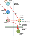
“The germline cycle in freshwater planarians. Freshwater planarians possess a population of stem cells, the so-called neoblasts, which represents the primordial stem cells in these organisms (PriSC). Neoblasts are able to give rise to the germ cells and to somatic cells. Germ cells give rise to oocytes and sperm, which jointly give rise to the zygote. The zygote gives rise to both somatic cells and the PriSCs. The planarian PriSCs have unlimited self-renewal and both germ potential (GP) and somatic potential (SP). Green dots represent the presence of nuage granules and germ plasm components. The vertical blue line represents the position of the germ-to-soma boundary, as classically understood. The horizontal green line represents the proposed position of the germ-to-soma boundary as postulated in the Primordial Stem Cells hypothesis (of Solana, 2013); this coincides with the Weismann barrier (solid black line) in freshwater planarians.” This figure is an original simplified drawing, based on Fig. 2 of Solana (2013; open access; see that paper and this review text for details; abbreviations and some label designations were changed in the quoted legend). Note that the Weismann barrier essentially prevents somatic cell mutations from affecting the germline and future generations. The PriSCs can allow some planaria to renew all of the cells of their bodies continuously, thus potentially avoiding senescence.
Figure 8
Figure 8
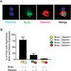
“Divergent cell fates associated with asymmetric segregation of template strands. (A) Pairs of cells were co-immunostained for BrdU and the myoblast marker Desmin; representative images are shown. (B) The data from experiments as in (A) were quantified. Pairs with Desmin expression and asymmetric BrdU labeling were distinguished based on the pattern of Desmin expression (asymmetric and coincident with BrdU, asymmetric and mutually exclusive with BRdU, or symmetric). The legend for individual cells is shown to the right, and the legends for the cell pairs are shown below. Data represent mean +/− SEM (n=4).” Figure and legend reprinted from Fig. 3 of the open-access review of Conboy et al. (2007; doi:10.1371/journal.pbio.0050102.g003.). In this study of dividing mouse muscle stem cells (satellite cells) we see that the daughter cells inheriting the older templates (no BrdU) usually retain the more immature phenotype, thus apparently remaining in the stem cell population, while the daughter cells inheriting the younger template (BrdU positive) show that they are forming the differentiated cell type, in this case, the Desmin-containing myoblast. This kind of asymmetric division is different from the examples shown in figure 3 of the present review. The strategy of asymmetric segregation of template strands may ensure that the stem cells remain as an immortal cell clone, while the fate of the differentiated cell is only eventual senescence.

Similar articles

Cited by

References

    1. Ackermann M, Stearns SC, Jenal U. Senescence in a bacterium with asymmetric division. Science. 2003;300:1920. - PubMed
    1. Aitlhadj L, Sturzenbaum SR. Caenorhabditis elegans in regenerative medicine: a simple model for a complex discipline. Drug discovery today. 2014 - PubMed
    1. Alié A, Leclere L, Jager M, Dayraud C, Chang P, Le Guyader H, Queinnec E, Manuel M. Somatic stem cells express Piwi and Vasa genes in an adult ctenophore: ancient association of “germline genes” with stemness. Developmental biology. 2011;350:183–197. - PubMed
    1. Arumugam TV, Gleichmann M, Tang SC, Mattson MP. Hormesis/preconditioning mechanisms, the nervous system and aging. Ageing research reviews. 2006;5:165–178. - PubMed
    1. Ashworth JH, Annandale N. Observations on some aged specimens of Sagartia troglodytes and on the duration of life in Coelenterates. Proc Roy Soc Edin. 1904;25:295–308.

Publication types