. 2014 Jun 26;7(6):1994-2005.
doi: 10.1016/j.celrep.2014.05.013. Epub 2014 Jun 6.

The DGCR8 RNA-binding heme domain recognizes primary microRNAs by clamping the hairpin

Affiliations

The DGCR8 RNA-binding heme domain recognizes primary microRNAs by clamping the hairpin

Jen Quick-Cleveland et al. Cell Rep. .

Abstract

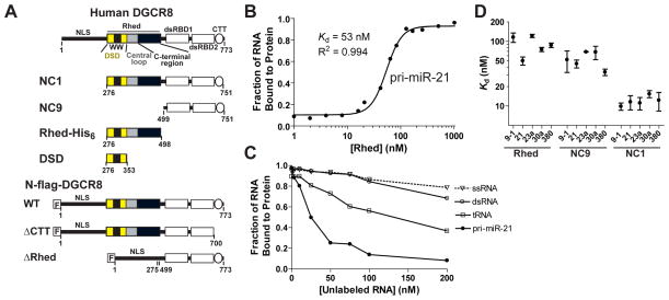
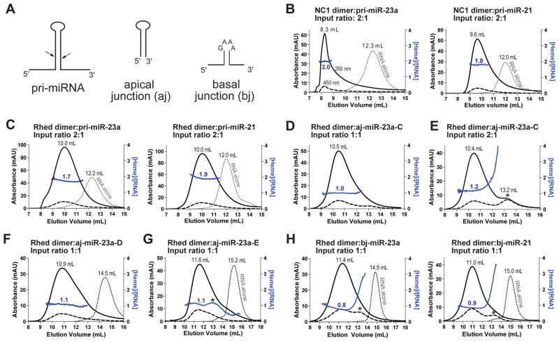
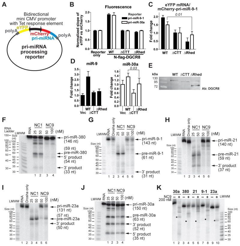
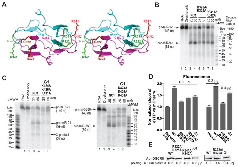
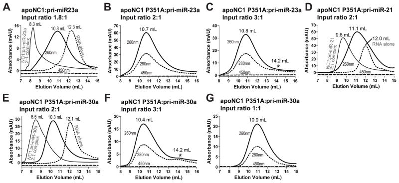
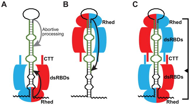
Canonical primary microRNA transcripts (pri-miRNAs) are characterized by a ∼30 bp hairpin flanked by single-stranded regions. These pri-miRNAs are recognized and cleaved by the Microprocessor complex consisting of the Drosha nuclease and its obligate RNA-binding partner DGCR8. It is not well understood how the Microprocessor specifically recognizes pri-miRNA substrates. Here, we show that in addition to the well-known double-stranded RNA-binding domains, DGCR8 uses a dimeric heme-binding domain to directly contact pri-miRNAs. This RNA-binding heme domain (Rhed) directs two DGCR8 dimers to bind each pri-miRNA hairpin. The two Rhed-binding sites are located at both ends of the hairpin. The Rhed and its RNA-binding surface are important for pri-miRNA processing activity. Additionally, the heme cofactor is required for formation of processing-competent DGCR8-pri-miRNA complexes. Our study reveals a unique protein-RNA interaction central to pri-miRNA recognition. We propose a unifying model in which two DGCR8 dimers clamp a pri-miRNA hairpin using their Rheds.


Conflict of interest statement

The authors declare that they have no conflict of interest.

Figures

Figure 1
Figure 1. The Rhed contributes to pri-miRNA recognition by directly binding these RNAs and by collaborating with the dsRBDs.
(A) Recombinant human DGCR8 proteins used. “F” represents a FLAG tag. (B) A representative curve from filter binding assays showing that the Rhed binds pri-miRNAs. The data were fit using a cooperative binding model. The Kd is defined as the Rhed dimer concentration at which half maximal RNA binding is achieved. (C) Competition filter binding assays using unlabeled ssRNA, siRNA duplex, yeast tRNAs or pri-miR-21 to compete with a trace amount of 32P-labeled pri-miR-21 for association with 150 nM of Rhed dimer. An average molecular mass of 25 kDa was assumed in calculating molar concentrations of tRNAs. (D) Comparison of the Kd values of Rhed, NC9 and NC1 for a panel of five pri-miRNAs. The average Kd values and SD are summarized in Table 1. Purity of the recombinant proteins is shown in Figure S1. The sequences and MFOLD-predicted secondary structures (Zuker, 2003) of these pri-miRNAs are shown in Table S1 and Figure S2.
Figure 2
Figure 2. RNA truncation and SEC analyses suggest that the Rhed binds to pri-miRNA junctions
(A) Schematics of pri-miRNA fragments. The arrows indicate the Drosha cleavage sites. The sequences and secondary structures are shown in Table S1 and Figures S2 and S3. (B–H) Size exclusion chromatograms of NC1 in complex with 2 μM pri-miRNAs (B), the Rhed with 2 μM pri-miRNAs (C), the Rhed with 4 μM of aj-miR-23a-C at varying input ratios (D–E), the Rhed with 4 μM of aj-miR-23a-D (F), the Rhed with 4 μM of aj-miR-23a-E (G), and the Rhed with 4 μM of indicated basal junctions (H). Solid black lines indicate A260, dashed lines show A450 and dotted lines are A260 of the RNA-only runs. Solid blue lines represent heme:RNA ratios calculated from A450 and A260, following the scale on the right y axis. The asterisk in (E, G, H) marks a peak of free Rhed. See also Figures S1–S4 and Table S1.
Figure 3
Figure 3. The DGCR8 Rhed is important for pri-miRNA processing
(A) Schematic of the reporter plasmids. (B–E) The reporters were transfected into HeLa cells either alone or with the indicated N-flag-DGCR8 expression plasmids. (B) Slopes of the eYFP and mCherry fluorescence intensities, after normalization to that of the reporter-only transfection, are plotted. Error bars represent 95% CI. (C) Ratios of eYFP mRNA and mCherry-pri-miRNA. (D) Abundance of mature miR-9 and miR-30a normalized by that of β-actin (mean ± SD, n = 3). Select P values are indicated in italic. miR-30a is highly expressed endogenously in HeLa cells and thus the relative changes are modest. (E) An anti-DGCR8 immunoblot of nuclear extracts from the transfected cells. Equal amount of total proteins was loaded in each lane, as estimated using a Coomassie-stained SDS gel. (F–K) Reconstituted pri-miRNA processing assays. Low molecular weight marker, LMWM. Relationship between LMWM and a true RNA ladder in 15% gels is shown in panel (F). In panel (K), the asterisks mark a pre-miRNA band and the dots mark the position expected for a pre-miRNA product. See also Figure S1.
Figure 4
Figure 4. The pri-miRNA-binding surfaces of the Rhed are important for processing
(A) Stereo diagram of the DSD crystal structure of human DGCR8 (PDB access code 3LE4) (Senturia et al., 2010), with the side chains of the mutated residues shown in sticks. The two subunits are drawn in cyan and magenta. (B, C) Reconstituted pri-miRNA processing assays. (D) Cellular assays using the pri-miR-9-1 reporter. The amounts of DGCR8 expression plasmids or the pCMV-Tag-2A vector are indicated on the graph. Error bars represent 95% CI. The presence of pCMV-Tag2A vector in the control transfection does not alter the fluorescence slope. (E). Anti-DGCR8 immunoblots of nuclear extracts from transfected cells. Equal amount of total proteins was loaded in each lane. See also Figures S1 and S5.
Figure 5
Figure 5. Fe(III) heme causes a large conformational change to DGCR8-pri-miRNA complexes
Size exclusion chromatograms of (A) apoNC1 in complex with 0.45 μM pri-miR-23a and (B–G) apoNC1 P351A with 2 μM pri-miRNAs at the indicated input ratios. The asterisk in (C) and (F) marks a potential free protein peak.
Figure 6
Figure 6. Models of how a pri-miRNA is recognized by the Microprocessor
(A) The basal junction anchoring model (Han et al., 2006). (B) The apical junction anchoring model (Zeng et al., 2005). (C) Our proposed molecular clamp model. See Discussion for details. The DGCR8 subunits in a dimer are shown in red and cyan. The thick avocado strands represent 5′ and 3′ mature miRNAs.

References

    1. Auyeung VC, Ulitsky I, McGeary SE, Bartel DP. Beyond secondary structure: primary-sequence determinants license pri-miRNA hairpins for processing. Cell. 2013;152:844–858. - PMC - PubMed
    1. Axtell MJ, Westholm JO, Lai EC. Vive la difference: biogenesis and evolution of microRNAs in plants and animals. Genome Biol. 2011;12:221. - PMC - PubMed
    1. Barr I, Guo F. Primary microRNA processing assay reconstituted using recombinant Drosha and DGCR8. Methods Mol Biol. 2014;1095:73–86. - PMC - PubMed
    1. Barr I, Smith AT, Chen Y, Senturia R, Burstyn JN, Guo F. Ferric, not ferrous, heme activates RNA-binding protein DGCR8 for primary microRNA processing. Proc Natl Acad Sci USA. 2012;109:1919–1924. - PMC - PubMed
    1. Barr I, Smith AT, Senturia R, Chen Y, Scheidemantle BD, Burstyn JN, Guo F. DiGeorge Critical Region 8 (DGCR8) is a double-cysteine-ligated heme protein. J Biol Chem. 2011;286:16716–16725. - PMC - PubMed

Publication types