Review
. 2014 May 30:5:199.
doi: 10.3389/fphys.2014.00199. eCollection 2014.

Origins and consequences of hyperosmolar stress in retinal pigmented epithelial cells

Affiliations
Review

Origins and consequences of hyperosmolar stress in retinal pigmented epithelial cells

François Willermain et al. Front Physiol. .

Abstract

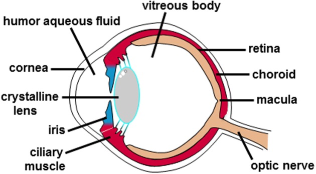
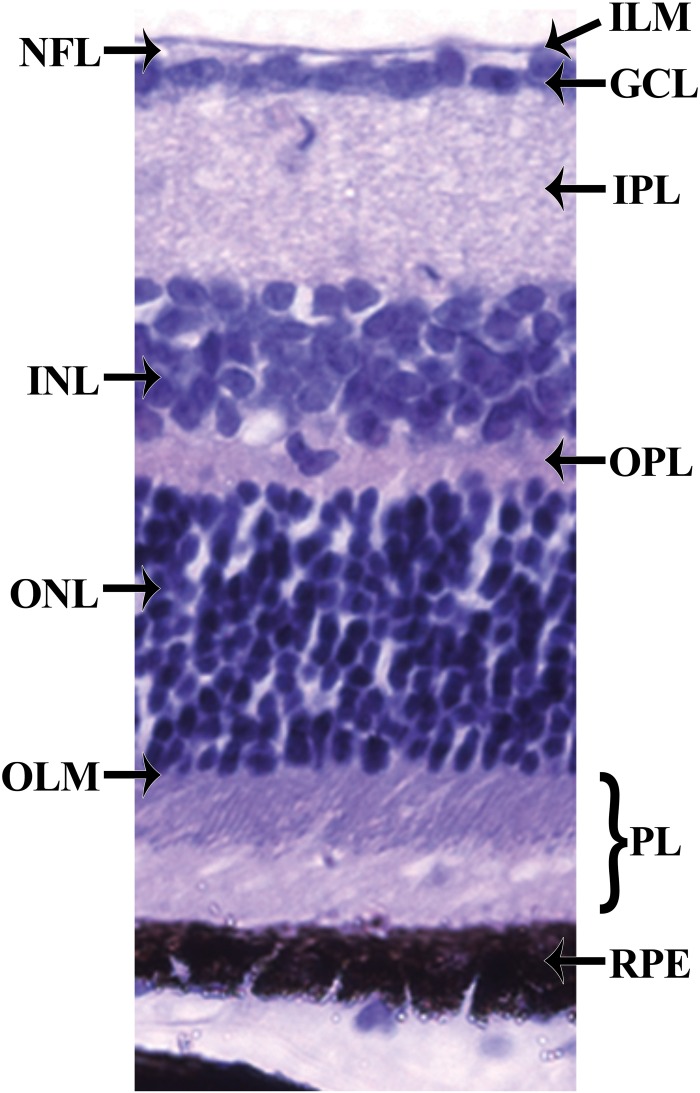
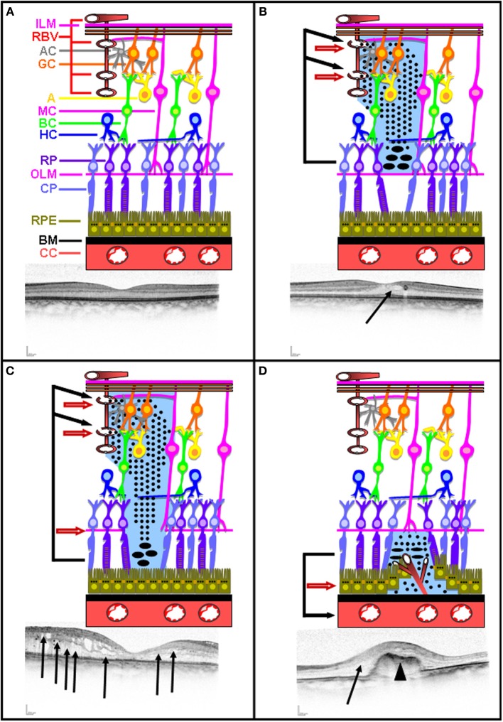
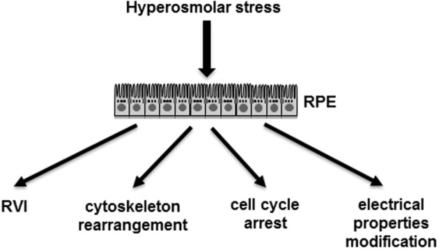
The retinal pigmented epithelium (RPE) is composed of retinal pigmented epithelial cells joined by tight junctions and represents the outer blood-retinal barrier (BRB). The inner BRB is made of endothelial cells joined by tight junctions and glial extensions surrounding all the retinal blood vessels. One of the functions of the RPE is to maintain an osmotic transepithelial gradient created by ionic pumps and channels, avoiding paracellular flux. Under such physiological conditions, transcellular water movement follows the osmotic gradient and flows normally from the retina to the choroid through the RPE. Several diseases, such as diabetic retinopathy, are characterized by the BRB breakdown leading to leakage of solutes, proteins, and fluid from the retina and the choroid. The prevailing hypothesis explaining macular edema formation during diabetic retinopathy incriminates the inner BRB breakdown resulting in increased osmotic pressure leading in turn to massive water accumulation that can affect vision. Under these conditions, it has been hypothesized that RPE is likely to be exposed to hyperosmolar stress at its apical side. This review summarizes the origins and consequences of osmotic stress in the RPE. Ongoing and further research advances will clarify the mechanisms, at the molecular level, involved in the response of the RPE to osmotic stress and delineate potential novel therapeutic targets and tools.

Keywords: age-related macular degeneration; blood-retinal barrier; diabetes; edema; hyperosmolar stress; retinal pigmented epithelium; uveitis.


Figures

Figure 1
Figure 1
Anatomical structure of the human eye. The different anatomical entities of the human eye are indicated.
Figure 2
Figure 2
Histology of the retina. The retina can be divided into 10 layers including (1) the inner limiting membrane (ILM); (2) the nerve fiber layer (NFL); (3) the ganglion cell layer (GCL); (4) the inner plexiform layer (IPL); (5) the inner nuclear layer (INL); (6) the outer plexiform layer (OPL); (7) the outer nuclear layer (ONL); (8) the outer limiting membrane (OLM); (9) the photoreceptor layer (PL), and (10) the retinal pigmented epithelium (RPE) monolayer.
Figure 3
Figure 3
Intact and disrupted BRB. Schematic representation of intact inner and outer BRB representation illustrated with a corresponding OCT picture (A). Schematic representation of inner BRB rupture without concomitant OLM rupture illustrated with a corresponding OCT picture (B). Schematic representation of inner BRB rupture with concomitant OLM rupture illustrated with a corresponding OCT picture (C). Schematic representation of outer BRB rupture following neovascularization occurring in AMD illustrated with a corresponding OCT picture (D). Plain arrow indicates edema. Arrow head indicates neovascularization.
Figure 4
Figure 4
Demonstrated effects of hyperosmolar stress on RPE cells. RPE, retinal pigmented epithelium monolayer; RVI, regulated volume increase.

References

    1. Adorante J. S., Miller S. S. (1990). Potassium-dependent volume regulation in retinal pigment epithelium is mediated by Na,K,Cl cotransport. J. Gen. Physiol. 96, 1153–1176 10.1085/jgp.96.6.1153 - DOI - PMC - PubMed
    1. Agre P. (2004). Aquaporin water channels (Nobel Lecture). Angew. Chem. Int. Ed. Engl. 43, 4278–4290 10.1002/anie.200460804 - DOI - PubMed
    1. Antonetti D. A., Klein R., Gardner T. W. (2012). Diabetic retinopathy. N. Engl. J. Med. 366, 1227–1239 10.1056/NEJMra1005073 - DOI - PubMed
    1. Arsenijevic T., Vujovic A., Libert F., Op de Beeck A., Hébrant A., Janssens S., et al. (2013). Hyperosmotic stress induces cell cycle arrest in retinal pigmented epithelial cells. Cell Death Dis. 4:e662 10.1038/cddis.2013.189 - DOI - PMC - PubMed
    1. Bok D. (1993). The retinal pigment epithelium: a versatile partner in vision. J. Cell. Sci. Suppl. 17, 189–195 10.1242/jcs.1993.Supplement_17.27 - DOI - PubMed

LinkOut - more resources