. 2014 Mar;3(1):1-13.
doi: 10.4236/cellbio.2014.31001.

Expression of PKC iota affects neuronal differentiation of PC12 cells at least partly independent of kinase function

Affiliations

Expression of PKC iota affects neuronal differentiation of PC12 cells at least partly independent of kinase function

Alana Doonachar et al. Cellbio (Irvine, Calif). 2014 Mar.

Abstract

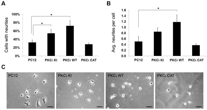
Atypical PKC (aPKC) plays a role in establishing cell polarity and has been indicated in neuronal differentiation and polarization, including neurite formation in rat pheochromocytoma PC12 cells, albeit by unclear mechanisms. Here, the role of the aPKC isoform, PKC iota (PKCι), in the early neuronal differentiation of PC12 cells was investigated. NGF-treated PC12 cells with stably expressed exogenous wild-type PKCι showed decreased expression of a neuroendocrine marker, increased expression of a neuronal marker, and increased neurite formation. Stable expression of a kinase- inactive PKCι, but not constitutively active PKCι lacking a regulatory domain, had similar although less potent effects. Pharmacological inhibition of endogenous aPKC kinase activity in parental PC12 cells did not inhibit neurite formation, suggesting that some of the observed effects of PKCι expression on neuronal differentiation are kinase- independent. Interestingly, exogenous expression of wild-type and kinase-inactive PKCι had little effect on overall PKCι activity, but caused a decrease in PKC zeta (PKCζ) kinase activity, suggesting an interplay between the two isoforms that may underlie the observed results. Overall, these findings suggest that in PC12 and perhaps other neuroendocrine precursor cells, PKCι influences an early differentiation decision between the neuroendocrine (chromaffin) and sympathetic neuron cell lineages, potentially by affecting PKCζ function.

Keywords: PC12; PKC iota; atypical PKC; neurite outgrowth; neuroendocrine; neuronal differentiation.


Figures

Figure 1
Figure 1
Creation of PC12 cell lines stably expressing PKCι. PC12 cells were stably infected with retroviruses directing expression of HA-tagged wild-type (WT) PKCι and kinase-inactive (KI) and constitutively active catalytic domain (CAT) mutants. Lysates from indicated cell lines grown to confluence were equally loaded and separated by SDS-PAGE. Lane 1 contains lysate from uninfected cells as a control. Western blots were performed using a rabbit anti-HA antibody (top panel), anti PKCι antibody (middle panel), and anti-α-tubulin antibody. NS indicates a non-specific band that appears in the top panel.
Figure 2
Figure 2
Exogenous expression of kinase-inactive PKCι decreases levels of certain neuronal markers. Lysates from indicated cell lines grown to confluence were equally loaded and separated by SDS-PAGE. Western blots were performed using antibodies to MAP2 (top panels), tyrosine hydroxylase (lower middle panel), and β1 integrin (lower panel).
Figure 3
Figure 3
Exogenous expression of either kinase-inactive or wild-type PKCι increases neurite outgrowth, with wild-type PKCι having the greatest effect. Indicated PC12 cell lines were plated on laminin-coated plates, and then treated with nerve growth factor (NGF) for 2 days. (A) The percentage of cells positive for neurites (displaying a neurite at least 2 times the body length was determined for each cell line) was determined for each cell line. * indicates statistically significant differences (P < 0.05). (B) The total number of neurites per cell that were at least 2 times the cell diameter was determined for each cell line. (C) Representative pictures of the neurite growth assays for each cell line are presented. The scale bars in the lower right corner or each picture represent 200 μm.
Figure 4
Figure 4
Increased MAP2 A/B expression is revealed in cells exogenously expressing kinase-inactive or wild-type PKCι after NGF treatment. The indicated PC12 cell lines were incubated in media containing NGF (100 ng/ml) for 5 days and then lysed. Lysates were equally loaded and separated by SDS-PAGE. Western blots were performed using antibodies to MAP2 (top panels), tyrosine hydroxylase (lower middle panel), and α-tubulin (bottom panel).
Figure 5
Figure 5
Neurite outgrowth can occur independently of aPKC kinase activity. (A) Indicated PC12 cell lines were treated with PMA for 2 days and then a neurite outgrowth assay was performed as previously, using NGF. The percentage of cells positive for neurites (displaying a neurite at least 2 times the body length) was determined for each cell line. * indicates statistically significant differences (P < 0.05). (B) Parental PC12 cells were treated with NGF only or with NGF plus the PKC inhibitor Gö6983 (200 nM). The percentage of cells positive for neurites was determined for each treatment. * indicates statistically significant differences (P < 0.05). (C) Parental PC12 cells were prepared as in (B) and then PAR-3 was immunoprecipitated. Half of the eluted immune complexes were loaded onto two lanes of a 6% SDS-PAGE gel for PAR-3 western blotting (top panel) and the remaining half of eluates were loaded on the same gel for phosphoserine western blotting (bottom panel). The lysates, representing 1/25th of the amount of lysate used for immunoprecipitation, were also separated to the left of one set of eluates. After transfer, the membrane was sliced vertically to perform the PAR-3 and phosphoserine western blots.
Figure 6
Figure 6
Both wild-type and kinase-inactive PKCι bind to PAR-6B and PAR-3 in cells. Indicated PC12 cell lines were lysed. Co-immunoprecipitation assays were performed by incubating lysates with anti-HA-agarose beads. Immune complexes were eluted and 1/3 of the eluate was loaded onto one gel for HA western blotting (top panel) and the remaining 2/3 of eluate was loaded onto a second gel for PAR-6B western blotting (middle panel). The lysates, representing 1/20th of the amount of lysate used for immunoprecipitation, were also separated on the left half of the gels, with immune complexes separated on the right side of the gels. The procedure was repeated for PAR-3 co-immunoprecipitation assay (bottom panels), except a lighter exposure was used to show PAR-3 in lysates (bottom left panel).
Figure 7
Figure 7
Expression of exogenous PKCι down-regulates PKCζ kinase activity. The set of PC12 cell lines were grown on laminin-coated culture dishes and incubated in serum-free media containing NGF (100 ng/ml) for 1 day and then lysed. Lysates were immunoprecipitated with antibodies to PKCι (in A) or PKCζ (in C) and the beads containing the immune complexes were tested using the PepTag Non-Radioactive Protein Kinase C Assay (Promega, Madison WI). (A and C) Fluorescent bands corresponding to the phosphorylated PKC substrates (indicative of PKC kinase activity) for one representative assay are shown in the top panel. The immunoprecipitated PKCι (in A) or PKCζ (in C) is shown in the middle panel and PKCι (in A) or PKCζ (in C) contained in the lysates is shown in the bottom panel. (B and D) Graphical representation of the PKC kinase assays in A and C, respectively, are shown. For D, the average of two separate PKC kinase assays is shown. Bars represent the percentage of phosphorylated PKC substrate as compared to the control assay performed with parental PC12 cells (in first column), which was set to 100%.

References

    1. Nishizuka Y. Protein kinase C and Lipid Signaling for Sustained Cellular Responses. The FASEB Journal. 1995;9(7):484–496. - PubMed
    1. Newton AC. Protein Kinase C: Poised to Signal. American Journal of Physiology Endocrinology and Metabolism. 2010;298(3):E395–402. http://dx.doi.org/10.1152/ajpendo.00477.2009. - DOI - PMC - PubMed
    1. Rosse C, Linch M, Kermorgant S, Cameron AJ, Boeckeler K, Parker PJ. PKC and the Control of Localized Signal Dynamics. Nature Reviews Molecular Cell Biology. 2010;11(2):103–112. http://dx.doi.org/10.1038/nrm2847. - DOI - PubMed
    1. Izumi Y, Hirose T, Tamai Y, Hirai S, Nagashima Y, Fujimoto T, et al. An Atypical PKC Directly Associates and Colocalizes at the Epithelial Tight Junction with ASIP, a Mammalian Homologue of Caenorhabditis elegans Polarity Protein PAR-3. Journal of Cell Biology. 1998;143(1):95–106. http://dx.doi.org/10.1083/jcb.143.1.95. - DOI - PMC - PubMed
    1. Shi SH, Jan LY, Jan YN. Hippocampal Neuronal Polarity Specified by Spatially Localized mPar3/mPar6 and PI 3-Kinase Activity. Cell. 2003;112(1):63–75. http://dx.doi.org/10.1016/S0092-8674(02)01249-7. - DOI - PubMed

LinkOut - more resources