. 2014 Jun 9;9(6):e99396.
doi: 10.1371/journal.pone.0099396. eCollection 2014.

Fucoidan can function as an adjuvant in vivo to enhance dendritic cell maturation and function and promote antigen-specific T cell immune responses

Affiliations

Fucoidan can function as an adjuvant in vivo to enhance dendritic cell maturation and function and promote antigen-specific T cell immune responses

Jun-O Jin et al. PLoS One. .

Abstract

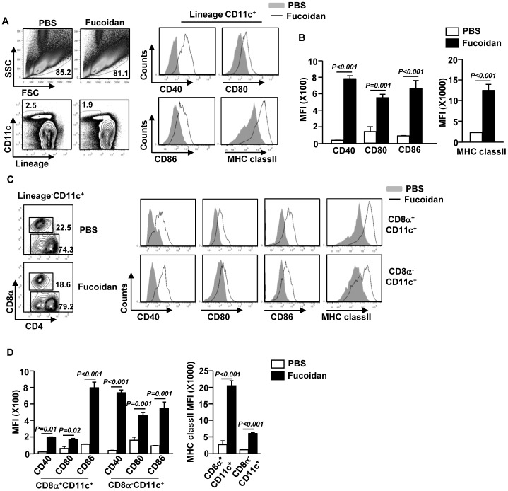
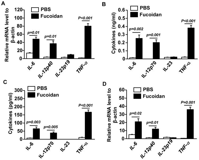
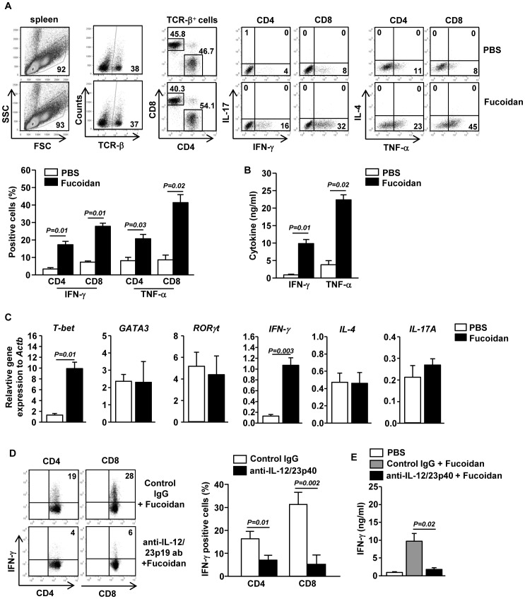
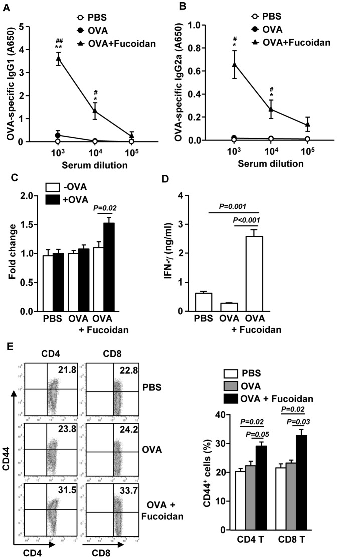
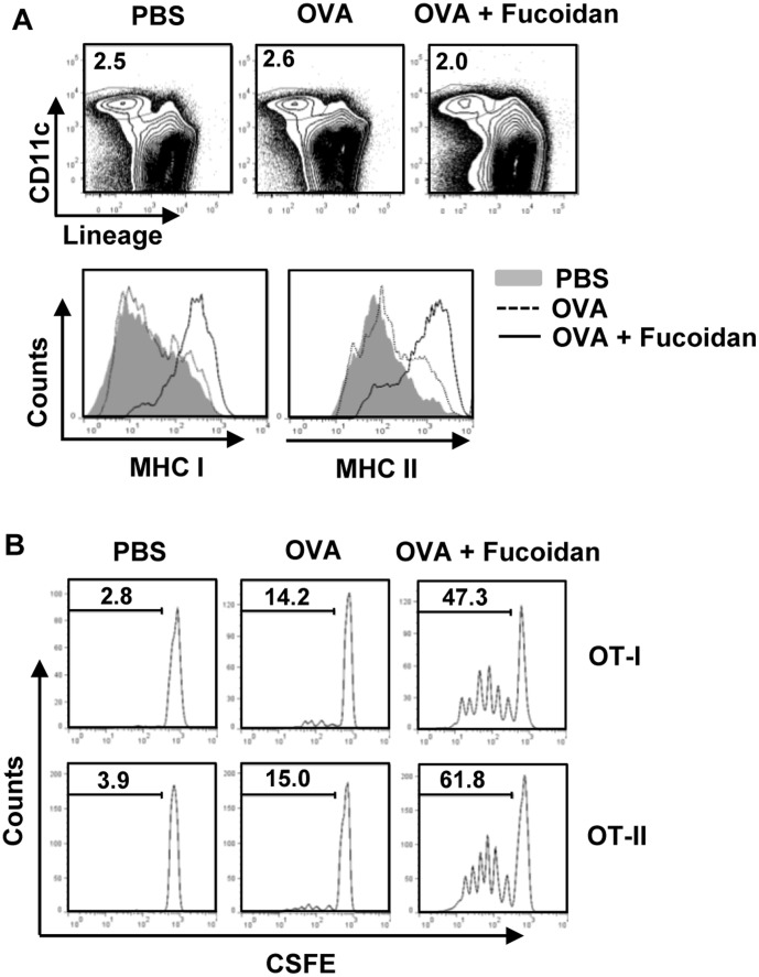
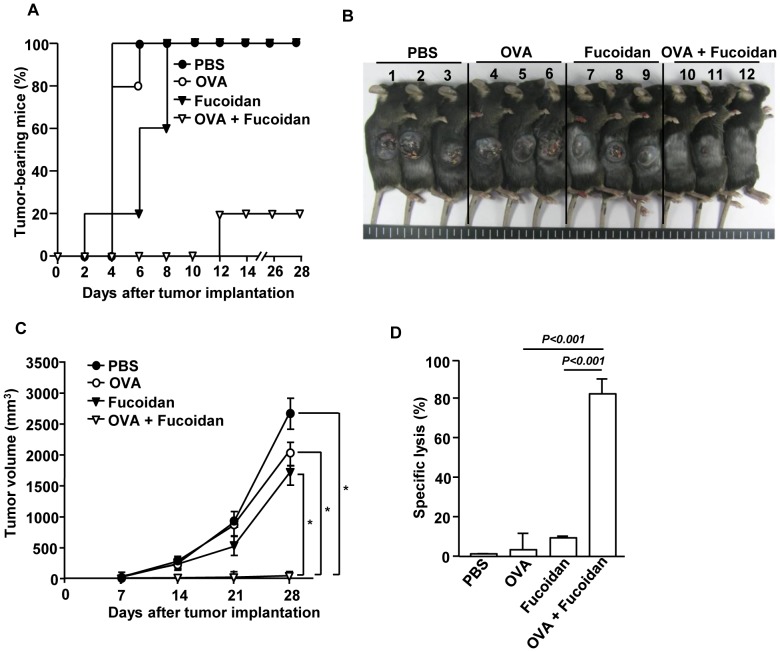
Fucoidan, a sulfated polysaccharide purified from brown algae, has a variety of immune-modulation effects, including promoting antigen uptake and enhancing anti-viral and anti-tumor effects. However, the effect of fucoidan in vivo, especially its adjuvant effect on in vivo anti-tumor immune responses, was not fully investigated. In this study, we investigated the effect of fucoidan on the function of spleen dendritic cells (DCs) and its adjuvant effect in vivo. Systemic administration of fucoidan induced up-regulation of CD40, CD80 and CD86 expression and production of IL-6, IL-12 and TNF-α in spleen cDCs. Fucoidan also promoted the generation of IFN-γ-producing Th1 and Tc1 cells in an IL-12-dependent manner. When used as an adjuvant in vivo with ovalbumin (OVA) antigen, fucoidan promoted OVA-specific antibody production and primed IFN-γ production in OVA-specific T cells. Moreover, fucoidan enhanced OVA-induced up-regulation of MHC class I and II on spleen cDCs and strongly prompted the proliferation of OVA-specific CD4 and CD8 T cells. Finally, OVA immunization with fucoidan as adjuvant protected mice from the challenge with B16-OVA tumor cells. Taken together, these results suggest that fucoidan can function as an adjuvant to induce Th1 immune response and CTL activation, which may be useful in tumor vaccine development.


Conflict of interest statement

Competing Interests: The authors have declared that no competing interests exist.

Figures

Figure 1
Figure 1. In vivo administration of fucoidan induces spleen cDC maturation.
C57BL/6 mice were treated with 10 mg/kg fucoidan for 24 hrs. (A) Flow cytometric analysis of CD40, CD80, CD86 and MHC class II expression on the gated lineageCD11c+ cDCs in splenocytes (upper panels). Lineage markers included CD3, Thy1.1, B220, Gr1, CD49b and TER-119. (B) Mean fluorescence intensity (MFI) of CD40, CD80, CD86 (left panel) and MHC class II (right panel) was shown. (C) LineageCD11c+ cDCs were further divided into CD8α+ and CD8α cDCs. Expression of CD40, CD80, CD86 and MHC class II was shown by histogram. (D) MFI of CD40, CD80, CD86 (right panel) and MHC class II (left panel) on CD8α+ and CD8α cDCs was shown. All data are representative of or the average of analyses of 6 independent samples (2 mice per experiment, total 3 independent experiments).
Figure 2
Figure 2. Fucoidan promotes production of pro-inflammation cytokines in cDCs.
Expression levels of IL-6, IL-12p40, IL-23p19 and TNF-α mRNA in spleens were measured 3 hrs after fucoidan injection. (A) mRNA levels of IL-6, IL-12p40, IL-23p19 and TNF-α in spleens. (B) IL-6, IL-12p70, IL-23 and TNF-α concentration in serum. (C) LineageCD11c+ cDCs were isolated by cell sorter 2 hrs after fucoidan injection. Isolated cDCs were incubated in culture medium for 4 hrs, and then analyzed for IL-6, IL-12p70, IL-23 and TNF-α levels in the culture supernatants were measured by ELISA. (D) mRNA levels of IL-6, IL-12p40, IL-23p19 and TNF-α from isolated cDCs. All data are representative of or the average of analyses of 6 independent samples (2 mice per experiment, total 3 independent experiments).
Figure 3
Figure 3. Fucoidan-induced cDC maturation promotes generation of IFN-γ-producing T cells in an IL-12 dependent manner.
C57BL/6 mice were injected i.p. with 10 mg/kg fucoidan and 3 days later, injected again with same amount of fucoidan. (A) Percentage of IFN-γ, IL-17, IL-4 and TNF-α positive cells within CD4 and CD8 T cells in spleen was assessed by flow cytometric analysis (upper panel). Percentage of IFN-γ+ or TNF-α+ cells (lower panel). (B) IFN-γ and TNF-α levels in serum. All data are representative of or the average of analyses of 6 independent samples (2 mice per experiment, total 3 independent experiments). (C) Gene expression in spleens was measured 24 hrs after fucoidan injection. Data are the average of analyses of 6 independent samples (2 mice per experiment, total 3 independent experiments). (D) Fucoidan was injected to C57BL/6 mice in conjunction with a neutralizing anti-IL-12/23p40 antibody or control rat IgG and the same injections were repeated 3 day later. Intracellular IFN-γ expressions in CD4 or CD8 T cells from these mice were analyzed on a flow cytometer. (E) Serum IFN-γ levels from mice described in (D). All data are representative of 6 samples from 3 independent experiments.
Figure 4
Figure 4. Fucoidan provides an adjuvant effect on OVA-induced B and T cell responses.
C57BL/6 mice were immunized i.p with PBS, OVA or OVA + fucoidan on days 0, 15, 30. On day 35, serum OVA-specific IgG1 (A) and IgG2a (B) concentrations were measured by ELISA. *P<0.05, **P<0.01 versus OVA group. # P<0.05, ## P<0.01 versus PBS group. (C) Splenocytes were harvested from immunized mice on day 35, and re-stimulated with or without OVA (50 µg/ml) for 4 days. Cell proliferation from re-stimulated splenocytes was measured. (D) IFN-γ concentrations in the above splenocytes culture supernatants were shown. (E) CD44 expression on CD4 or CD8 T cells was analyzed on a flow cytometer (left panel). Percentage of CD44+ cells in CD4 or CD8 T cells was shown (right panel). All data are representative of 6 samples from 3 independent experiments.
Figure 5
Figure 5. Fucoidan promotes antigen presentation and antigen-specific T cell proliferation in vivo.
(A) C57BL/6 mice were injected with PBS, OVA or OVA + fucoidan for 24 hrs, and the expression levels of MHC class I and II on the gated LineageCD11c+ cDCs in splenocytes from these mice were analyzed. (B) Purified CD8 T cells from OT-I or CD4 T cells from OT-II mice were labeled with CFSE and transferred into CD45.1 congenic mice, and 24 hrs later, mice were injected with PBS, OVA or OVA + fucoidan. After 3 day treatment, splenocytes from these mice were stained for CD45.2 to identify the donor OT-I or OT-II cells and the proliferation of these cells was determined by CFSE dilution. All data are from analyses of 6 individual mice each group (2 mice per experiment, total 3 independent experiments).
Figure 6
Figure 6. Immunization with OVA and fucoidan protects mice from challenge with B16-OVA tumor cells.
C57BL/6 mice were immunized with PBS, OVA, fucoidan or OVA + fucoidan on days 0, 15 and 30. On day 35 of immunization, the mice were challenged s.c. with 1×10⁶ B16-OVA (melanoma) tumor cells. (A) The percentage of tumor-bearing mice and (B) the picture of tumor bearing mice are shown. (C) Tumor growth curves are shown. All data are representative of or the average of analyses of 5 independent samples (2 or 3 mice per experiment, total 2 independent experiments). *, statistically significant values, defined as P<0.01 and determined with paired Student's t test, compared with corresponding groups. (D) On day 35, in vivo killing of adoptively transferred SIINFEK-coated and CFSE-labeled target cells by CTLs in the immunized mice was measured. Data are from analyses of 6 individual mice each group (2 mice per experiment, total 6 independent experiments).

Similar articles

Cited by

References

    1. Banchereau J, Steinman RM (1998) Dendritic cells and the control of immunity. Nature 392: 245–252. - PubMed
    1. Banchereau J, Palucka AK (2005) Dendritic cells as therapeutic vaccines against cancer. Nature reviews Immunology 5: 296–306. - PubMed
    1. Pooley JL, Heath WR, Shortman K (2001) Cutting edge: intravenous soluble antigen is presented to CD4 T cells by CD8- dendritic cells, but cross-presented to CD8 T cells by CD8+ dendritic cells. Journal of immunology 166: 5327–5330. - PubMed
    1. Schnorrer P, Behrens GM, Wilson NS, Pooley JL, Smith CM, et al. (2006) The dominant role of CD8+ dendritic cells in cross-presentation is not dictated by antigen capture. Proceedings of the National Academy of Sciences of the United States of America 103: 10729–10734. - PMC - PubMed
    1. Shortman K, Heath WR (2010) The CD8+ dendritic cell subset. Immunological reviews 234: 18–31. - PubMed

Publication types

MeSH terms