. 2014 Jun 9;9(6):e99656.
doi: 10.1371/journal.pone.0099656. eCollection 2014.

Delayed fracture healing and increased callus adiposity in a C57BL/6J murine model of obesity-associated type 2 diabetes mellitus

Affiliations

Delayed fracture healing and increased callus adiposity in a C57BL/6J murine model of obesity-associated type 2 diabetes mellitus

Matthew L Brown et al. PLoS One. .

Abstract

Introduction: Impaired healing and non-union of skeletal fractures is a major public health problem, with morbidity exacerbated in patients with diabetes mellitus (DM). DM is prevalent worldwide and affects approximately 25.8 million US adults, with >90% having obesity-related type 2 DM (T2DM). While fracture healing in type 1 DM (T1DM) has been studied using animal models, an investigation into delayed healing in an animal model of T2DM has not yet been performed.

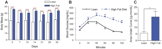
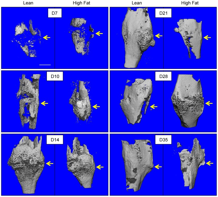
Methods: Male C57BL/6J mice at 5 weeks of age were placed on either a control lean diet or an experimental high-fat diet (HFD) for 12 weeks. A mid-diaphyseal open tibia fracture was induced at 17 weeks of age and a spinal needle was used for intra-medullary fixation. Mice were sacrificed at days 7, 10, 14, 21, 28, and 35 for micro-computed tomography (μCT), histology-based histomorphometry and molecular analyses, and biomechanical testing.

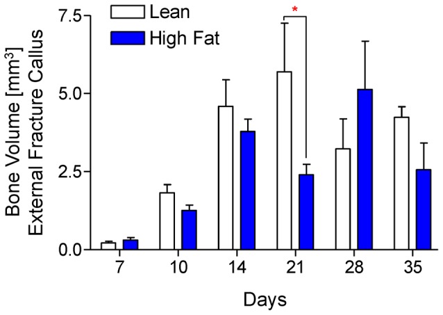
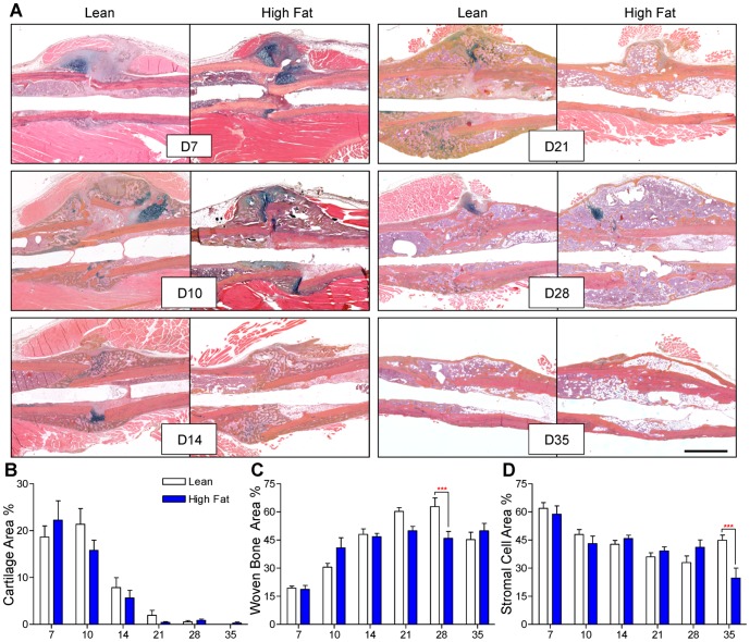
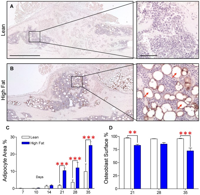
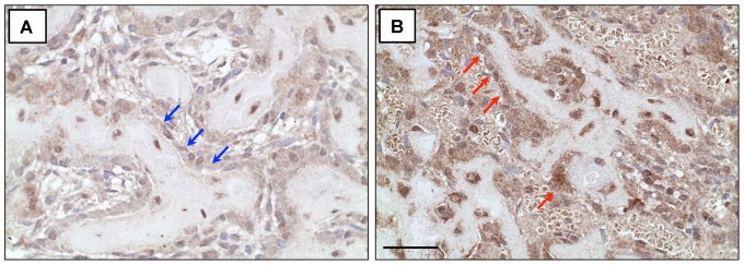
Results: HFD-fed mice displayed increased body weight and impaired glucose tolerance, both characteristic of T2DM. Compared to control mice, HFD-fed mice with tibia fractures showed significantly (p<0.001) decreased woven bone at day 28 by histomorphometry and significantly (p<0.01) decreased callus bone volume at day 21 by μCT. Interestingly, fracture calluses contained markedly increased adiposity in HFD-fed mice at days 21, 28, and 35. HFD-fed mice also showed increased PPARγ immunohistochemical staining at day 14. Finally, calluses from HFD-fed mice at day 35 showed significantly (p<0.01) reduced torsional rigidity compared to controls.

Discussion: Our murine model of T2DM demonstrated delayed fracture healing and weakened biomechanical properties, and was distinctly characterized by increased callus adiposity. This suggests altered mesenchymal stem cell fate determination with a shift to the adipocyte lineage at the expense of the osteoblast lineage. The up-regulation of PPARγ in fracture calluses of HFD-fed mice is likely involved in the proposed fate switching.


Conflict of interest statement

Competing Interests: The authors have declared that no competing interests exist.

Figures

Figure 1
Figure 1. HFD-fed mice are obese and glucose intolerant.
(A) Body weights of HFD-fed and control lean diet-fed mice at various time points after surgical fracture. Bars represent means ± SEM (n≥5). ***p<0.001 using a two-way, unpaired ANOVA with a Bonferroni post-test. (B) Glucose tolerance testing (GTT) was performed immediately prior to fracture on lean and HFD-fed mice. Plotted data points represent means ± SEM (n = 5). (C) Net area under the curve (AUC) was calculated for each mouse that underwent GTT (n = 5). *p<0.05 compared to respective lean diet controls using an unpaired, two-tailed Student's t test. Bars represent means ± SEM (n = 5).
Figure 2
Figure 2. Callus neovascularization in lean- and HFD-fed mice is similar.
μCT scans of fractured tibiae that were perfused with lead chromate paint after decalcification were performed in lean and HFD-fed mice at 10 and 14 days post-fracture. Representative three-dimensional reconstructions are presented in panel (A). Vascular volume (B) and vascular volume normalized to callus bone volume (C) was quantified from μCT data. Bars represent means ± SEM (n≥5). Scale bar (white line)  = 1 mm.
Figure 3
Figure 3. μCT imaging reveals a trend toward decreased bone volume in HFD-fed mice.
μCT scans were performed on tibiae at 7, 10, 14, 21, 28, and 35 days post-fracture. Representative three-dimensional reconstructions, from groups of 5–7 tibiae, show bone volume within the external fracture callus. Scale bar (white line)  = 1 mm. Fracture sites are denoted with yellow arrows.
Figure 4
Figure 4. Quantitative analysis confirms decreased and delayed accrual of peak bone volume in fracture callus of HFD-fed mice.
Using μCT scans that were used create the reconstructions shown Fig. 3, bone volume was quantified from lean- and HFD-fed mice at 7, 10, 14, 21, 28, and 35 days post-fracture. Bars represent means ± SEM (n≥5). *p<0.05 compared to time-matched lean diet controls using two-way, unpaired ANOVA with Bonferroni post-tests.
Figure 5
Figure 5. Woven bone content is decreased at later stages of fracture healing in HFD-fed mice.
(A) Representative Alcian Blue Hematoxylin/Orange G stained histologic sections of fracture callus in lean- and HFD-fed mice at 7, 10, 14, 21, 28, and 35 days post-fracture. Quantification of cartilage (B), woven bone (C), and stromal cell areas (D), expressed as percentages of total callus area, were determined using a point-counting histomorphometric method. Bars represent means ± SEM (n≥5). ***p<0.001 compared to time-matched controls using two-way, unpaired ANOVA with Bonferroni post-tests. The black size marker in the lower right panel  = 1 mm.
Figure 6
Figure 6. Adiposity is increased and osteoblast-occupied bone surface is decreased within the external fracture callus during late stage healing in HFD-fed mice.
Immunohistochemical staining for perilipin was performed to confirm the presence of adipocytes within the fracture callus. Representative sections from day 21 post-fracture demonstrate an increased number of adipocytes within the fracture callus of the HFD-fed mouse (B) compared to the lean diet control (A). Higher magnifications of selected regions from lean and HFD-fed calluses illustrate specificity of perilipin staining, with several typical perilipin-positive adipocytes denoted with red arrows. (C) Adipocyte area as a percentage of total callus area and percent of osteoblast occupied bone surface were determined using histomorphometric methods in lean- and HFD-fed mice at the indicated time points. Bars represent the means ± SEM (n≥5). **p<0.01 and ***p<0.001 compared to time-matched controls using an unpaired, two-way ANOVA with Bonferroni post-test. The black size marker in the low magnification images (left panels of A and B)  = 1 mm. The black size marker for the high magnification images (right panels of A and B)  = 100 µm.
Figure 7
Figure 7. PPARγ expression is increased in fracture callus of HFD-fed mice.
Sections were subjected to immunohistochemical staining for PPARγ and representative fracture calluses at day 14 show increased PPARγ expression in the fracture callus of HFD-fed mice (B) compared to lean controls (A), particularly in cells adjacent to trabecular bone elements (marked by blue arrows in callus from lean-fed mice and red arrows in callus from HFD-fed mice). The black size marker (panel B)  = 20 µm.
Figure 8
Figure 8. Healed tibiae from HFD-fed mice are biomechanically weaker.
(A) Surgically fractured right tibiae from lean- and HFD-fed mice (n≥8) were subjected to biomechanical torsional testing at days 14 and 35 post-fracture, with a significant decrease in strength observed in the HFD-fed group at day 35. Bars represent the means ± SEM (n≥8). **p<0.01 compared to time-matched controls using an unpaired, two-way ANOVA with Bonferroni post-tests.

References

    1. Alarcon T, Gonzalez-Montalvo JI, Gotor P, Madero R, Otero A (2011) Activities of daily living after hip fracture: profile and rate of recovery during 2 years of follow-up. Osteoporos Int 22: 1609–1613. - PubMed
    1. Brenneman SK, Barrett-Connor E, Sajjan S, Markson LE, Siris ES (2006) Impact of recent fracture on health-related quality of life in postmenopausal women. J Bone Miner Res 21: 809–816. - PubMed
    1. Ding R, McCarthy ML, Houseknecht E, Ziegfeld S, Knight VM, et al. (2006) The health-related quality of life of children with an extremity fracture: a one-year follow-up study. J Pediatr Orthop 26: 157–163. - PubMed
    1. Giannoudis PV, Harwood PJ, Kontakis G, Allami M, Macdonald D, et al. (2009) Long-term quality of life in trauma patients following the full spectrum of tibial injury (fasciotomy, closed fracture, grade IIIB/IIIC open fracture and amputation). Injury 40: 213–219. - PubMed
    1. Lonnroos E, Kautiainen H, Sund R, Karppi P, Hartikainen S, et al. (2009) Utilization of inpatient care before and after hip fracture: a population-based study. Osteoporos Int 20: 879–886. - PubMed

Publication types

MeSH terms