Review
. 2014 Oct;61(4):912-24.
doi: 10.1016/j.jhep.2014.05.047. Epub 2014 Jun 6.

Vascular pathobiology in chronic liver disease and cirrhosis - current status and future directions

Affiliations
Review

Vascular pathobiology in chronic liver disease and cirrhosis - current status and future directions

Yasuko Iwakiri et al. J Hepatol. 2014 Oct.

Abstract

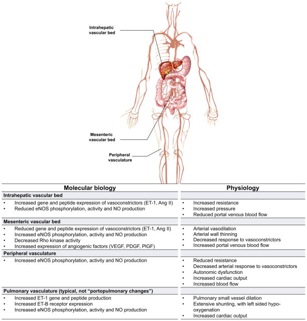
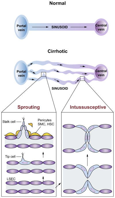
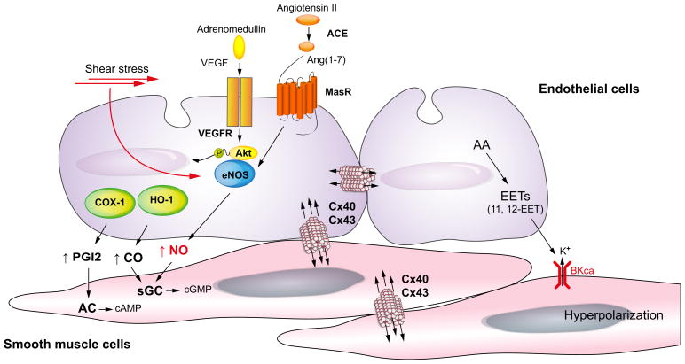
Chronic liver disease is associated with remarkable alterations in the intra- and extrahepatic vasculature. Because of these changes, the fields of liver vasculature and portal hypertension have recently become closely integrated within the broader vascular biology discipline. As developments in vascular biology have evolved, a deeper understanding of vascular processes has led to a better understanding of the mechanisms of the dynamic vascular changes associated with portal hypertension and chronic liver disease. In this context, hepatic vascular cells, such as sinusoidal endothelial cells and pericyte-like hepatic stellate cells, are closely associated with one another, where they have paracrine and autocrine effects on each other and themselves. These cells play important roles in the pathogenesis of liver fibrosis/cirrhosis and portal hypertension. Further, a variety of signaling pathways have recently come to light. These include growth factor pathways involving cytokines such as transforming growth factor β, platelet derived growth factor, and others as well as a variety of vasoactive peptides and other molecules. An early and consistent feature of liver injury is the development of an increase in intra-hepatic resistance; this is associated with changes in hepatic vascular cells and their signaling pathway that cause portal hypertension. A critical concept is that this process aggregates signals to the extrahepatic circulation, causing derangement in this system's cells and signaling pathways, which ultimately leads to the collateral vessel formation and arterial vasodilation in the splanchnic and systemic circulation, which by virtue of the hydraulic derivation of Ohm's law (pressure = resistance × flow), worsens portal hypertension. This review provides a detailed review of the current status and future direction of the basic biology of portal hypertension with a focus on the physiology, pathophysiology, and signaling of cells within the liver, as well as those in the mesenteric vascular circulation. Translational implications of recent research and the future directions that it points to are also highlighted.

Keywords: Endothelial cell; Pericyte; Pressure; Resistance; Sinusoid; Therapy.


Figures

Fig. 1
Fig. 1. Heterogeneity of vascular beds in portal hypertension
Shown are prominent vascular beds and their associated pathophysiology. The intrahepatic vascular bed is typified by an increase in resistance to flow. Liver injury leads to abnormal endothelial function, with a reduction in the production of vasodilators by sinusoidal endothelial cells such as nitric oxide (NO); concomitantly, there is an increase in the synthesis of vasoconstrictors such as endothelin-1 (ET1) and angiotensin II (Ang II) by other cells in the sinusoid (see also Fig. 2). The mesenteric vascular bed is characterised by vasodilation by reduced resistance caused by upregulation of vasodilators such as NO, leading to increased flow to the portal vein. The net result of this physiology is an increase in intrahepatic resistance and portal blood flow from the splanchnic circulation, leading to increased portal pressure and portal hypertension. In peripheral vascular beds, increased eNOS activity and NO production typically leads to reduced resistance, low systemic pressure, and a hyperdynamic state typical of patients with cirrhosis. Abnormalities also exist in other vascular beds such as the pulmonary, brain, renal, and likely even others. Of note, increased intrahepatic resistance is only for the portal system, and the resistance of the hepatic artery is decreased. Therefore, due to the hepatic arterial buffer response, the total blood flow into the liver may be the same in cirrhosis [130]. ET-1, endothelin-1; Ang II, angiotensin II; NO, nitric oxide; eNOS, endothelial nitric oxide synthase; VEGF, vascular endothelial growth factor; PDGF, platelet derived growth factor; PlGF, placental growth factor.
Fig. 2
Fig. 2. Changes in the hepatic sinusoid in response to liver injury
(A) shows a simplified version of a portal tract (left), central vein (right), and hepatic sinusoidal morphology with associated cell types within the sinusoid. (B) shows the sinusoid in response to liver injury; the upper panel depicts a cross sectional image, while the lower panel shows a 3-dimensional view. In the normal state (left), sinusoidal endothelial cells produce NO supporting a low resistance state. In response to liver injury, extensive changes occur in the sinusoid. Liver injury (right) leads to both morphological as well as molecular abnormalities in sinusoidal endothelial cells and stellate cells. For example, endothelial cells become defenestrated, develop a defect in NO production, and concurrently increase production of other proteins such as endothelin and fibronectin that can contribute to stellate cell activation. Upon their activation, stellate cells develop an enhanced contractile phenotype and produce increased extracellular matrix. A number of paracrine and autocrine interactions occur between sinusoidal endothelial cells and stellate cells with some of the putative molecules and their associated functions shown (gray box). HSC, hepatic stellate cells; LSEC, liver sinusoidal endothelial cell.
Fig. 3
Fig. 3. Sprouting and intussusceptive angiogenesis in cirrhosis and portal hypertension
Normal architecture of sinusoidal vasculature is shown in left panel with normal flow from portal venules, through sinusoidal microcirculation, to central veins. The sinusoidal vasculatures in the cirrhotic livers (right panel) are altered significantly, with increased number of sinusoidal vessels (angiogenesis) of various diameters and flow pattern. This is a simplified diagram of the sinusoidal microcirculation in liver cirrhosis. In cirrhosis, however, the relationship between the portal and central veins is not maintained because of fibrous septa and regenerative nodules, which disrupt the normal vascular architecture of portal venules, sinusoid and central vein. In sprouting angiogenesis, it is proposed that an endothelial ‘tip cell’ leads the vessel sprouts at the forefront and a trailing endothelial ‘stalk cell’ elongates behind the tip cell of a growing branch of vessels. Intussusceptive angiogenesis involves splitting vessels by the formation of translumenal pillars. Both forms of angiogenesis require the recruitment of pericytes or smooth muscle cells (SMCs). In cirrhotic livers, activated hepatic stellate cells (HSCs) are thought to serve as a role of pericytes and SMCs.
Fig. 4
Fig. 4. Mechanisms of increased vasodilation in arteries of the splanchnic circulation in cirrhosis with portal hypertension
In the splanchnic arterial circulation, agonists such as vascular endothelial growth factor (VEGF) and Ang(1-7) or physical stimuli such as shear stress activate endothelial nitric oxide (NO) synthase (eNOS). In cirrhosis, Ang(1-7) levels are significantly increased by upregulation of angiotensin-converting enzyme (ACE)-2. In addition, MasR, a receptor of Ang(1-7) is up-regulated in cirrhosis, resulting in an increase in eNOS activity and NO production. Besides NO, carbon monoxide (CO) produced by hemeoxygenase-1 (HO-1) causes vasodilation by activating soluble guanylate cyclase (sGC) to generate cyclic guanosine monophosphate (cGMP) in vascular smooth muscle cells. Prostacyclin (PGI2) is synthesised by cyclooxygenase (COX) and relaxes smooth muscle cells by stimulating adenylate cyclase (AC) to generate cyclic adenosine monophosphate (cAMP). Arachidonic acid (AA) metabolites [epoxyeicosatrienoic acids (EETs)] cause hyperpolarisation/relaxation, acting through voltage-gated potassium channel (BKCa) and gap junction [in particular connexins (Cx) 40 and 43].

Similar articles

Cited by

References

    1. Iwakiri Y, Grisham M, Shah V. Vascular biology and pathobiology of the liver: report of a single-topic symposium. Hepatology. 2008;47:1754–1763. - PMC - PubMed
    1. Wisse E, De Zanger RB, Jacobs R, McCuskey RS. Scanning electron microscope observations on the structure of portal veins, sinusoids and central veins in rat liver. Scanning Microsc. 1983:1441–1452. - PubMed
    1. Bhunchet E, Fujieda K. Capillarization and venularization of hepatic sinusoids in porcine serum-induced rat liver fibrosis: a mechanism to maintain liver blood flow. Hepatology. 1993;18:1450–1458. - PubMed
    1. Svistounov D, Warren A, McNerney GP, Owen DM, Zencak D, Zykova SN, et al. The relationship between fenestrations, sieve plates and rafts in liver sinusoidal endothelial cells. PLoS One. 2012;7:e46134. - PMC - PubMed
    1. Shah V, Toruner M, Haddad F, Cadelina G, Papapetropoulos A, Choo K, et al. Impaired endothelial nitric oxide synthase activity associated with enhanced caveolin binding in experimental cirrhosis in the rat. Gastroenterology. 1999;117:1222–1228. - PubMed

Publication types

MeSH terms