. 2014 Jun 9;9(6):e99545.
doi: 10.1371/journal.pone.0099545. eCollection 2014.

HIV-1 Nef is transferred from expressing T cells to hepatocytic cells through conduits and enhances HCV replication

Affiliations

HIV-1 Nef is transferred from expressing T cells to hepatocytic cells through conduits and enhances HCV replication

In-Woo Park et al. PLoS One. .

Abstract

HIV-1 infection enhances HCV replication and as a consequence accelerates HCV-mediated hepatocellular carcinoma (HCC). However, the precise molecular mechanism by which this takes place is currently unknown. Our data showed that infectious HIV-1 failed to replicate in human hepatocytic cell lines. No discernible virus replication was observed, even when the cell lines transfected with HIV-1 proviral DNA were co-cultured with Jurkat T cells, indicating that the problem of liver deterioration in the co-infected patient is not due to the replication of HIV-1 in the hepatocytes of the HCV infected host. Instead, HIV-1 Nef protein was transferred from nef-expressing T cells to hepatocytic cells through conduits, wherein up to 16% (average 10%) of the cells harbored the transferred Nef, when the hepatocytic cells were co-cultured with nef-expressing Jurkat cells for 24 h. Further, Nef altered the size and numbers of lipid droplets (LD), and consistently up-regulated HCV replication by 1.5∼2.5 fold in the target subgenomic replicon cells, which is remarkable in relation to the initially indolent viral replication. Nef also dramatically augmented reactive oxygen species (ROS) production and enhanced ethanol-mediated up-regulation of HCV replication so as to accelerate HCC. Taken together, these data indicate that HIV-1 Nef is a critical element in accelerating progression of liver pathogenesis via enhancing HCV replication and coordinating modulation of key intra- and extra-cellular molecules for liver decay.


Conflict of interest statement

Competing Interests: The authors have declared that no competing interests exist.

Figures

Figure 1
Figure 1. Replication of HIV-1 in human hepatocytes.
(A) Replication of HIV-1 in Huh7.5.1. Culture supernatants of RLuc and Jurkat T cells were collected for RT assay, and RT activity in the supernatants was measured at the indicated time points. Jurkat T cells were included as a positive control. (B) Entry of pseudotype HIV-1 into Huh7.5.1. HIV-1 pseudotyped with Env of HXBc2 or 89.6 strain and VSV with G protein were used to infect target Huh7.5.1 cells, and a Luc assay was performed with the cell lysates prepared from the infected cells, as described in the text. (C) Replication of HIV-1 from transfected cells. Proviral DNA of the indicated strain of HIV-1 was transfected into Huh7.5.1 cells (closed bar) or 293T cells (open bar), and accumulation of infectious progeny viruses from the transfected cells was monitored by measuring RT activity in the culture supernatants. (D) Expression of p24. Huh7.5.1 cells were transfected with proviral DNA of the HXBc2 strain of HIV-1, and expression of p24 in the transfected cells was examined by fluorescence microscopy. Green and blue colors indicate p24 protein and nucleus, respectively.
Figure 2
Figure 2. Transfer of Nef protein from Jurkat T cells into hepatocytes.
(A) Formation of conduits in nef.GFP-expressing Jurkat T cells. gfp- or nef.GFP-expressing Jurkat T cells were adhered to the poly-lysine coated cover slip, and cell morphology was investigated by confocal microscopy. Arrows indicate conduits. (B) Extension of conduits from nef.GFP-expressing Jurkat cells to hepatocytes. RLuc replicon cells grown onto a poly-lysine coated cover slip were stained with SP-DilC18(3), and the stained cells (red) were co-cultured with nef.GFP-expressing Jurkat cells (green) for 24 hr in complete DMEM media. Extension of conduits from Jurkat cells to RLuc cells (arrows) was then analyzed by confocal microscopy. (C) Quantification of Nef transferred from Jurkat to RLuc. (Co-cultivation) RLuc (1×10⁶ cells) stained with SP-DilC18(3) was co-cultivated with the same number of gfp- (closed bar) or nef.GFP (open bar)-expressing Jurkat cells for 24 hr in a 6 well plate, as described above. After Jurkat cells were removed by gentle wash with PBS, RLuc cells were detached from the plate by trypsinization, washed twice, and subjected to flow cytomery to quantify the transfer of GFP to the target RLuc cells. (Exosomes) The same number of RLuc cells was cultured with exosomes in the culture supernatants of gfp- or nef.GFP-expressing Jurkat cells, and the transfer of Nef in the exosomes to the hepatocytes was quantified in a similar manner, as described. Bar graph indicates % presentation of green positive cells to total red positive cells (RLuc cells).
Figure 3
Figure 3. Confocal analyses.
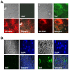
(A) Transfer of Nef. Transfer of Nef protein from wild type- (right panel) or Δnef-HIV-1 infected (left panel) Jurkat cells to the target, RLuc, was investigated by confocal microscopy. The transferred Nef protein (green) from the infected Jurkat cells to RLuc was visualized using anti-nef antibody followed by anti-mouse Alexa488. Red indicated Nef protein and RLuc cells stained with SD-DilC18. (B) Immunohistochemistry of primary hepatocytes. Left and right panels showed liver specimen from HCV- and HIV-1/HCV co-infected patients, and blue and green indicated nucleus and Nef protein, respectively.
Figure 4
Figure 4. Effect of Nef on HCV replication.
(A) Effect of co-cultivation of nef-expressing Jurkat with RLuc on the expression of replicon. The indicated number of nef.GFP-expressing Jurkat was co-cultured with RLuc, and the effect of Nef on replicon expression was assayed by measuring RLuc activity. “0” in the X-axis represents the control of GFP-expressing Jurkat cells (10∧6), and the total number of Jurkat was adjusted with GFP-expressing Jurkat to be 10∧6 cells. The data were expressed as mean +/− SEM of triplicates. (B) (upper panel) Effect of Nef on the expression of HCV subgenomic replicon. Replicon cells were transfected with the indicated amount of nef.GFP-expressing plasmid. “0” in the X-axis indicates the amount of GFP-expressing plasmid (1.6 µg), and GFP-expressing plasmid input was adjusted to equalize total plasmid transfected. Forty-eight hours post-transfection, cells were harvested and Renillar luciferase (RLuc) activity was assayed. Means and standard deviation values of triplicate experiments are depicted as bars and lines. (Lower panel) Western blot analysis. RLuc replicon cells were cultured onto a 6 well plate and transfected with the indicated amount of nef.GFP-expressing plasmid under similar conditions as above. Cell lysates were prepared, and expression of GFP or Nef.GFP fusion protein was detected by Western blot analysis using anti-rabbit anti-GFP antibody. GFP and Nef.GFP are indicated with arrows. (C) Subcellular localization of Nef. Huh7.5.1 or RLuc cells were co-transfected with plasmids to examine the expression of nef.GFP fusion protein and an ER marker, and subcellular localization of Nef was analyzed by immunofluorescence microscopy. A portion of Nef protein (green) was localized to the ER (red), depicted in yellow when the two colors were merged.
Figure 5
Figure 5. Effect of Nef on the expression of lipids and the formation of LD.
(A) Formation of LD. RLuc cells cultured on a cover slip were transfected with gfp- or nef.GFP-expressing plasmids, and the transfected cells were stained with ORO (red). (B) Effect of MβCD on Nef-mediated alteration of replicon expression. RLuc cells transfected with nef.GFP-expressing plasmid were treated with the SF-DMEM for 24 hr, followed by a 1 hr treatment with the indicated concentration of MβCD in the SF-DMEM. RLuc activity was then determined in the cells. Means and standard deviation values of triplicate experiments are depicted as bars and lines. (C) Effect of Nef on the promoter activities for the lipid synthesis genes. Each promoter-reporter construct, pGL2B-FAS-1500 (FAS) or LDLRLuc (LDLR), was co-transfected with the indicated amount of gfp- or nef.GFP-expressing plasmids, and at 48 hr post-transfection, a FLuc assay was performed with the cell lysates from the transfected cells using a Firefly Luciferase assay kit.
Figure 6
Figure 6. Effect of ethanol on nef-mediated alteration of HCV replicon expression.
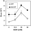
RLuc cells were transfected with gfp- or nef.GFP-expressing plasmids, and the cells were treated with the indicated concentration of ethanol for 48 hr. Renillar luciferase activity in each transfected lysate was then measured. Means and standard deviation values of triplicate experiments are depicted as lines.
Figure 7
Figure 7. Analysis of ROS.
(A) MitoSox Red staining. Huh7.5.1 (left) or RLuc (right) cells transfected with 0.1 µg of isotype (mock) and nef-expressing plasmid (Nef) were stained with MitoSox Red and visualized by fluorescence microscopy (x5). (B) Quantification of the ROS. Huh7.5.1 (left) and RLuc (right) were transfected with the indicated amount of nef-expressing plasmid in sextuplicate and loaded with DCFDA after 48 h culture. Fluorescence was measured at 495 nm excitation and 530 nm emission wavelengths, and the relative amount of ROS was shown.
Figure 8
Figure 8. Schematic presentation of Nef role.
The transferred Nef from HIV-1-infected cells to hepatocytes through conduits could exacerbate liver decay by enhancing HCV replication independently or together with ethanol and by producing ROS independently or synergistically with ethanol/HCV replication cycle. The dashed arrows indicate newly identified Nef role in hepatocytes.

Similar articles

Cited by

References

    1. Koziel MJ, Peters MG (2007) Viral hepatitis in HIV infection. N Engl J Med 356: 1445–1454. - PMC - PubMed
    1. Monforte A, Abrams D, Pradier C, Weber R, Reiss P, et al. (2008) HIV-induced immunodeficiency and mortality from AIDS-defining and non-AIDS-defining malignancies. AIDS 22: 2143–2153. - PMC - PubMed
    1. Alter MJ (2006) Epidemiology of viral hepatitis and HIV co-infection. J Hepatol 44: S6–9. - PubMed
    1. Salmon-Ceron D, Lewden C, Morlat P, Bevilacqua S, Jougla E, et al. (2005) Liver disease as a major cause of death among HIV infected patients: role of hepatitis C and B viruses and alcohol. J Hepatol 42: 799–805. - PubMed
    1. Thein HH, Yi Q, Dore GJ, Krahn MD (2008) Natural history of hepatitis C virus infection in HIV-infected individuals and the impact of HIV in the era of highly active antiretroviral therapy: a meta-analysis. AIDS 22: 1979–1991. - PubMed

Publication types

Substances