

SOD1(G93A) transgenic mouse CD4(+) T cells mediate neuroprotection after facial nerve axotomy when removed from a suppressive peripheral microenvironment
- PMID: 24911596
- PMCID: PMC4131730
- DOI: 10.1016/j.bbi.2014.05.019
SOD1(G93A) transgenic mouse CD4(+) T cells mediate neuroprotection after facial nerve axotomy when removed from a suppressive peripheral microenvironment
Abstract
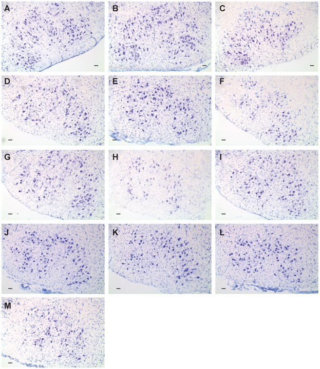
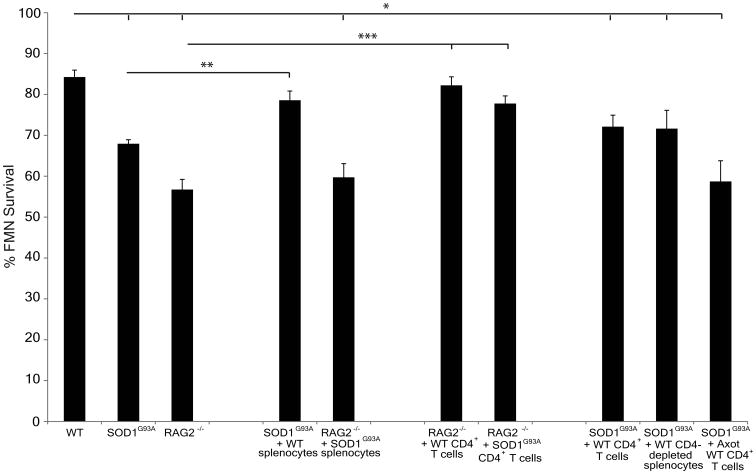
Amyotrophic lateral sclerosis (ALS) is a fatal neurodegenerative disease involving motoneuron (MN) axonal withdrawal and cell death. Previously, we established that facial MN (FMN) survival levels in the SOD1(G93A) transgenic mouse model of ALS are reduced and nerve regeneration is delayed, similar to immunodeficient RAG2(-/-) mice, after facial nerve axotomy. The objective of this study was to examine the functionality of SOD1(G93A) splenic microenvironment, focusing on CD4(+) T cells, with regard to defects in immune-mediated neuroprotection of injured MN. We utilized the RAG2(-/-) and SOD1(G93A) mouse models, along with the facial nerve axotomy paradigm and a variety of cellular adoptive transfers, to assess immune-mediated neuroprotection of FMN survival levels. We determined that adoptively transferred SOD1(G93A) unfractionated splenocytes into RAG2(-/-) mice were unable to support FMN survival after axotomy, but that adoptive transfer of isolated SOD1(G93A) CD4(+) T cells could. Although WT unfractionated splenocytes adoptively transferred into SOD1(G93A) mice were able to maintain FMN survival levels, WT CD4(+) T cells alone could not. Importantly, these results suggest that SOD1(G93A) CD4(+) T cells retain neuroprotective functionality when removed from a dysfunctional SOD1(G93A) peripheral splenic microenvironment. These results also indicate that the SOD1(G93A) central nervous system microenvironment is able to re-activate CD4(+) T cells for immune-mediated neuroprotection when a permissive peripheral microenvironment exists. We hypothesize that a suppressive SOD1(G93A) peripheral splenic microenvironment may compromise neuroprotective CD4(+) T cell activation and/or differentiation, which, in turn, results in impaired immune-mediated neuroprotection for MN survival after peripheral axotomy in SOD1(G93A) mice.
Keywords: ALS; APC; Axotomy; Immune; Motoneuron; SOD1; T cell.
Copyright © 2014 Elsevier Inc. All rights reserved.
Figures
References
-
- Alexianu ME, Kozovska M, Appel SH. Immune reactivity in a mouse model of familial ALS correlates with disease progression. Neurology. 2001;57:1282–1289. - PubMed
Publication types
MeSH terms
Substances
Grants and funding
LinkOut - more resources
Full Text Sources
Other Literature Sources
Medical
Research Materials
Miscellaneous
