. 2014 Jun 9;9(6):e99211.
doi: 10.1371/journal.pone.0099211. eCollection 2014.

Fludarabine downregulates indoleamine 2,3-dioxygenase in tumors via a proteasome-mediated degradation mechanism

Affiliations

Fludarabine downregulates indoleamine 2,3-dioxygenase in tumors via a proteasome-mediated degradation mechanism

Laïla-Aïcha Hanafi et al. PLoS One. .

Abstract

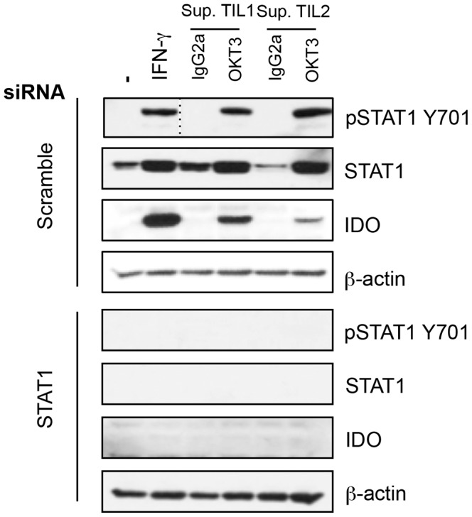
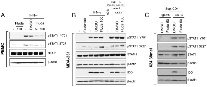
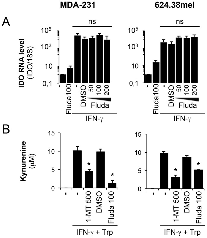
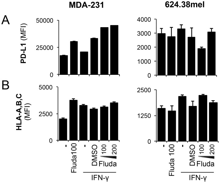
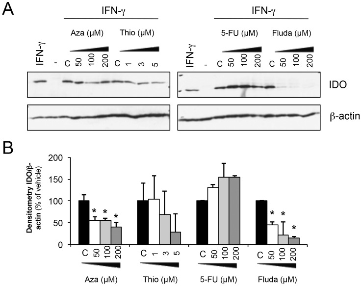
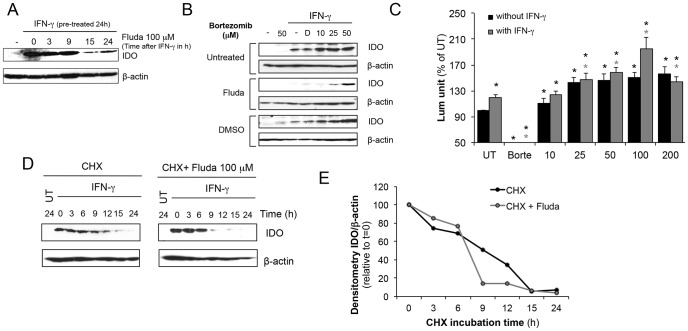
Indoleamine 2,3-dioxygenase (IDO) is found in multiple malignancies and exerts immunosuppressive effects that are central in protecting tumors from host T lymphocyte rejection. IDO is an enzyme involved in the catabolism of tryptophan resulting in inhibition of T lymphocyte function. While inhibition of IDO enzymatic activity results in tumor rejection, it is still unknown how we can directly target IDO expression within tumors using drugs. We have chosen to interfere with IDO expression by targeting the key-signaling event signal transducer and activator of transcription 1 (STAT1). We evaluated the efficacy of fludarabine, previously described to inhibit STAT1 phosphorylation. Interestingly, fludarabine was efficient in suppressing protein expression and consequently IDO activity in two different cell lines derived from breast cancer and melanoma when IDO was activated with interferon-gamma (IFN-γ) or supernatants prepared from activated T lymphocytes. However, fludarabine had no inhibitory effect on STAT1 phosphorylation. Other IFN-γ-responsive genes were only marginally inhibited by fludarabine. The level of IDO transcript was unaffected by this inhibitor, suggesting the involvement of post-transcriptional control. Strikingly, we have found that the inhibition of proteasome partially protected IDO from fludarabine-induced degradation, indicating that fludarabine induces IDO degradation through a proteasome-dependent pathway. Currently used in the clinic to treat some malignancies, fludarabine has the potential for use in the treatment of human tumors through induction of IDO degradation and consequently, for the promotion of T cell-mediated anti-tumor response.


Conflict of interest statement

Competing Interests: The authors have declared that no competing interests exist.

Figures

Figure 1
Figure 1. STAT1 is involved in IDO expression in response to T lymphocyte-derived factors.
MDA-231 were transfected with siRNA against STAT1 or scrambled siRNA before activation with IFN-γ or supernatants of cultured TIL for 30 minutes (pSTAT1) or 24 h. Protein extracts were prepared for STAT1 (phosphorylated and total), IDO and β-actin immunoblot analysis. Results are representative of three independent experiments.
Figure 2
Figure 2. Fludarabine inhibits IDO protein independently of STAT1 phosphorylation on Y710 and S727.
A- PBMC were pre-treated with the indicated concentrations of fludarabine or DMSO (vehicle) for 24 h. Cells were washed and activated for 30 min (pSTAT1) or 24 h (total STAT1 and β-actin) with 50 U/ml of IFN-γ. B- MDA-231 were pre-treated with 100 µM fludarabine before activation with 50 U/ml of IFN-γ, anti-CD3 (OKT3) or IgG2a-activated TIL supernatants (Sup.). C- 624.38mel were pre-treated with 50 µM fludarabine, and cultured with anti-CD3 (OKT3) or IgG2a-activated CD4+ T lymphocyte supernatants. B-C Cells were harvested after 30 min (pSTAT1, STAT1 and β-actin) or 24 h (IDO and β-actin). A-C Proteins were extracted for immunoblot analysis. Results are representative of three independent experiments.
Figure 3
Figure 3. Fludarabine inhibits IDO activity independently of the mRNA level.
A- MDA-231 and 624.38 pre-treated with 100 µM of fludarabine or DMSO prior to IFN-γ activation with 50 U/ml for 24 h. RNA was extracted from activated cells. cDNA was prepared and IDO expression was evaluated by quantitative real-time RT-PCR and normalized to 18S rRNA. Error bars represent standard deviation. Results are representative of three independent experiments B- MDA-231 and 624.38 were pre-treated with 100 µM of fludarabine or DMSO prior to IFN-γ activation with 50 U/ml for 24 h. Cells were resuspended in HBSS with tryptophan with or without 1-MT and incubated for 4 h. Kynurenine was quantified by HPLC. Errors bars represent standard deviation of triplicates of one experiment. * p<0.05 t-test compared to IFN-© without inhibitor. Results are representative of two independent experiments.
Figure 4
Figure 4. MHC I and PD-L1 expression levels remain unchanged following fludarabine treatment.
MDA-231 and 624.38mel were pre-treated with the indicated concentrations of fludarabine or DMSO prior to IFN-γ activation with 50 U/ml for 24 h. Cells were harvested for flow cytometry analysis. MFI was assessed on viable populations for A- PD-L1 and B- HLA-ABC. Error bars represent standard deviation from one experiment. Results are representative of three independent experiments.
Figure 5
Figure 5. Downregulation of IDO by nucleoside analogs.
A- MDA-231 were pre-treated with various nucleoside analogs at indicated concentrations or with vehicle of each inhibitor for 24 h. Cells were washed and 50 U/ml of IFN-γ were added. Proteins were extracted after 24 h for immunoblot analysis. B- Compilation of densitometry analysis of three different immunoblots as described in A. * significantly lower expression of IDO compared to vehicle (p<0.05 t-test).
Figure 6
Figure 6. Fludarabine inhibits IDO via a proteasome-mediated degradation pathway.
A- MDA-231 cells were first activated with IFN-γ (50 U/ml) to induce IDO expression for 24 h. Cells were then washed and treated with 100 µM of fludarabine for 3-24 h. Proteins were extracted for Immunoblot analysis. Immunoblots are representative of three independent experiments B- To evaluate the role of the proteasome, MDA-231 cells were pre-treated with 100 µM of fludarabine for 24 h. Cells were washed and treated with indicated concentrations of bortezomib before IFN-γ (50 U/ml) activation. Proteins were extracted after 24 h for immunoblot analysis. D: DMSO control (bortezomib vehicle). Bortezomib concentrations are expressed in nM. Immunoblots are representative of four independent experiments C- Cells were pretreated with indicated concentration of fludarabine with or without 50 U/ml of IFN-γ and proteasomal activity was assessed using Proteasome-Glo kit. Results are represented as % of activity of untreated cells. Results combine data from 6 independent experiments and 3 independent experiments with IFN-γ. Black * p<0.01 t-test compared to untreated; grey * p<0.01 t-test compared to untreated with IFN-γ. D-E 50 U/ml of IFN-γ were used to activate the cells to express IDO. 24 h later, cells were washed and incubated with 100 µM of cycloheximide with or without 100 µM fludarabine. IDO protein stability was assessed at indicated time by immunoblot. Results are representative of three independent experiments. E- Densitometry of IDO/actin by immunoblot in D.

References

    1. Munn DH, Zhou M, Attwood JT, Bondarev I, Conway SJ, et al. (1998) Prevention of allogeneic fetal rejection by tryptophan catabolism. Science 281: 1191–1193. - PubMed
    1. Prendergast GC, Chang MY, Mandik-Nayak L, Metz R, Muller AJ (2011) Indoleamine 2,3-dioxygenase as a modifier of pathogenic inflammation in cancer and other inflammation-associated diseases. Curr Med Chem 18: 2257–2262. - PMC - PubMed
    1. Godin-Ethier J, Hanafi LA, Piccirillo CA, Lapointe R (2011) Indoleamine 2,3-dioxygenase expression in human cancers: clinical and immunologic perspectives. Clin Cancer Res 17: 6985–6991. - PubMed
    1. Godin-Ethier J, Pelletier S, Hanafi LA, Gannon PO, Forget MA, et al. (2009) Human activated T lymphocytes modulate IDO expression in tumors through Th1/Th2 balance. J Immunol 183: 7752–7760. - PubMed
    1. Sharma MD, Hou DY, Baban B, Koni PA, He Y, et al. (2010) Reprogrammed foxp3(+) regulatory T cells provide essential help to support cross-presentation and CD8(+) T cell priming in naive mice. Immunity 33: 942–954. - PMC - PubMed

Publication types

MeSH terms