. 2014 Jun 9;9(6):e90572.
doi: 10.1371/journal.pone.0090572. eCollection 2014.

Mutant human FUS Is ubiquitously mislocalized and generates persistent stress granules in primary cultured transgenic zebrafish cells

Affiliations

Mutant human FUS Is ubiquitously mislocalized and generates persistent stress granules in primary cultured transgenic zebrafish cells

Jamie Rae Acosta et al. PLoS One. .

Abstract

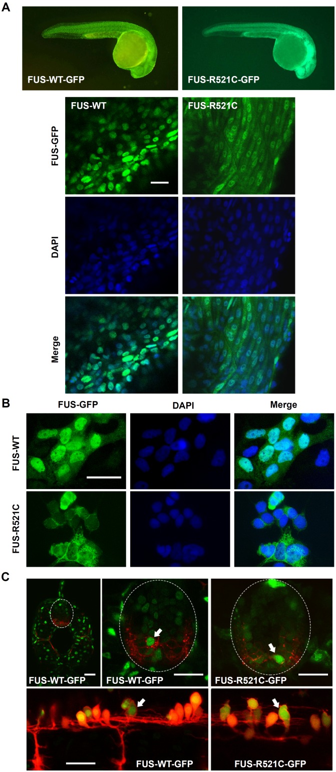
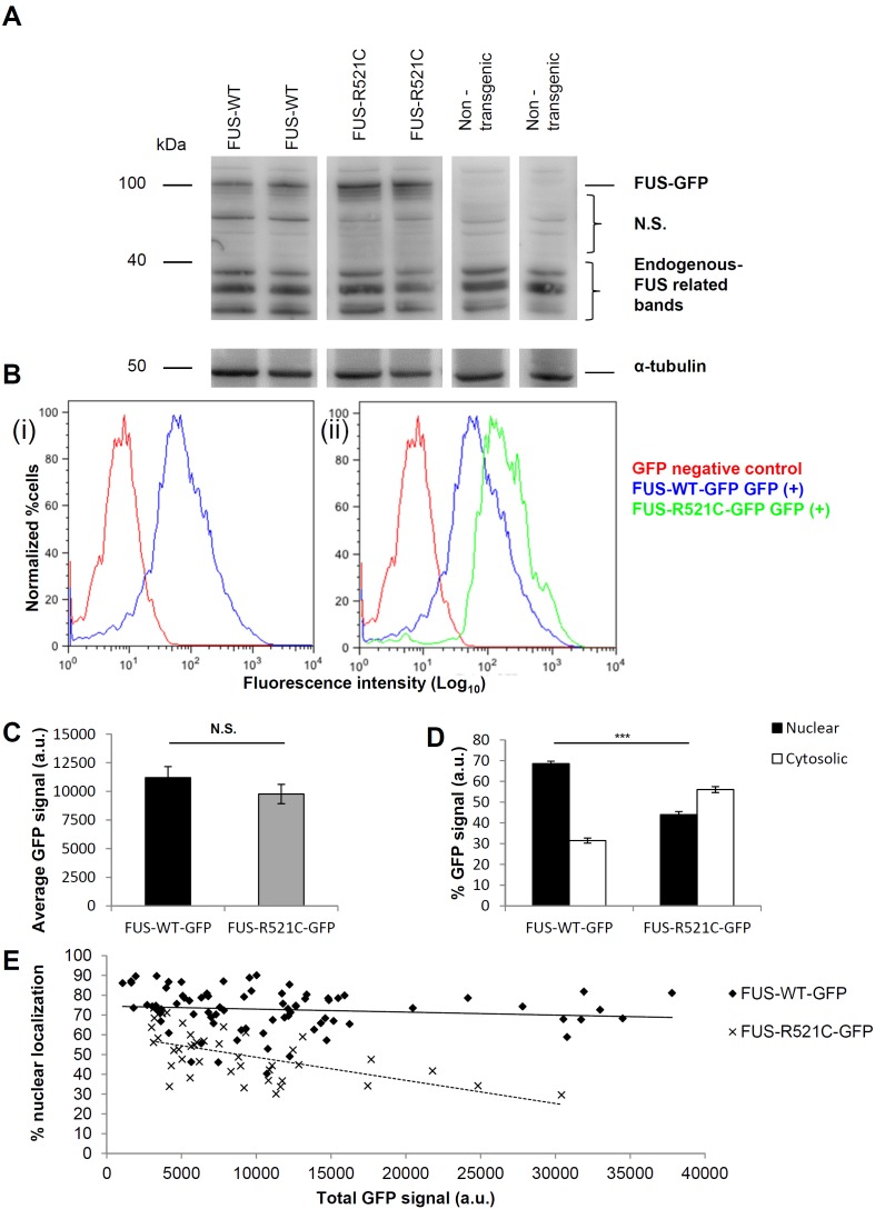
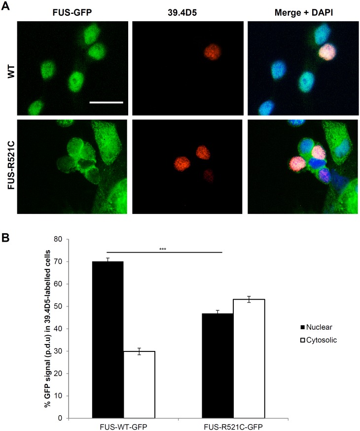
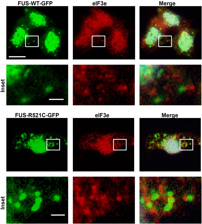
FUS mutations can occur in familial amyotrophic lateral sclerosis (fALS), a neurodegenerative disease with cytoplasmic FUS inclusion bodies in motor neurons. To investigate FUS pathology, we generated transgenic zebrafish expressing GFP-tagged wild-type or fALS (R521C) human FUS. Cell cultures were made from these zebrafish and the subcellular localization of human FUS and the generation of stress granule (SG) inclusions examined in different cell types, including differentiated motor neurons. We demonstrate that mutant FUS is mislocalized from the nucleus to the cytosol to a similar extent in motor neurons and all other cell types. Both wild-type and R521C FUS localized to SGs in zebrafish cells, demonstrating an intrinsic ability of human FUS to accumulate in SGs irrespective of the presence of disease-associated mutations or specific cell type. However, elevation in relative cytosolic to nuclear FUS by the R521C mutation led to a significant increase in SG assembly and persistence within a sub population of vulnerable cells, although these cells were not selectively motor neurons.


Conflict of interest statement

Competing Interests: The authors have declared that no competing interests exist.

Figures

Figure 1
Figure 1. Whole mount and cell cultures of FUS-GFP transgenic zebrafish.
(A) Transgenic zebrafish larvae whole mounts showed cytosolic mislocalization of mutant human FUS in FUS-R521C-GFP in comparison to FUS-WT-GFP which was restricted to cell nuclei. (B) FUS-R521C-GFP showed greater cytosolic distribution in comparison to FUS-WT-GFP in zebrafish primary cell cultures. (C) Confocal images of 48 hpf transgenic zebrafish spinal cord further demonstrate mislocalization of mutant FUS-521C-GFP (green) in motor neurons (red) (arrows). Images are maximum projections captured using a Leica SPE5 confocal microscope. Sagittal sections (upper images) of Tg(s1020tGAL4: UASmCherry) (Scott and Baier, 2009) and transverse sections (lower images) of Tg(HB9: mK02caax) membrane localised mk02 expressed in motorneurons by HB9 promoter (Flanagan-Steet et al 2005) with either FUS-WT-GFP or FUS-521C-GFP as indicated. Scale bar  = 20 µm.
Figure 2
Figure 2. Universal cytosolic mislocalization of mutant FUS-GFP protein expressed in zebrafish cells.
(A) A band at ∼100 kDa was seen uniquely in transgenic lysates corresponding to full length human FUS (75 kDa) conjugated to GFP (25 kDa) but not non-transgenic lysates. Lower MW bands (∼30–40 kDa) consistent with endogenous FUS were detected in all including non-transgenic zebrafish lysates. Multiple bands for endogenous FUS may indicate some degradation. N.S. denotes non-specific bands. Alpha-tubulin was used as a loading control. (B) Flow cytometric analysis demonstrated GFP expression in dissociated transgenic larvae. Y-axis units are normalized to the number of cells analyzed (5000–10000 cells per sample) and GFP intensity readings are presented in a log scale on the x-axis. (i) Cell suspensions of GFP positive vs. GFP negative larvae siblings derived from human FUS-WT-GFP transgenic fish. Clear separation of cells positive and negative for GFP is shown by the GFP +ve peak on the right and the GFP –ve peak on the left. (ii) Mean GFP fluorescence per cell in fresh cell suspensions from FUS-R521C-GFP and FUS-WT-GFP lines. (C) There was no significant difference in total GFP intensity per cell between lines after plating and culturing cells. Error bars represent SE. (D) Quantification of GFP fluorescence (A.U.) in nucleus and cytosol in individual cells in cultures from each fish line (n = 50–100 cells) demonstrated that there was a significant elevation in cytosolic and reduction in nuclear GFP fluorescence in FUS-R521C-GFP compared to FUS-WT-GFP cells (post-hoc Tukey HSD. *** = P<0.001). (E) To address the question whether elevated levels of FUS fusion protein expression could in itself cause mislocalization, we measured the total GFP fluorescence intensity versus the % nuclear GFP fluorescence in 50 to 100 individual cells of each genotype. Data were collected from 3 independent experiments for each line. There was no significant correlation between level of total GFP expressed in a cell and its subcellular distribution based on the R2 values for each line (FUS-WT-GFP: R2 = 0.0145 and FUS-R521C-GFP: R2 = 0.3458) (n = 79 and 41 respectively).
Figure 3
Figure 3. Mislocalization of mutant FUS-GFP was also found in motor neurons.
(A) FUS-R521C-GFP was similarly mislocalized to the cytosol in motor neurons (labeled with 39.4D5 for islet1 and islet2 homeodomain marker). (B) Quantification of FUS-GFP signal in nucleus vs. cytosol in 39.4D5 marked motor neurons demonstrated that FUS-R521C-GFP was significantly more cytosolic compared to FUS-WT-GFP (post-hoc Tukey HSD. ** = P<0.01, N.S. = >0.05, n = 40 for all samples. Error bars represent SE. Scale bars  = 20 µm.
Figure 4
Figure 4. Ubiquitous FUS-GFP SG assembly in zebrafish cells.
(A) FUS-GFP SGs formed in cultured transgenic zebrafish cells after heat-shock at 40°C for 30 mins (arrows). However, SGs were more abundant in mutant FUS-R521C-GFP cultures (right panels) than in FUS-WT-GFP. (B) Motor neurons (39.4D5-labeled cells) were not particularly susceptible to SG assembly. (C) SGs were reversible when cells were allowed to recover at 37°C for another 30 mins. Some persistent SGs were still present particularly in FUS-R521C-GFP cultures. Motor neurons labeled with 39.4D5 readily reversed SGs. (D) Stress granules (SGs) were also induced by sodium arsenite (Na3AsO3; 0.2 mM) treatment. SGs formed in mutant (arrows) but not in the FUS-WT-GFP line after Na3AsO3 treatment for 1 hr. Similar to heat-shocked cells, 39.4D5-labeled cells were no more susceptible to chemical-induced SG formation. (E) Chemical-induced FUS-GFP containing SGs were reversible in both lines. Reversibility also occurred readily in 39.4D5 labelled motor neurons.
Figure 5
Figure 5. Punctuate staining with SG marker eIF3e was commonly found adjacent to or surrounding FUS-GFP SGs.
Scale bars  = 10 µm; Insets  = 1 µm.
Figure 6
Figure 6. Quantification of SG assembly and reversibility
(A) FUS-R521C-GFP generated SGs in almost double the number of cells compared to FUS-WT-GFP after 40 minutes heat shock. Further, FUS-R521C-GFP cells were less able to reverse SGs compared to FUS-WT-GFP cells. (B) Quantification of the number of SGs per cell in SG containing cells after heat shock and recovery in three experiments showed that FUS-WT-GFP generated ∼5 FUS-containing SGs per cell and recovered to ∼2 SGs per cell. By contrast, FUS-R521C-GFP SG-containing cells remained at ∼12 SGs per cell before and after heat shock recovery despite recovery of many surrounding cells. *P≤0.05 and ***P≤0.005. Error bars indicate SE.

References

    1. Robberecht W, Philips T (2013) The changing scene of amyotrophic lateral sclerosis. Nature Reviews Neuroscience 14(4): 248–64. - PubMed
    1. Sreedharan J, Blair IP, Tripathi VB, Hu X, Vance C, et al. (2008) TDP-43 Mutations in Familial and Sporadic Amyotrophic Lateral Sclerosis. Science 319(5870): 1668–1672. - PMC - PubMed
    1. Kwiatkowski TJ Jr, Bosco DA, Leclerc AL, Tamrazian E, Vanderburg CR, et al. (2009) Mutations in the FUS/TLS Gene on Chromosome 16 Cause Familial Amyotrophic Lateral Sclerosis. Science 323(5918): 1205–1208. - PubMed
    1. DeJesus-Hernandez M, Mackenzie IR, Boeve BF, Boxer AL, Baker M, et al. (2011) Expanded GGGGCC hexanucleotide repeat in noncoding region of C9ORF72 causes chromosome 9p-linked FTD and ALS. Neuron 72(2): 245–256. - PMC - PubMed
    1. Vance C, Rogelj B, Hortobágyi T, De Vos KJ, Nishimura AL, et al. (2009) Mutations in FUS, an RNA processing protein, cause familial amyotrophic lateral sclerosis type 6. Science 323(5918): 1208–1211. - PMC - PubMed

Publication types

Substances