. 2014 Jul 8;111(27):9792-7.
doi: 10.1073/pnas.1402569111. Epub 2014 May 27.

Father's brain is sensitive to childcare experiences

Affiliations

Father's brain is sensitive to childcare experiences

Eyal Abraham et al. Proc Natl Acad Sci U S A. .

Abstract

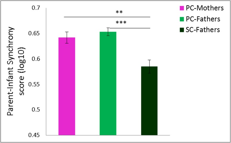
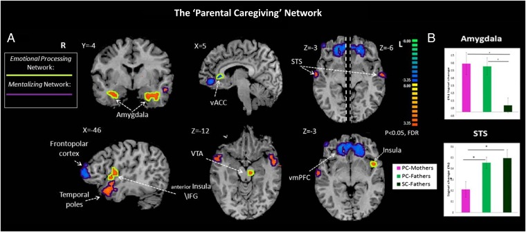
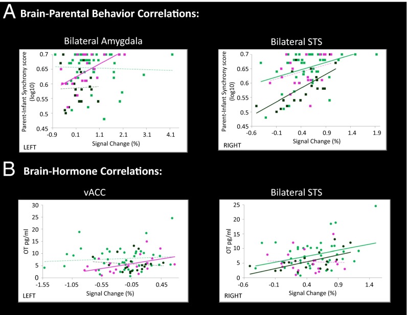
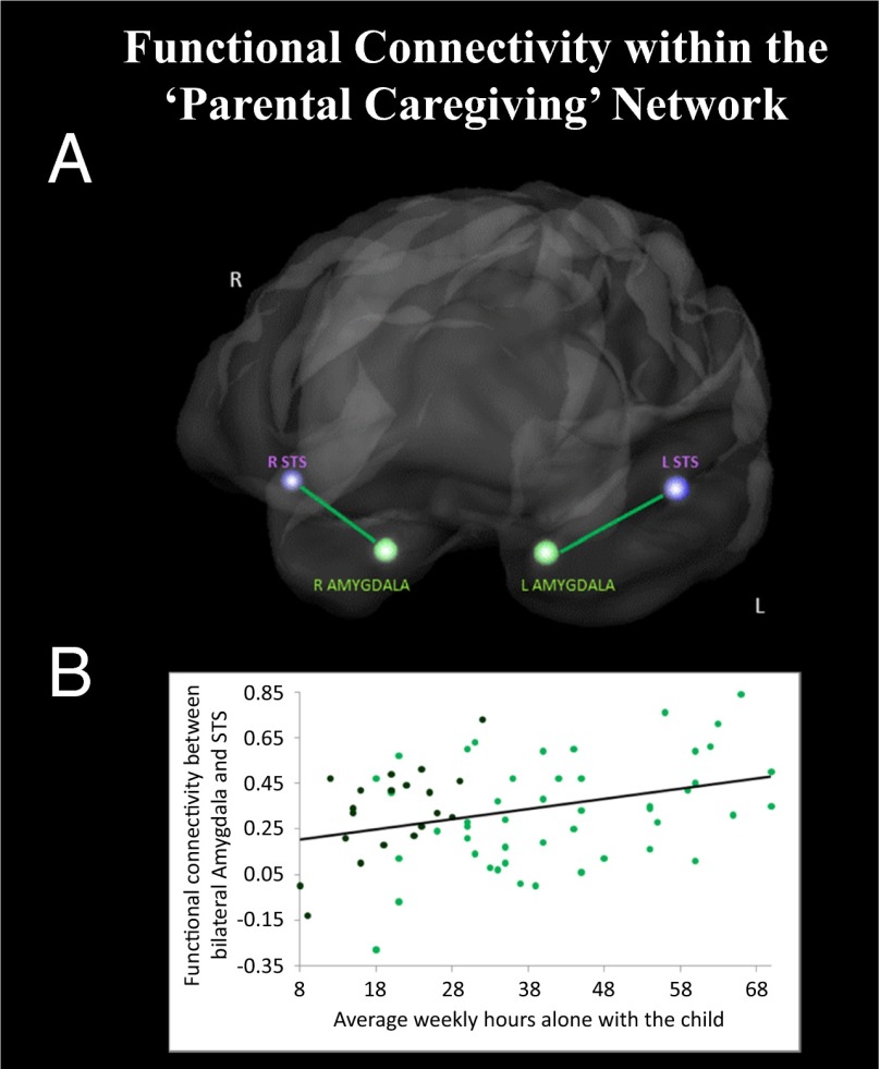
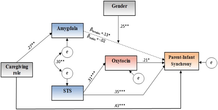
Although contemporary socio-cultural changes dramatically increased fathers' involvement in childrearing, little is known about the brain basis of human fatherhood, its comparability with the maternal brain, and its sensitivity to caregiving experiences. We measured parental brain response to infant stimuli using functional MRI, oxytocin, and parenting behavior in three groups of parents (n = 89) raising their firstborn infant: heterosexual primary-caregiving mothers (PC-Mothers), heterosexual secondary-caregiving fathers (SC-Fathers), and primary-caregiving homosexual fathers (PC-Fathers) rearing infants without maternal involvement. Results revealed that parenting implemented a global "parental caregiving" neural network, mainly consistent across parents, which integrated functioning of two systems: the emotional processing network including subcortical and paralimbic structures associated with vigilance, salience, reward, and motivation, and mentalizing network involving frontopolar-medial-prefrontal and temporo-parietal circuits implicated in social understanding and cognitive empathy. These networks work in concert to imbue infant care with emotional salience, attune with the infant state, and plan adequate parenting. PC-Mothers showed greater activation in emotion processing structures, correlated with oxytocin and parent-infant synchrony, whereas SC-Fathers displayed greater activation in cortical circuits, associated with oxytocin and parenting. PC-Fathers exhibited high amygdala activation similar to PC-Mothers, alongside high activation of superior temporal sulcus (STS) comparable to SC-Fathers, and functional connectivity between amygdala and STS. Among all fathers, time spent in direct childcare was linked with the degree of amygdala-STS connectivity. Findings underscore the common neural basis of maternal and paternal care, chart brain-hormone-behavior pathways that support parenthood, and specify mechanisms of brain malleability with caregiving experiences in human fathers.

Keywords: alloparental care; mothering; parent–infant interaction; social brain; transition to parenthood.


Conflict of interest statement

The authors declare no conflict of interest.

Figures

Fig. 1.
Fig. 1.
Bars present mean log-transformed levels of parent–infant synchrony scores as indicated by pink (PC-Mothers, n = 20), bright green (PC-Fathers, n = 48), and dark green (SC-Fathers, n = 21) bars. PC-Mother and PC-Father groups showed higher synchrony than SC-Fathers (Tukey post hoc comparisons; **P < 0.01; ***P < 0.001).
Fig. 2.
Fig. 2.
(A) Whole-brain conjunction analysis (Self–Infant Interaction > Self ∩ Self–Infant Interaction > Unfamiliar Parent–Infant Interaction) revealed two brain systems: the emotional processing network (yellow line around activation) included bilateral amygdala, vACC, left insular cortex and IFG, and VTA, and the mentalizing network (purple line around activation) included bilateral STS, lateral frontopolar cortex, vmPFC, and temporal poles. Random, n = 87, P < 0.05 FDR-corrected, cluster size > 3 × 33. (B) Bar plots present averaged percent signal change for Self–Infant Interaction minus Unfamiliar parent–infant interaction contrast for PC-Mothers (pink, n = 20), PC-Fathers (bright green, n = 47), and SC-Fathers (dark green, n = 20). (Tukey post hoc comparisons; *P < 0.05). vmPFC, ventromedial prefrontal cortex; vACC, ventral anterior cingulate cortex; IFG, inferior frontal gyrus; VTA, ventral tegmental area; L, left; R, right.
Fig. 3.
Fig. 3.
Regression lines indicated by pink (PC-Mothers, n = 20), bright green (PC-Fathers, n = 47), and dark green (SC-Fathers, n = 20) lines. Solid lines indicate significant correlations and broken lines nonsignificant correlations. (A) Scatter plots show significant correlations between brain activity and parent–infant synchrony scores for PC-Mothers in bilateral amygdala (A, Left, r = 0.579, P < 0.01), but not for PC-Fathers and SC-Fathers (A, Left, r = −0.043, P > 0.7; and r = 0.057, P > 0.8, respectively). For both PC-Fathers and SC-Fathers, significant correlation was found in the bilateral STS (A, Right, r = 0.400, P = 0.005; and r = 0.667, P = 0.001, respectively), but not for PC-Mothers (A, Right, r = 0.30, P = 0.2). (B) Scatter plots show correlations between brain and oxytocin levels for PC-Mothers in vACC (B, Left, r = 0.477, P < 0.05), but not for PC-Fathers and SC-Fathers (B, Left, r = 0.09, P > 0.5; and r = −0.119, P > 0.6, respectively), and for PC-Fathers and SC-Fathers in bilateral STS (B, Right, r = 0.337, P < 0.05; and r = 0.603, P < 0.01, respectively), but not for PC-Mothers (B, Right, r = −0.039, P > 0.8).
Fig. 4.
Fig. 4.
Presented are correlation lines as indicated by bright green (PC-Fathers, n = 47) and black (all fathers, n = 67) lines and dots. Solid lines indicate significant correlations. (A) Functional connectivity between amygdala and STS. Amygdala and STS are significantly more interconnected during Self–Infant Interaction compared with baseline only among PC-Fathers (P < 0.05, Bonferroni-corrected), but not for PC-Mothers or SC-Fathers (P > 0.05, uncorrected). (B) Scatter plot shows that functional connectivity between amygdala and STS, measured by the correlation between blood-oxygen–level dependent (BOLD) signal in bilateral amygdala and STS during Self–Infant Interaction condition, is predicted by father’s average weekly hours alone with the infant for both fathers’ groups (r = 0.330, P = 0.005).
Fig. 5.
Fig. 5.
Path model leading from the parents' role in caregiving to parent–infant synchrony as mediated by brain activation and oxytocin levels. *P < 0.05; **P < 0.01; ***P < 0.001.

Comment in

  • Flexibility of the father's brain.
    Saturn SR. Saturn SR. Proc Natl Acad Sci U S A. 2014 Jul 8;111(27):9671-2. doi: 10.1073/pnas.1408807111. Epub 2014 Jun 30. Proc Natl Acad Sci U S A. 2014. PMID: 24982185 Free PMC article. No abstract available.

References

    1. Geary DC. Evolution and proximate expression of human paternal investment. Psychol Bull. 2000;126(1):55–77. - PubMed
    1. Hrdy SB. Mother Nature: A History of Mothers, Infants, and Natural Selection. New York: Pantheon Books; 1999. - PubMed
    1. Lamb ME, Lewis C. In: The Role of the Father in Child Development. 5th Ed. Lamb ME, editor. Hoboken, NJ: Wiley; 2010.
    1. Wynne-Edwards KE. Hormonal changes in mammalian fathers. Horm Behav. 2001;40(2):139–145. - PubMed
    1. Gordon I, Zagoory-Sharon O, Leckman JF, Feldman R. Oxytocin and the development of parenting in humans. Biol Psychiatry. 2010;68(4):377–382. - PMC - PubMed

Publication types