. 2014 Jun 10;111(23):8661-6.
doi: 10.1073/pnas.1313222111. Epub 2014 May 27.

Encoding and storage of spatial information in the retrosplenial cortex

Affiliations

Encoding and storage of spatial information in the retrosplenial cortex

Rafał Czajkowski et al. Proc Natl Acad Sci U S A. .

Abstract

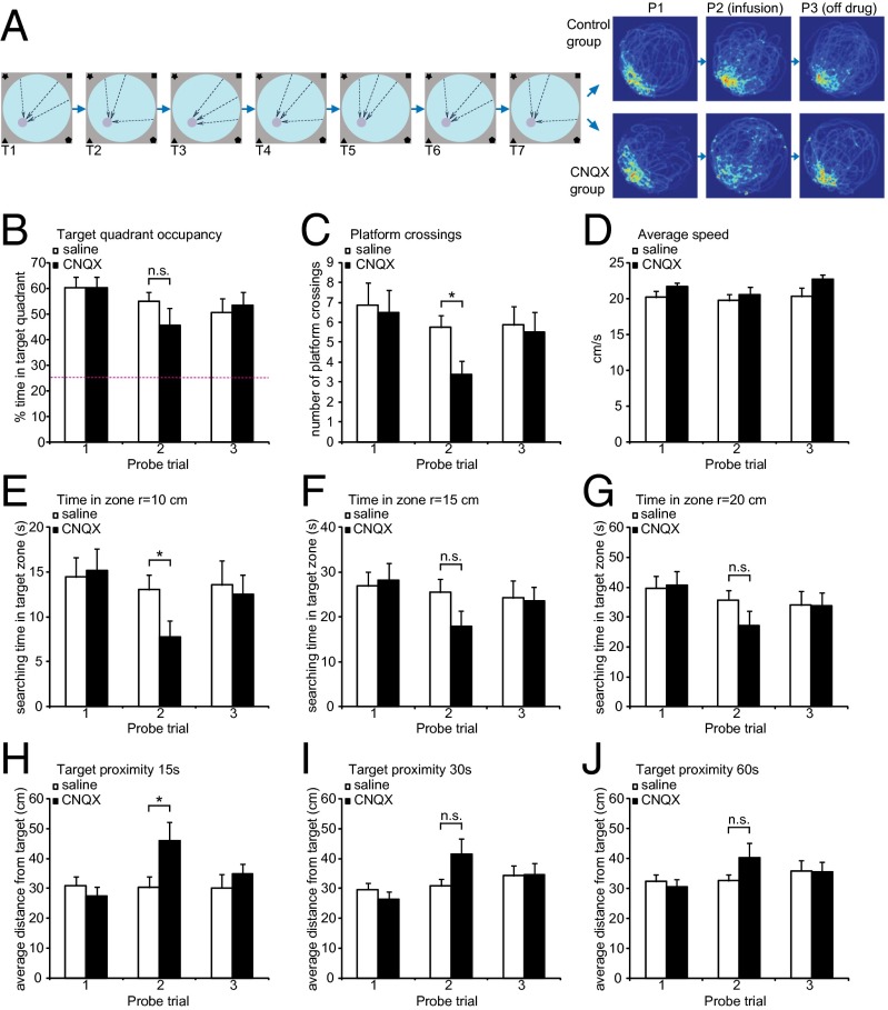
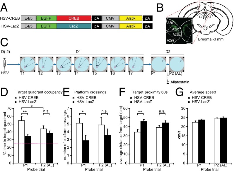
The retrosplenial cortex (RSC) is part of a network of interconnected cortical, hippocampal, and thalamic structures harboring spatially modulated neurons. The RSC contains head direction cells and connects to the parahippocampal region and anterior thalamus. Manipulations of the RSC can affect spatial and contextual tasks. A considerable amount of evidence implicates the role of the RSC in spatial navigation, but it is unclear whether this structure actually encodes or stores spatial information. We used a transgenic mouse in which the expression of green fluorescent protein was under the control of the immediate early gene c-fos promoter as well as time-lapse two-photon in vivo imaging to monitor neuronal activation triggered by spatial learning in the Morris water maze. We uncovered a repetitive pattern of cell activation in the RSC consistent with the hypothesis that during spatial learning an experience-dependent memory trace is formed in this structure. In support of this hypothesis, we also report three other observations. First, temporary RSC inactivation disrupts performance in a spatial learning task. Second, we show that overexpressing the transcription factor CREB in the RSC with a viral vector, a manipulation known to enhance memory consolidation in other circuits, results in spatial memory enhancements. Third, silencing the viral CREB-expressing neurons with the allatostatin system occludes the spatial memory enhancement. Taken together, these results indicate that the retrosplenial cortex engages in the formation and storage of memory traces for spatial information.


Conflict of interest statement

The authors declare no conflict of interest.

Figures

Fig. 1.
Fig. 1.
Two-photon (2P) in vivo imaging of FosGFP reporter during a modified water maze task. (A) Behavioral setup. Days 1, 3, 5, 7, and 9 show unmarked platform task, with platform location indicated by extra maze cues on the walls of the room. Days 2, 4, 6, 8, and 10 show marked platform task, with room cues obscured by curtain and the platform location marked by vertical rod attached to the center of the platform. Arrows indicate the release points. On each of the last 4 d (marked by a dashed rectangle), 90 min after the behavioral session, FosGFP was imaged using a 2P microscope. (B) Sample images of the same area of the RSC acquired after the last four behavioral sessions. Dashed circle marks the cell shown in C. (C) Example of typical changes in FosGFP level of a single RSC cell during the course of the experiment. For each corresponding UP/MP pair, ΔF was calculated to estimate FosGFP level related to the UP-dependent component of the task (ΔF4 = 4UP − 4MP, ΔF5 = 5UP − 5MP). (D) Summary of FosGFP activation in the RSC after alternating UP and MP sessions. UP sessions evoked higher expression of the reporter than the corresponding MP sessions. Nanimals = 9, NROIs = 25,127. (E) Summary of activation changes displayed by RSC cells during the imaging experiment. Each cell could follow one of eight patterns of response. Approximately 30% of the cells show higher FosGFP level in both UP sessions and lower in the MP sessions (pattern 6, marked in red) and therefore are potentially specific for the UP version of the task. Nanimals = 9, NROIs = 25,127. (F) High Fos level in session 4 is a good predictor of activity in session 5. Spatially specific cells that follow pattern 6 in E were divided into four quartiles according to the ΔF4 and ΔF5 values and a colocalization matrix was created. Cells that show highest ΔF in session 4 (Q1) are 50% more likely than chance to remain in the Q1 also for session 5. Matrix element [1,1] contains 9.27% of the entire population, statistically different from random distribution (*Z score = 2.37, P < 0.01). Nanimals = 9.
Fig. 2.
Fig. 2.
The effect of RSC inactivation on retrieval of spatial memory in Morris water maze. (A) Experimental design and density maps of performance during probe trials. Mice (n = 16) were implanted with cannuli and trained for 7 d with three trials per day. Dashed arrows represent release points during training sessions. Twenty-four hours after last training session animals were tested for memory retention. On the following day, mice were infused with CNQX (n = 8) or saline (n = 8) and probed again. Twenty-four hours later, another probe trial was performed. Heat maps depict time spent by the animals in each pixel during probe trials. (BF) Performance measures during probe trials 1–3. (B) Target quadrant occupancy (percentage of time spent in the quadrant with the target). CNQX-infused animals are not significantly worse in general measure of memory retention. (C) Average number of platform crossings (swims over former platform location). CNQX group is significantly impaired when assessed by a specific measure of search specificity. (*P < 0.05). (D) Average speed (centimeters per second). (EG) Percentage of time spent in the target zone, defined as circular region of r = 10 cm (H), 15 cm (I), or 30 cm (J) centered on the former platform location. (*P < 0.05). (HJ) Average distance from the target during the first 15 s (E), 30 s (F), and the entire probe trial (G). Statistical significance: *P < 0.05, paired t test.
Fig. 3.
Fig. 3.
HSV-CREB in the RSC affects learning in water maze. (A) Schematic diagram of vectors used for HSV production. (B) Expression of GFP in retrosplenial cortex following HSV infection. (Scale bar, 200 µm.) (C) Experimental procedure. Animals were injected with HSV expressing either CREB (n = 10) or LacZ (n = 10) and allowed to recover for 2 d. On the training day, mice were subjected to seven training sessions (T1–T7, each consisting of three trials), separated by 30 min. After 24 h, mice were probed for memory retention (probe trial 1, P1), infused with allatostatin, and then probed again (P2). (DF) Performance measures during P1 and P2. (D) Target quadrant occupancy (percentage of time spent in the quadrant centered on former platform location). Mice infected with HSV-CREB spent more time in the target quadrant than HSV-LacZ animals (**P < 0.01). This effect is abolished by allatostatin infusion (P2). Inactivation of AlstR-expressing neurons impairs the performance of HSV-CREB animals (P1 vs. P2, *P < 0.05) whereas HSV-LacZ mice remain unaffected. (E) Target crossings (number of passes over the former location of the platform) during P1 and P2. CREB-HSV animals are more precise than CREB-LacZ mice (*P < 0.05). This effect is eliminated by allatostatin infusion. (F) Target proximity (average distance from former platform location) during P1 and P2. CREB-HSV animals search more closely to the target than CREB-LacZ mice (*P < 0.05). This effect is eliminated by allatostatin infusion. (G) Average swimming velocity during probe trials (centimeters per second).

References

    1. Morris RG, Garrud P, Rawlins JN, O’Keefe J. Place navigation impaired in rats with hippocampal lesions. Nature. 1982;297(5868):681–683. - PubMed
    1. Hafting T, Fyhn M, Molden S, Moser MB, Moser EI. Microstructure of a spatial map in the entorhinal cortex. Nature. 2005;436(7052):801–806. - PubMed
    1. Cho J, Sharp PE. Head direction, place, and movement correlates for cells in the rat retrosplenial cortex. Behav Neurosci. 2001;115(1):3–25. - PubMed
    1. Jones BF, Witter MP. Cingulate cortex projections to the parahippocampal region and hippocampal formation in the rat. Hippocampus. 2007;17(10):957–976. - PubMed
    1. Czajkowski R, et al. Superficially projecting principal neurons in layer V of medial entorhinal cortex in the rat receive excitatory retrosplenial input. J Neurosci. 2013;33(40):15779–15792. - PMC - PubMed

Publication types

MeSH terms

Substances