. 2014 Jul 8;111(27):9798-803.
doi: 10.1073/pnas.1402597111. Epub 2014 May 27.

Direct observation of R-loop formation by single RNA-guided Cas9 and Cascade effector complexes

Affiliations

Direct observation of R-loop formation by single RNA-guided Cas9 and Cascade effector complexes

Mark D Szczelkun et al. Proc Natl Acad Sci U S A. .

Abstract

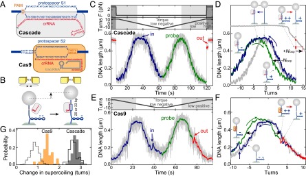
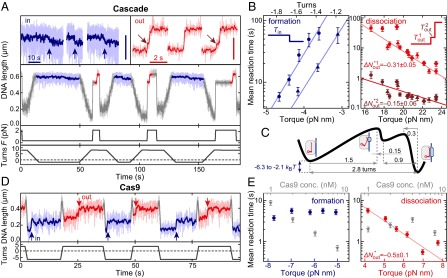
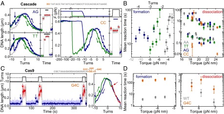
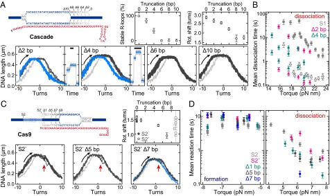
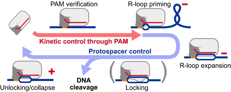
Clustered, regularly interspaced, short palindromic repeats (CRISPR)/CRISPR-associated (Cas) systems protect bacteria and archaea from infection by viruses and plasmids. Central to this defense is a ribonucleoprotein complex that produces RNA-guided cleavage of foreign nucleic acids. In DNA-targeting CRISPR-Cas systems, the RNA component of the complex encodes target recognition by forming a site-specific hybrid (R-loop) with its complement (protospacer) on an invading DNA while displacing the noncomplementary strand. Subsequently, the R-loop structure triggers DNA degradation. Although these reactions have been reconstituted, the exact mechanism of R-loop formation has not been fully resolved. Here, we use single-molecule DNA supercoiling to directly observe and quantify the dynamics of torque-dependent R-loop formation and dissociation for both Cascade- and Cas9-based CRISPR-Cas systems. We find that the protospacer adjacent motif (PAM) affects primarily the R-loop association rates, whereas protospacer elements distal to the PAM affect primarily R-loop stability. Furthermore, Cascade has higher torque stability than Cas9 by using a conformational locking step. Our data provide direct evidence for directional R-loop formation, starting from PAM recognition and expanding toward the distal protospacer end. Moreover, we introduce DNA supercoiling as a quantitative tool to explore the sequence requirements and promiscuities of orthogonal CRISPR-Cas systems in rapidly emerging gene-targeting applications.

Keywords: crRNA; genome engineering; magnetic tweezers.


Conflict of interest statement

The authors declare no conflict of interest.

Figures

Fig. 1.
Fig. 1.
R-loop formation and dissociation by Cascade and Cas9 observed in single-molecule twisting experiments. (A) Schematics of the anticipated R-loops formed by Cascade (33 bp) and Cas9 (20 bp). (B) Magnetic tweezers-based twisting assay. R-loop formation on supercoiled DNA molecules at fixed rotation causes local DNA untwisting. Compensatory overtwisting of the DNA changes the supercoiling, resulting in a DNA length change (see also SI Appendix, Fig. S1A). (C and D) R-loop cycle experiment in the presence of 10 nM Cascade. DNA with matching protospacer/PAM (A) is negatively supercoiled at 0.31 pN to induce R-loop formation (blue area of trace), followed by positive supercoiling to probe: the presence of the R-loop (green area of trace); and R-loop dissociation at an increased force of 3.0 pN (red area of trace). Blue and red arrows indicate the positions of R-loop formation and dissociation, respectively. In D, the lower and upper gray supercoiling curves were taken on the same DNA molecule at 0.31 and 3.0 pN, respectively, before Cascade addition. (E and F) R-loop cycle experiment in the presence of 1 nM Cas9 on DNA containing a matching protospacer/PAM (A) at a constant force of 0.31 pN. R-loop dissociation occurs readily at low positive torque. (G) Cas9-induced shift of the supercoiling curve (orange bars). For Cascade, shifts of the right part of the supercoiling curve after R-loop formation (gray bars; −Nloop in D) and after full R-loop dissociation (bars with solid black outline; +Nloop in D; also see SI Appendix, SI Methods) are shown. Bars with dashed black outline show the shift of the first R-loop dissociation substep for Cascade (Fig. 2A).
Fig. 2.
Fig. 2.
Torque dependence of R-loop formation and dissociation by Cascade and Cas9. (A) Repetitive cycles of R-loop formation (at −1.6 turn, 0.36 pN) and R-loop dissociation (at +8 turns, 2.2 pN) by Cascade. R-loop formation and dissociation is seen as a DNA length decrease or increase, respectively (enlarged views on top). (Vertical scale bars: 100 nm.) Blue arrows indicate R-loop formation, and brown arrows indicate a short-lived intermediate state upon R-loop dissociation. (B) Mean R-loop formation times (blue scatter plots) and dissociation times (red and brown scatter plots for the first and second dissociation step, respectively) as a function of torque. Different symbols indicate measurements on different DNAs to show experimental variation. Solid lines are exponential fits to the data according to SI Appendix, Eqs. S8 and S9. Numbers indicate distances to the transitions states as obtained from the fits. (C) Energy landscape of the R-loop formation process by Cascade based on the torque dependency. (D) Repetitive cycles of R-loop formation (at −6 turns; blue stretches) and R-loop dissociation (at +3 turns; red stretches) by Cas9. A constant force of 0.31 pN was applied. Red arrows indicate dissociation events. (E) Mean R-loop formation and dissociation times as a function of torque (at 1 nM Cas9) and of Cas9 concentration (at a torque of −6.2 and +6.2 pN nm).
Fig. 3.
Fig. 3.
PAM mutations affect primarily R-loop formation and not dissociation. (A) Repetitive R-loop cycles in the presence of Cascade on DNA with the matching protospacer S1 but modified dinucleotide PAMs (see sketch above and labels in the graphs). Curve coloring and experimental conditions are as in Fig. 1 C and D. For the CC PAM, R-loop formation requires −200 turns (at 2.0 pN; blue curve). The shift in the probe curve (green) and the dissociation step (red curve) reveals the presence of the R-loop. (Scale bars: 10 s.) (B) Mean R-loop formation and dissociation times as a function of torque for the different PAMs (colors given in the key). Circles and triangles indicate the first and second dissociation step, respectively. Gray torque values indicate the phase where the torque is no longer proportional to turns. (C) Repetitive cycles of R-loop formation and dissociation by Cas9 (as in Fig. 2D) at 0.31 pN using a DNA with protospacer S2 and G4C PAM (see sketch). (D) Mean R-loop formation and dissociation times as a function of torque for the G4C PAM compared with the canonical PAM.
Fig. 4.
Fig. 4.
Protospacer end truncations affect the R-loop stability and reveal a locking mechanism for Cascade but not for Cas9. (A) R-loop probe and dissociation curves in the presence of Cascade for protospacer S1 truncated by 2, 4, 6, and 10 bp (see sketch above), revealing stable R-loops (blue) and unstable R-loops (dark gray). Percentages of stable R-loops and the sizes of the rotational shifts are shown in the upper graphs. (B) Mean dissociation times as function of torque (from experiments in A) for the stable R-loops of 0-, 2-, and 4-bp truncations. Filled circles and open triangles indicate the first and second dissociation step, respectively. (C) R-loop probe curves in the presence of Cas9 for protospacer S2′ and truncations of S2′ by 5 and 7 bp (see sketch) revealing R-loops with fast dissociation kinetics (dark gray; red arrows indicate dissociation event) and, in the case of Δ7 bp, additional slow dissociation kinetics (blue). Light gray curves were taken in absence of Cascade or Cas9. The sizes of the rotational shifts are shown in the upper graph. (D) Mean formation and dissociation times as a function of torque (from experiments in C) for different S2′ protospacer truncations.
Fig. 5.
Fig. 5.
Unified model for the differential control of R-loop formation and dissociation by PAM and protospacer sequences. The locking step is only observed for Cascade and not for Cas9. Red positive (+) and negative (–) symbols indicate where supercoiling of the respective sign can accelerate the step.

Comment in

References

    1. Garneau JE, et al. The CRISPR/Cas bacterial immune system cleaves bacteriophage and plasmid DNA. Nature. 2010;468(7320):67–71. - PubMed
    1. Hale CR, et al. RNA-guided RNA cleavage by a CRISPR RNA-Cas protein complex. Cell. 2009;139(5):945–956. - PMC - PubMed
    1. Zhang J, et al. Structure and mechanism of the CMR complex for CRISPR-mediated antiviral immunity. Mol Cell. 2012;45(3):303–313. - PMC - PubMed
    1. Brouns SJJ, et al. Small CRISPR RNAs guide antiviral defense in prokaryotes. Science. 2008;321(5891):960–964. - PMC - PubMed
    1. Carte J, Wang R, Li H, Terns RM, Terns MP. Cas6 is an endoribonuclease that generates guide RNAs for invader defense in prokaryotes. Genes Dev. 2008;22(24):3489–3496. - PMC - PubMed

Publication types

MeSH terms