. 2014 Jun 10:4:5240.
doi: 10.1038/srep05240.

A novel proteasome inhibitor suppresses tumor growth via targeting both 19S proteasome deubiquitinases and 20S proteolytic peptidases

Affiliations

A novel proteasome inhibitor suppresses tumor growth via targeting both 19S proteasome deubiquitinases and 20S proteolytic peptidases

Ningning Liu et al. Sci Rep. .

Abstract

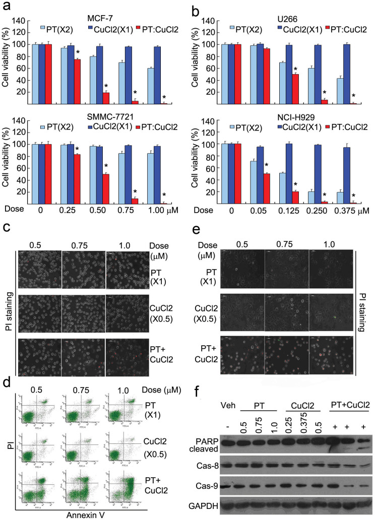
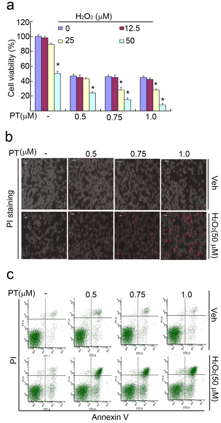
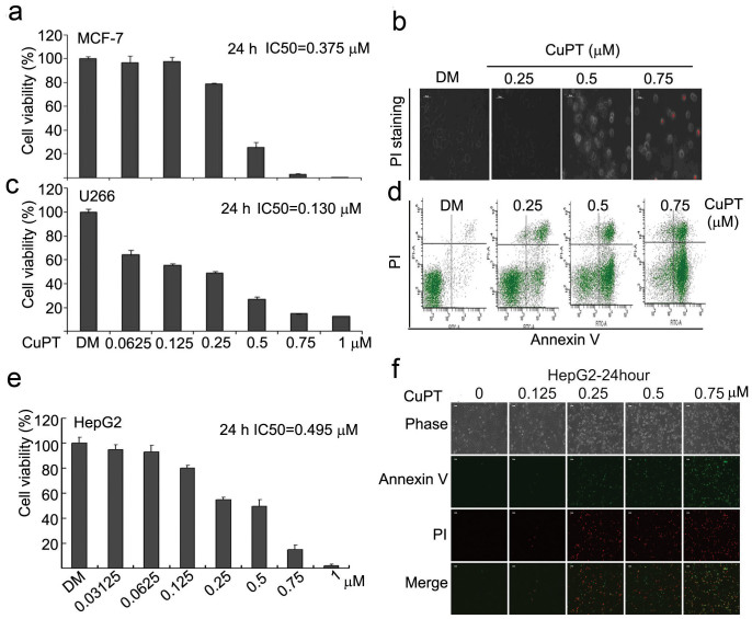
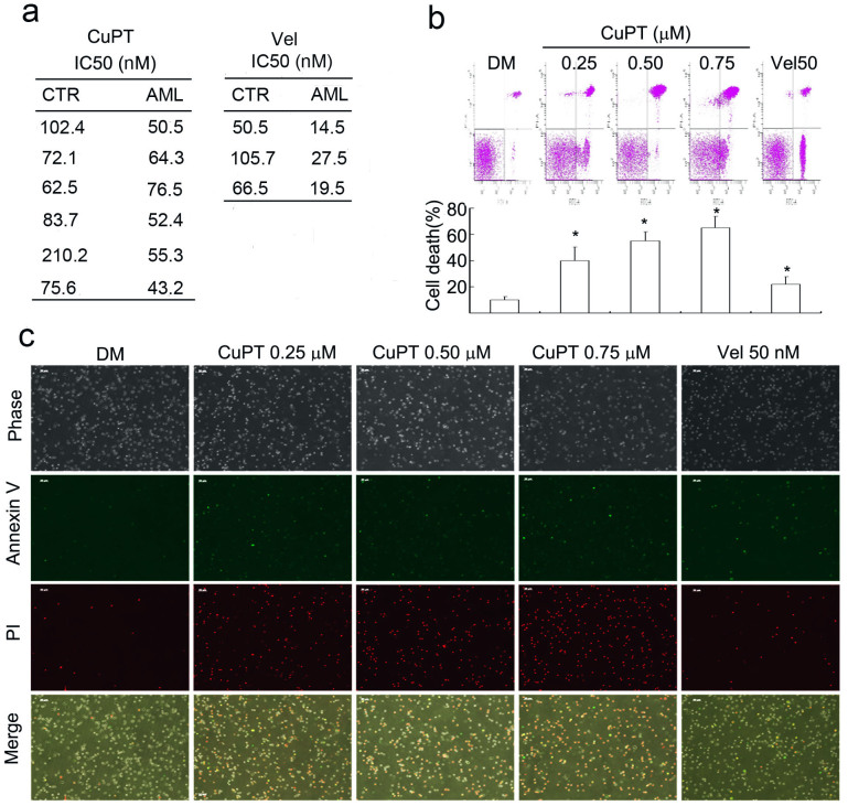
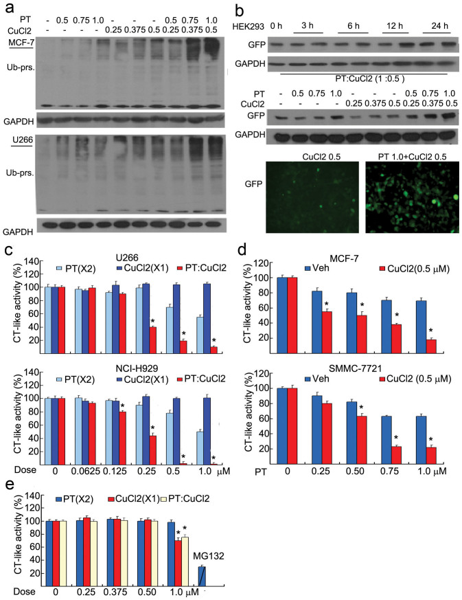
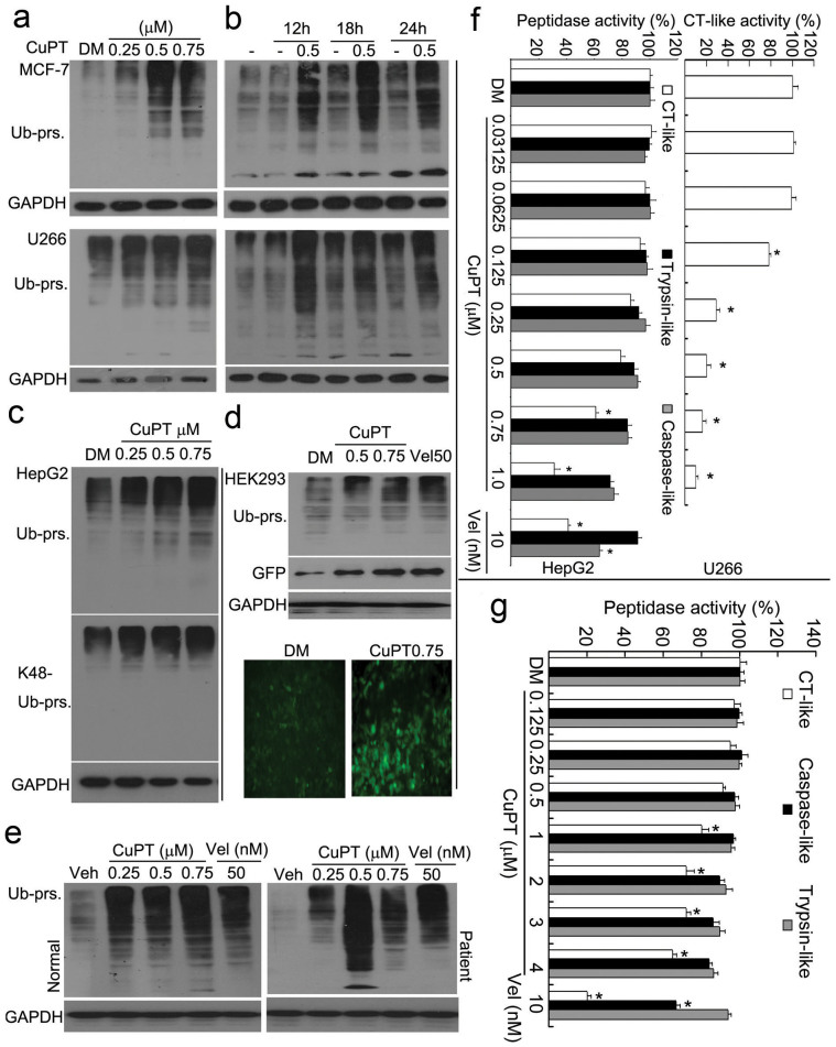
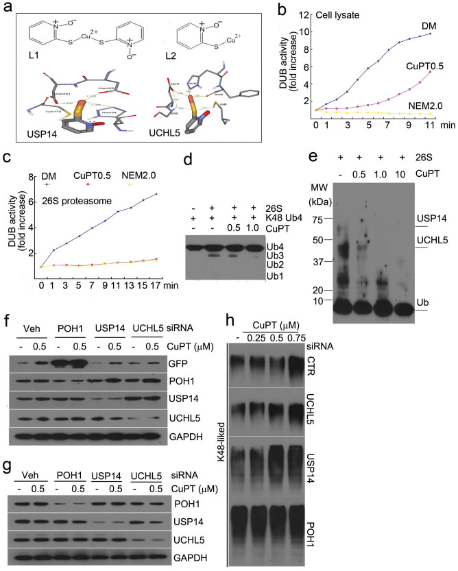
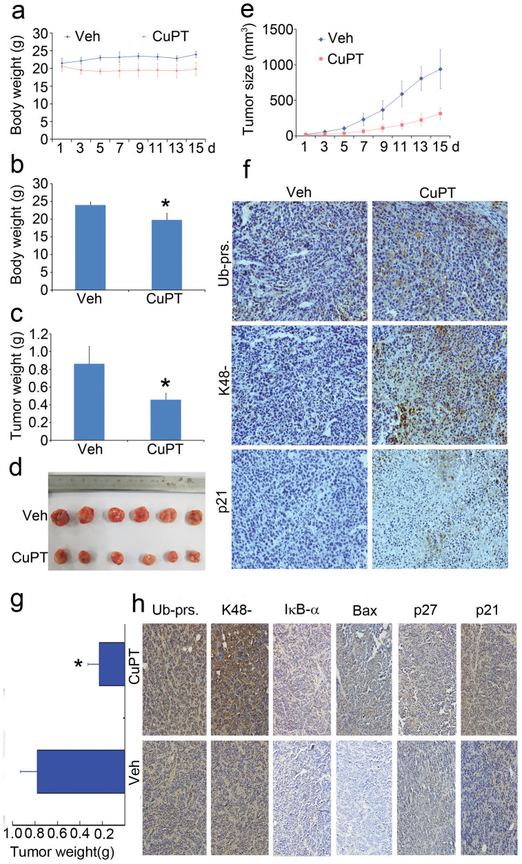
The successful development of bortezomib-based therapy for treatment of multiple myeloma has established proteasome inhibition as an effective therapeutic strategy, and both 20S proteasome peptidases and 19S deubiquitinases (DUBs) are becoming attractive targets of cancer therapy. It has been reported that metal complexes, such as copper complexes, inhibit tumor proteasome. However, the involved mechanism of action has not been fully characterized. Here we report that (i) copper pyrithione (CuPT), an alternative to tributyltin for antifouling paint biocides, inhibits the ubiquitin-proteasome system (UPS) via targeting both 19S proteasome-specific DUBs and 20S proteolytic peptidases with a mechanism distinct from that of the FDA-approved proteasome inhibitor bortezomib; (ii) CuPT potently inhibits proteasome-specific UCHL5 and USP14 activities; (iii) CuPT inhibits tumor growth in vivo and induces cytotoxicity in vitro and ex vivo. This study uncovers a novel class of dual inhibitors of DUBs and proteasome and suggests a potential clinical strategy for cancer therapy.


Figures

Figure 1
Figure 1. Pyrithione (PT) and CuCl2 in combination enhanced cytotoxicity.
(a and b) PT and CuCl2 synergistically decreased cell viability. Cancer cells (MCF-7, HepG2, U266, NCI-H929) were treated with PT, CuCl2 alone and their combination (PT/CuCl2: 2:1) at the indicated doses for 24 hours, cell viability was detected by MTS assay. Mean ± SD (n = 3). *P < 0.05, versus each treatment alone. (c and d) PT and CuCl2 in combination accelerated cell apoptosis and cell death in U266 cells. U266 cells were exposed to PT, CuCl2 and their combination at the indicated doses for 24 hours, cell death and cell apoptosis were detected by either PI staining with an inverted fluorescence microscope in live cells (c) or by Annexin V/propidium (PI) staining with flow cytometer (d). Scale bar = 50 μm. (e and f) PT and CuCl2 in combination accelerated cell death, PARP cleavage and caspase activation in MCF-7 cells. MCF-7 cells were incubated with various doses of PT, CuCl2 and their combination, then cell death was detected with PI staining in live cells (24 hours), and caspase-8, -9, PARP cleavage were detected by Western blot (12 hours). GAPDH: loading control. Scale bar = 50 μm.
Figure 2
Figure 2. Pyrithione (PT) and H2O2 in combination enhanced cytotoxicity.
(a) PT and H2O2 synergistically decreased cell viability. U266 cancer cells were treated with PT, H2O2 alone and their combination at the indicated doses for 24 hours, cell viability was detected by MTS assay. Mean ± SD (n = 3). *P < 0.05, versus each PT treatment alone. (b and c) PT and H2O2 in combination accelerated cell apoptosis and cell death in U266 cells. U266 cells were exposed to PT, H2O2 and their combination at the indicated doses for 24 hours, cell death and cell apoptosis were detected by either PI staining with an inverted fluorescence microscope in live cells (b) or by Annexin V/propidium (PI) staining with flow cytometer (c). Scale bar = 50 μm.
Figure 3
Figure 3. Copper pyrithione (CuPT), the chelating product of PT and CuCl2 induced cytotoxicity in multiple cancer cells.
(a and b) CuPT decreased cell viability and induced cell death in MCF-7 cells. MCF-7 cells were treated with CuPT for 24 hours, and then cell viability and cell death were detected as described above. IC50 was calculated and shown in (a) and representative PI-positive morphological images were shown in (b). Scale bar = 50 μm. (c and d) CuPT decreased cell viability and induced cell apoptosis in U266 cells. U266 cells were treated with CuPT for 24 hours, and then cell viability and cell apoptosis were detected. IC50 was calculated and shown in (c), and representative cell apoptosis flow images were shown in (d). (e and f) CuPT decreased cell viability and induced cell apoptosis in HepG2 cells. HepG2 cells were treated with CuPT for 24 hours, and then cell viability and cell apoptosis were detected. IC50 was calculated and shown in (e), and representative Annexin V/PI-positive and morphological images were shown in (f). Scale bar = 50 μm.
Figure 4
Figure 4. CuPT induces cytotoxicity in primary cancer cells from patients with acute myeloid leukemia (AML).
(a) Cancer cells from 6 AML patients and peripheral blood mononuclear cells from 6 healthy volunteers were treated with CuPT (0.0625, 0.125, 0.25, 0.5 μM) or bortezomib (Vel, 50 nM) for 24 hours and the cell viability was detected by MTS assay. Mean ± SD (n = 3). *P < 0.05, versus DMSO control. (b and c) Cancer cells from 3 AML patients were isolated and incubated with CuPT (0.25, 0.5, 0.75 μM) or Vel (50 nM) for 12 hours. Cell apoptosis was analyzed by flow cytometry and by imaging under a fluorescence microscope. Representative flow images were shown in and the cell death data summarized (b). Mean ± SD (n = 3). *P < 0.05, versus DMSO control. The phase contrast and fluorescent images were taken and merged. Typical images were shown (c). Red indicates PI-positive and green indicates Annexin V-positive. Scale bar = 50 μm.
Figure 5
Figure 5. PT plus CuCl2 but not H2O2 enhanced the inhibition of the ubiquitin-proteasome system.
(a) PT plus CuCl2 enhanced the accumulation of ubiquitinated proteins. MCF-7 and U266 cells were treated with various doses of PT, CuCl2 and their combination for 12 hours, and then ubiquitinated proteins were detected by Western blot. GAPDH: loading control. (b) PT plus CuCl2 induced GFPu accumulation. HEK-293 cells stably harboring GFPu, a surrogate proteasome substrate, were treated with PT, CuCl2 and their combination for 12 hours, and then GFPu was detected either by Western blot or by inverted fluorescence microscope. (c) U266 and NCI-H929 cancer cells were exposed to PT, CuCl2 and the combination (PT/CuCl2: 2:1) for 6 hours, and then CT-like substrate was added to the treated cells and the CT-like activity was measured by a multiple plate reader. Mean ± SD (n = 3). *P < 0.05, versus each treatment alone. (d) MCF-7 and SMMC-7721 cancer cells were treated with increasing doses of PT in the absence or presence of 0.5 μM CuCl2 for 6 hours, CT-like activity was measured as in (c). Mean ± SD (n = 3). *P < 0.05, versus PT treatment alone. (e) 20S proteasome was incubated with PT, CuCl2 and their combination (2:1), and then CT-like activity was measured. MG132 (3 μM) was used as a positive control. Mean ± SD (n = 3). *P < 0.05, versus PT treatment alone. (f) PT and H2O2 in combination did not induce the accumulation of ubiquitinated proteins. U266 cells were incubated with various doses of PT, H2O2 (50 μM) and their combination for 12 hours, then ubiquitinated proteins were detected by Western blot. GAPDH: loading control.
Figure 6
Figure 6. Therapeutic doses of CuPT inhibits the proteasome function in cultured cells.
(a and b) CuPT induced the accumulation of ubiquitinated proteins. MCF-7 and U266 cells were treated either with CuPT (0.5, 0.75, 1.0 μM) for 12 hours or CuPT (0.5 μM) for various times (12, 18, 24 hours), and then ubiquitinated proteins were detected by Western blot. Dose- or time-dependent effect of CuPT was shown in (a) and (b), respectively. (c) CuPT induced the accumulation of K48-linked poly-ubiquitinated proteins. HepG2 cells were exposed to CuPT as indicated for 12 h, ubiquitinated and K48-linked proteins were detected by Western blot. (d) CuPT induced the accumulation of GFPu, a surrogate proteasome substrate. HEK-293 cells harboring GFPu were treated with CuPT for 12 hours, and then ubiquitinated proteins and GFP protein were detected by Western blot or imaged under an inverted fluorescence microscope. Bortezomib/Velcade (Vel) was used a positive control. (e) CuPT induced the accumulation of ubiquitinated proteins in primary cultured cancer cells. Cancer cells from AML patients or normal controls were cultured and treated with various doses of CuPT and Vel for 6 hours, and then the ubiquitinated proteins were detected by Western blot. (f) The effect of CuPT on proteasome peptidase activity in situ. HepG2 cells or U266 cells were treated with increasing doses of CuPT and Vel (10 nM) for 6 hours, followed by addition of proteasome substrates to the treated cells, and then peptidase activities including CT-like, caspase-like and trypsin-like were detected in situ. Mean ± SD (n = 3). *P < 0.05, versus control-treated. (g) The effect of CuPT on proteasome peptidase activities in vitro. 20S proteasome was treated with increasing doses of CuPT or Vel in vitro and then proteasome peptidase activities were detected.
Figure 7
Figure 7. CuPT inhibits the proteasome deubiquitinase function.
(a) Computational molecular docking of Cu2+ with POH1, UCHL5 and USP14 of 19S proteasomes. The following data were shown: the structure of copper pyrithione (L1); the structure of copper pyrithione intermediate (L2); the binding modes of compound L2 at the active site of USP14; the binding models of compound L2 at the active site of UCHL5. (b) HepG2 cell lysates were treated with CuPT (0.5 μM) and N-ethylmaleimide (NEM, 2 mM) and DUB activity was measured. Each point represents the average of 3 wells. (c) Therapeutic dose of CuPT directly inhibits 26S deubiquitinase activity. 26S proteasome was incubated with CuPT (0.5 μM), and then DUB activity was measured. Time-dependent activities were shown. Each point represents the average of 3 wells. (d) CuPT inhibits the ubiquitin chain disassembly. K48-linked ubiquitin tetramers was incubated with 26S proteasomes in the absence or presence of CuPT (0.5, 1.0 μM) for 30 min, and then ubiquitins were detected by Western blot. (e) Active-site-directed labeling of proteasomal deubiquitinases. 26S proteasomes were treated with CuPT (0.5, 1.0, 10 μM) and then labeled with HA-UbVS. Labeled HA was detected by Western blot. (f) Knockdown of proteasome DUBs affected the efficiency of CuPT on GFPu protein degradation. HEK-293 cells harboring GFPu protein were transfected with siRNA of POH1, UCHL5 and USP14 for 48 hours, and then were exposed to 0.5 μM of CuPT for 9 hours. POH1, UCHL5, USP14 and GFPu were detected by Western blot. (g and h) The effect of proteasome DUB knockdown on the accumulation of K48-linked ubiquitinated proteins. HepG2 cells were transfected with DUB siRNA for 48 hours, followed by CuPT treatment for 6 hours. DUB proteins (g) and K48-linked ubiquitinated proteins (h) were detected by Western blot.
Figure 8
Figure 8. CuPT inhibits tumor growth and the ubiquitin-proteasome system in vivo.
(a–f) Nude mice bearing HepG2 xenograft tumors were treated with vehicle and CuPT (2.5 mg/kg/d) for totally 15 days (Day 7 interval) after inoculation of HepG2 cells. On day 18 after inoculation, the mice were sacrificed, and the tumor tissues were weighed, imaged and summarized. Body weight was recorded everyday. (a) Body weight changes; (b) Tumor weight and image. *P < 0.05, versus CuPT-treated group; (c) Proteasome substrate protein changes. Proteasome-related proteins in tumor tissues were detected by immunohistological analysis. All the immunostaining was repeated in three mouse tumor tissues and the most typical images were shown. (g and h) Nude mice bearing NCI-H929 xenograft tumors were treated with vehicle and CuPT (2.5 mg/kg/d), for 5 days after inoculation of NCI-H929 cells. On day 8 after inoculation, the mice were sacrificed. The tumor tissues were weighed and proteasome-related proteins in tumor tissues were detected by immunohistological analysis.

References

    1. Richardson P. G. et al. A phase 2 study of bortezomib in relapsed, refractory myeloma. N. Engl. J. Med. 348, 2609–2617 (2003). - PubMed
    1. Adams J. The development of proteasome inhibitors as anticancer drugs. Cancer Cell 5, 417–421 (2004). - PubMed
    1. Richardson P. G. et al. Bortezomib or high-dose dexamethasone for relapsed multiple myeloma. N. Engl. J. Med. 352, 2487–2498 (2005). - PubMed
    1. Galanski M., Arion V. B., Jakupec M. A. & Keppler B. K. Recent developments in the field of tumor-inhibiting metal complexes. Curr. Pharm. Des. 9, 2078–2089 (2003). - PubMed
    1. Galanski M., Jakupec M. A. & Keppler B. K. Update of the preclinical situation of anticancer platinum complexes: novel design strategies and innovative analytical approaches. Curr. Med. Chem. 12, 2075–2094 (2005). - PubMed

Publication types

MeSH terms