Epigenetic silencing of DACH1 induces the invasion and metastasis of gastric cancer by activating TGF-β signalling
- PMID: 24912879
- PMCID: PMC4302654
- DOI: 10.1111/jcmm.12325
Epigenetic silencing of DACH1 induces the invasion and metastasis of gastric cancer by activating TGF-β signalling
Abstract
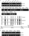
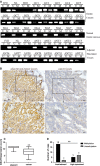
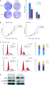
Gastric cancer (GC) is the fourth most common malignancy in males and the fifth most common malignancy in females worldwide. DACH1 is frequently methylated in hepatic and colorectal cancer. To further understand the regulation and mechanism of DACH1 in GC, eight GC cell lines, eight cases of normal gastric mucosa, 98 cases of primary GC and 50 cases of adjacent non-tumour tissues were examined. Methylation-specific PCR, western blot, transwell assay and xenograft mice were used in this study. Loss of DACH1 expression correlated with promoter region methylation in GC cells, and re-expression was induced by 5-Aza-2'-deoxyazacytidine. DACH1 is methylated in 63.3% (62/98) of primary GC and 38% (19/50) of adjacent non-tumour tissues, while no methylation was found in normal gastric mucosa. Methylation of DACH1 correlated with reduced expression of DACH1 (P < 0.01), late tumour stage (stage III/IV) (P < 0.01) and lymph node metastasis (P < 0.05). DACH1 expression inhibited epithelial-mesenchymal transition and metastasis by inhibiting transforming growth factor (TGF)-β signalling and suppressed GC cell proliferation through inducing G2/M phase arrest. The tumour size is smaller in DACH1-expressed BGC823 cell xenograft mice than in unexpressed group (P < 0.01). Restoration of DACH1 expression also sensitized GC cells to docetaxel. These studies suggest that DACH1 is frequently methylated in human GC and expression of DACH1 was controlled by promoter region methylation. DACH1 suppresses GC proliferation, invasion and metastasis by inhibiting TGF-β signalling pathways both in vitro and in vivo. Epigenetic silencing DACH1 may induce GC cells' resistance to docetaxel.
Keywords: DACH1; DNA methylation; chemosensitive marker; docetaxel; gastric cancer; invasion; metastasis.
© 2014 The Authors. Journal of Cellular and Molecular Medicine published by John Wiley & Sons Ltd and Foundation for Cellular and Molecular Medicine.
Figures


Similar articles
-
Epigenetic regulation of DACH1, a novel Wnt signaling component in colorectal cancer.Epigenetics. 2013 Dec;8(12):1373-83. doi: 10.4161/epi.26781. Epub 2013 Oct 22. Epigenetics. 2013. PMID: 24149323 Free PMC article.
-
Silencing DACH1 promotes esophageal cancer growth by inhibiting TGF-β signaling.PLoS One. 2014 Apr 17;9(4):e95509. doi: 10.1371/journal.pone.0095509. eCollection 2014. PLoS One. 2014. PMID: 24743895 Free PMC article.
-
KRAB zinc-finger protein 382 regulates epithelial-mesenchymal transition and functions as a tumor suppressor, but is silenced by CpG methylation in gastric cancer.Int J Oncol. 2018 Sep;53(3):961-972. doi: 10.3892/ijo.2018.4446. Epub 2018 Jun 19. Int J Oncol. 2018. PMID: 29956735 Free PMC article.
-
Promoter methylated microRNAs: potential therapeutic targets in gastric cancer.Mol Med Rep. 2015 Feb;11(2):759-65. doi: 10.3892/mmr.2014.2780. Epub 2014 Oct 27. Mol Med Rep. 2015. PMID: 25351138 Free PMC article. Review.
-
Epigenetic modulation of cytokine expression in gastric cancer: influence on angiogenesis, metastasis and chemoresistance.Front Immunol. 2024 Feb 22;15:1347530. doi: 10.3389/fimmu.2024.1347530. eCollection 2024. Front Immunol. 2024. PMID: 38455038 Free PMC article. Review.
Cited by
-
DACH1 inhibits breast cancer cell invasion and metastasis by down-regulating the transcription of matrix metalloproteinase 9.Cell Death Discov. 2021 Nov 12;7(1):351. doi: 10.1038/s41420-021-00733-4. Cell Death Discov. 2021. PMID: 34772908 Free PMC article.
-
Interplay of retinal determination gene network with TGF-β signaling pathway in epithelial-mesenchymal transition.Stem Cell Investig. 2015 Jun 9;2:12. doi: 10.3978/j.issn.2306-9759.2015.05.03. eCollection 2015. Stem Cell Investig. 2015. PMID: 27358880 Free PMC article. Review.
-
The regulation of cytokine signaling by retinal determination gene network pathway in cancer.Onco Targets Ther. 2018 Oct 4;11:6479-6487. doi: 10.2147/OTT.S176113. eCollection 2018. Onco Targets Ther. 2018. PMID: 30323623 Free PMC article. Review.
-
Effect of DACH1 on proliferation and invasion of laryngeal squamous cell carcinoma.Head Face Med. 2018 Sep 27;14(1):20. doi: 10.1186/s13005-018-0177-1. Head Face Med. 2018. PMID: 30261897 Free PMC article.
-
BMP6 Regulates Proliferation and Apoptosis of Human Sertoli Cells Via Smad2/3 and Cyclin D1 Pathway and DACH1 and TFAP2A Activation.Sci Rep. 2017 Apr 7;7:45298. doi: 10.1038/srep45298. Sci Rep. 2017. PMID: 28387750 Free PMC article.
References
-
- Jemal A, Bray F, Center MM, et al. Global cancer statistics. CA Cancer J Clin. 2011;61:69–90. - PubMed
-
- Massague J, Blain SW, Lo RS. TGFbeta signaling in growth control, cancer, and heritable disorders. Cell. 2000;103:295–309. - PubMed
-
- Azuma H, Ehata S, Miyazaki H, et al. Effect of Smad7 expression on metastasis of mouse mammary carcinoma JygMC(A) cells. J Natl Cancer Inst. 2005;97:1734–46. - PubMed
Publication types
MeSH terms
Substances
Grants and funding
LinkOut - more resources
Full Text Sources
Other Literature Sources
Medical
Miscellaneous

