. 2014 Jun 30;211(7):1307-14.
doi: 10.1084/jem.20132413. Epub 2014 Jun 9.

Neutrophils recruited by chemoattractants in vivo induce microvascular plasma protein leakage through secretion of TNF

Affiliations

Neutrophils recruited by chemoattractants in vivo induce microvascular plasma protein leakage through secretion of TNF

Michaela Finsterbusch et al. J Exp Med. .

Abstract

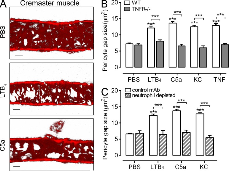
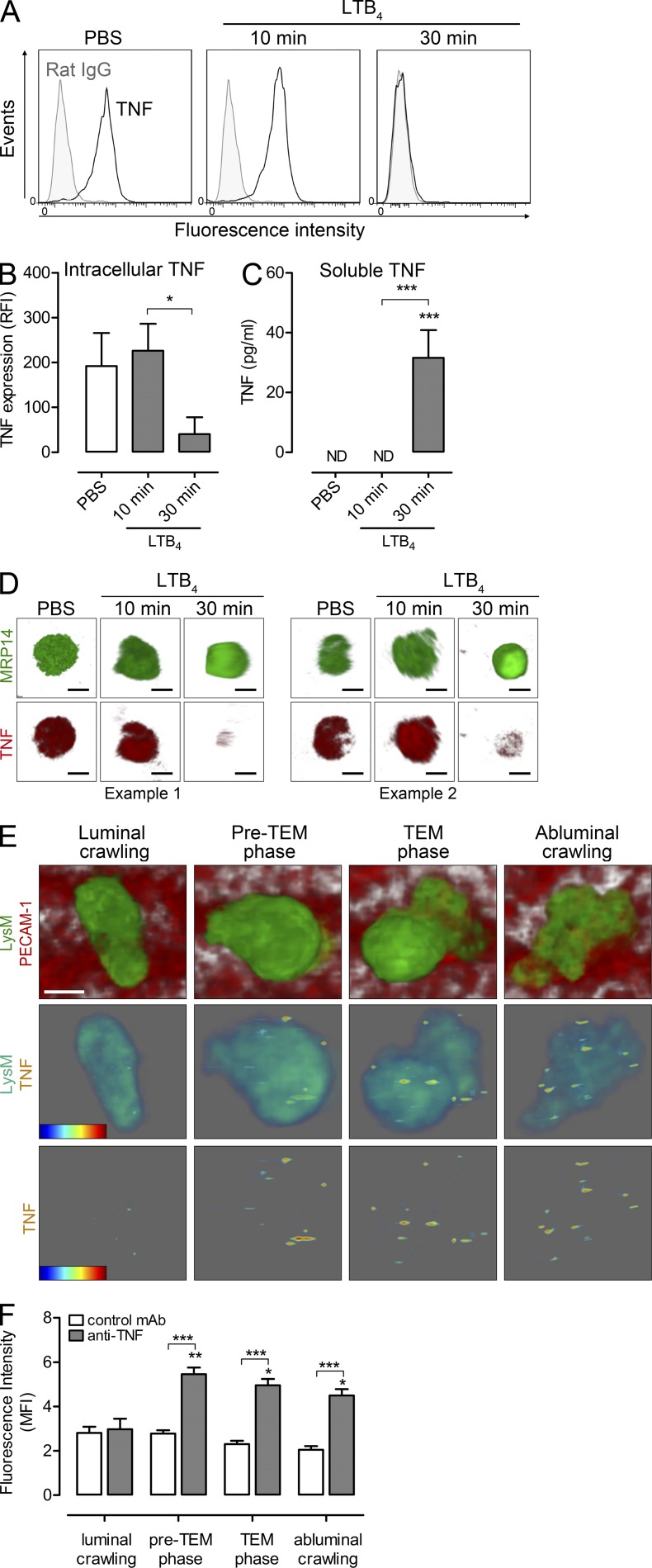
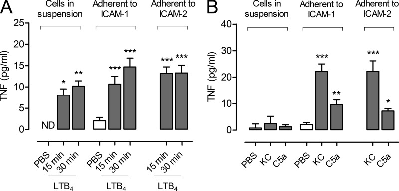
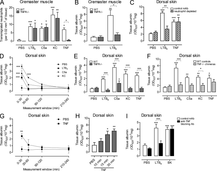
Microvascular plasma protein leakage is an essential component of the inflammatory response and serves an important function in local host defense and tissue repair. Mediators such as histamine and bradykinin act directly on venules to increase the permeability of endothelial cell (EC) junctions. Neutrophil chemoattractants also induce leakage, a response that is dependent on neutrophil adhesion to ECs, but the underlying mechanism has proved elusive. Through application of confocal intravital microscopy to the mouse cremaster muscle, we show that neutrophils responding to chemoattractants release TNF when in close proximity of EC junctions. In vitro, neutrophils adherent to ICAM-1 or ICAM-2 rapidly released TNF in response to LTB4, C5a, and KC. Further, in TNFR(-/-) mice, neutrophils accumulated normally in response to chemoattractants administered to the cremaster muscle or dorsal skin, but neutrophil-dependent plasma protein leakage was abolished. Similar results were obtained in chimeric mice deficient in leukocyte TNF. A locally injected TNF blocking antibody was also able to inhibit neutrophil-dependent plasma leakage, but had no effect on the response induced by bradykinin. The results suggest that TNF mediates neutrophil-dependent microvascular leakage. This mechanism may contribute to the effects of TNF inhibitors in inflammatory diseases and indicates possible applications in life-threatening acute edema.


Figures

Figure 1.
Figure 1.
TNF mediates chemoattractant-induced changes in venular morphology. (A) Representative confocal images of postcapillary venules from five independently conducted control (PBS) or chemoattractant-stimulated cremaster muscles (4 h) labeled for pericytes (αSMA). Bar, 20 µm. (B) Pericyte gap size was quantified in cremasteric postcapillary venules of WT and TNFR−/− mice injected with PBS (n = 10 and 5, respectively), LTB4 (n = 12 and 4, respectively), C5a (n = 5 and 4, respectively), KC (n = 5 and 4, respectively), or TNF (n = 4 and 3, respectively) for 2-4 h, involving 12 independent experiments. (C) As above but using control mice (n = 3 for PBS and LTB4, n = 5 for C5a, n = 6 for KC) or mice depleted of their circulating neutrophils (n = 3 mice/group), involving 5 independent experiments. Six vessel segments per mouse were analyzed. Data are means ± SEM. Significant differences from PBS-treated tissues or other statistical comparisons (indicated by lines) are shown by asterisks. ***, P < 0.001.
Figure 2.
Figure 2.
Chemoattractants stimulate release of TNF from neutrophils in vitro and in vivo. Mixed mouse blood leukocytes were treated with PBS (control) or stimulated with LTB4 (10 or 30 min). Supernatants were assayed for soluble TNF by ELISA and cells were permeabilized and immunostained for analysis of intracellular TNF by flow cytometry and confocal microscopy. (A) Representative flow cytometry histograms from 4 independent experiments showing the binding of anti-TNF mAb (black lines) or control IgG (filled). (B) Quantification of intracellular TNF by flow cytometry (expressed as RFI; n = 6 blood samples for PBS and 30 min LTB4, n = 5 for 10 min LTB4) from 4 independent experiments. (C) Quantification of released TNF (n = 4) from 4 independent experiments. ND, not detected. (D) Representative confocal images of neutrophils from two independent experiments show cells treated with PBS or LTB4 (10 and 30 min) and stained for the neutrophil marker MRP-14 (green) or TNF (red). Bar, 5 µm. (E) Representative 3D-reconstructed images of LTB4-stimulated cremasteric postcapillary venule (luminal side) showing GFP-neutrophils and PECAM-1–labeled ECs (red). The top panel shows neutrophils at different steps of the transmigration response, i.e., luminal crawling, preTEM phase (6 min before breaching ECs), TEM phase, and abluminal crawling. The associated TNF detected on the leukocyte surface for each step (intensity rainbow color code from blue [low intensity] to red [high intensity]) is shown in the bottom panels with and without the associated neutrophil. Bar, 5 µm. (F) Mean fluorescent intensity of signals on neutrophils was quantified in mice injected with anti-TNF or control mAbs (n = 4 mice/group) from 4 independent experiments. A total of ∼40 neutrophils were tracked and analyzed for each group. All data are means ± SEM. Statistical differences from PBS (B and C), signals from luminal crawling cells (F), or other comparisons (indicated by lines) are shown by asterisks. *, P < 0.05; **, P < 0.01; ***, P < 0.001.
Figure 3.
Figure 3.
Neutrophils in suspension or adherent to ICAM-1 and ICAM-2 release TNF in response to chemoattractants. Purified neutrophils in suspension or after adhesion to ICAM-1– or ICAM-2–coated plates were stimulated with LTB4 for 15 (n = 5, 7, and 7 samples, respectively) or 30 min (n = 4, 6, and 6 samples, respectively; A) or KC and C5a (n = 3) for 30 min (B). Control samples (in suspension or adherent to ICAM-1 o ICAM-2) were treated with PBS (n = 6, 7, and 7, respectively). Supernatants were assayed for TNF by ELISA. Graphs show means ± SEM of 3 independent experiments. ND, not detected. Significant differences from PBS control are indicated by asterisks. *, P < 0.05; **, P < 0.01; ***, P < 0.001.
Figure 4.
Figure 4.
TNF mediates chemoattractant-induced microvascular leakage. (A) Neutrophil transmigration was quantified in cremasteric postcapillary venules of WT and TNFR−/− mice injected with PBS (n = 11 and 5 mice, respectively), LTB4 (n = 25 and 4, respectively), C5a (n = 8 and 4, respectively), KC (n = 5 and 4, respectively), or TNF (n = 4 and 3, respectively) for 2-4 h involving 23 independent experiments. (B) Vascular leakage was analyzed in the cremaster muscle of WT and TNFR−/− mice treated with PBS (n = 4 and 3 mice, respectively) or LTB4 (4 h; n = 4 and 3 mice, respectively) from 4 independent experiments. (C) Vascular leakage in the dorsal skin of WT or neutrophil-depleted mice after i.d. injection of PBS (n = 9 and 8 mice, respectively), LTB4 (n = 3 mice), or TNF (n = 6 and 5 mice, respectively; 4h) from 3 independent experiments. (D) Kinetics of vascular leakage in the dorsal skin of WT mice injected i.d. with PBS (n = 9), LTB4 (n = 3), C5a (n = 3), or KC (n = 3) from 3 independent experiments. (E) Vascular leakage in the dorsal skin of WT or TNFR−/− mice injected i.d. with PBS (n = 9 mice/group), or the indicated stimuli (30 min; n = 3 mice/group) from 3 independent experiments. (F) Vascular leakage in the dorsal skin of TNF−/− chimeric mice or control mice injected i.d. with PBS or the indicated stimuli (30 min; n = 8 mice/group) from 3 independent experiments. (G) Kinetics of vascular leakage in the dorsal skin of WT mice injected i.d. with PBS (n = 9) or TNF (n = 3) from 3 independent experiments. (H) Time course of vascular leakage in the dorsal skin of WT mice injected i.d. with PBS or TNF (n = 4 mice/group) from 3 independent experiments. (I) Vascular leakage in the dorsal skin of WT mice was analyzed after i.d. injection of PBS, LTB4, or BK when co-injected with a control mAb (n = 10) or an anti-TNF blocking mAb (n = 8) using a 30 min reaction time from 3 independent experiments. All datasets are means ± SEM. Statistically significant differences from PBS treatment or other comparisons (indicated by lines) are shown by asterisks. *, P < 0.05; **, P < 0.01; ***, P < 0.001.

References

    1. Arfors K.E., Lundberg C., Lindbom L., Lundberg K., Beatty P.G., Harlan J.M. 1987. A monoclonal antibody to the membrane glycoprotein complex CD18 inhibits polymorphonuclear leukocyte accumulation and plasma leakage in vivo. Blood. 69:338–340 - PubMed
    1. Björk J., Hedqvist P., Arfors K.E. 1982. Increase in vascular permeability induced by leukotriene B4 and the role of polymorphonuclear leukocytes. Inflammation. 6:189–200 10.1007/BF00916243 - DOI - PubMed
    1. Brett J., Gerlach H., Nawroth P., Steinberg S., Godman G., Stern D. 1989. Tumor necrosis factor/cachectin increases permeability of endothelial cell monolayers by a mechanism involving regulatory G proteins. J. Exp. Med. 169:1977–1991 10.1084/jem.169.6.1977 - DOI - PMC - PubMed
    1. Cain R.J., Vanhaesebroeck B., Ridley A.J. 2010. The PI3K p110alpha isoform regulates endothelial adherens junctions via Pyk2 and Rac1. J. Cell Biol. 188:863–876 10.1083/jcb.200907135 - DOI - PMC - PubMed
    1. Colom B., Poitelon Y., Huang W., Woodfin A., Averill S., Del Carro U., Zambroni D., Brain S.D., Perretti M., Ahluwalia A., et al. 2012. Schwann cell-specific JAM-C-deficient mice reveal novel expression and functions for JAM-C in peripheral nerves. FASEB J. 26:1064–1076 10.1096/fj.11-196220 - DOI - PMC - PubMed

Publication types

MeSH terms