Review
. 2014 Aug;128(2):161-75.
doi: 10.1007/s00401-014-1305-z. Epub 2014 Jun 10.

The node of Ranvier in CNS pathology

Affiliations
Review

The node of Ranvier in CNS pathology

I Lorena Arancibia-Carcamo et al. Acta Neuropathol. 2014 Aug.

Abstract

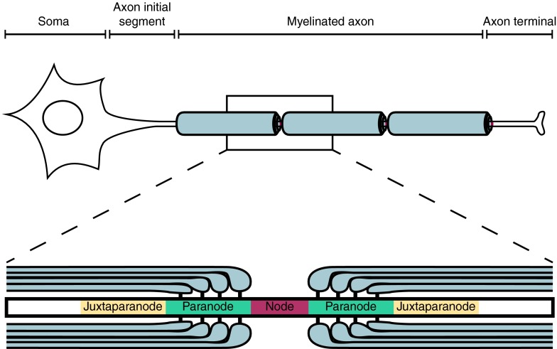
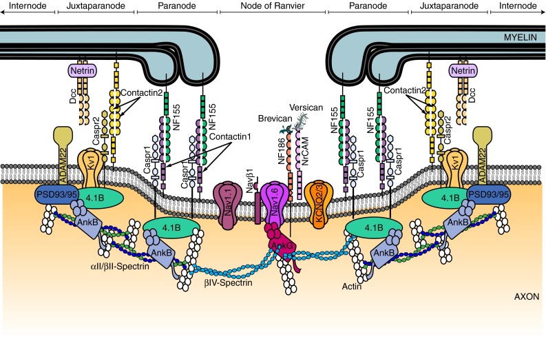
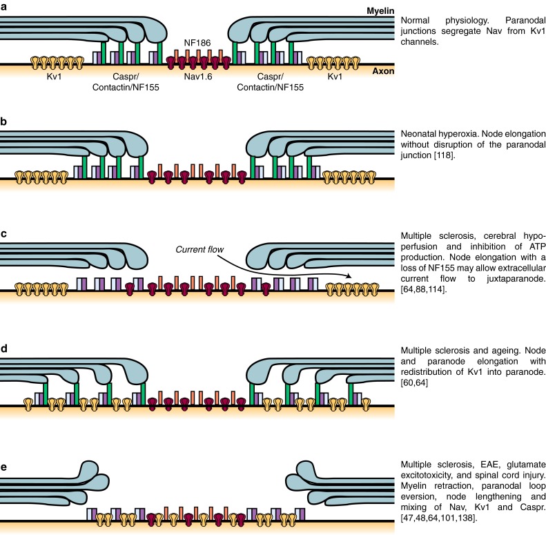
Healthy nodes of Ranvier are crucial for action potential propagation along myelinated axons, both in the central and in the peripheral nervous system. Surprisingly, the node of Ranvier has often been neglected when describing CNS disorders, with most pathologies classified simply as being due to neuronal defects in the grey matter or due to oligodendrocyte damage in the white matter. However, recent studies have highlighted changes that occur in pathological conditions at the node of Ranvier, and at the associated paranodal and juxtaparanodal regions where neurons and myelinating glial cells interact. Lengthening of the node of Ranvier, failure of the electrically resistive seal between the myelin and the axon at the paranode, and retraction of myelin to expose voltage-gated K(+) channels in the juxtaparanode, may contribute to altering the function of myelinated axons in a wide range of diseases, including stroke, spinal cord injury and multiple sclerosis. Here, we review the principles by which the node of Ranvier operates and its molecular structure, and thus explain how defects at the node and paranode contribute to neurological disorders.


Figures

Fig. 1
Fig. 1
Schematic diagram showing the different domains of a myelinated neuron. The axonal region around the node of Ranvier is expanded to show the different axonal domains: the node of Ranvier where voltage-gated Na+ channels are expressed, the paranode where the myelin is attached to the axon, and the juxtaparanode where most voltage-gated K+ channels are located. Each of these domains is characterised by the expression of specific proteins (shown in Fig. 2)
Fig. 2
Fig. 2
Schematic diagram of the proteins at the node of Ranvier, paranode and juxtaparanode. These domains are the location of ion channels (Nav1.6 and Nav1.1, KCNQ2/3, Kv3.1 and Kv1.1/1.2), cell adhesion molecules (neurofascin 155 (NF155), neurofascin 186 (NF186), contactin 1 and 2, contactin-associated protein (Caspr 1 and 2), cytoskeletal scaffolding proteins [Ankyrin (Ank) G and B, protein 4.1B, and postsynaptic density protein 93/95 (PSD93/95)], cytoskeletal proteins (βII- and βIV-spectrin), and extracellular matrix proteins (brevican, versican and a secreted form of NrCAM). Targeting and scaffolding mechanisms ensure that each protein is segregated to its specific subdomain
Fig. 3
Fig. 3
Cartoon illustrating potential mechanisms underlying enlongated nodes of Ranvier in different pathologies. a Diagram of the nodal region in control conditions. Nav channels and Kv1 channels are segregated by paranodes. b In neonatal hyperoxia, nodes of Ranvier are enlarged without any observed disruption of the paranodal junction. c In multiple sclerosis, cerebral hypoperfusion and in an experimental model of energy deprivation, the node of Ranvier is enlarged, there is a loss of NF155, and Nav channels slightly overlap with the paranodal protein Caspr. The disruption of the paranodal junction may allow current flow to under the myelin sheath to promote the activation of Kv1 channels at the juxtaparanode, thereby compromising action potential firing. d In the ageing brain and in multiple sclerosis, an elongation of the paranode can be caused by a separation of the paranodal loops. This is accompanied by a redistribution of Kv1 channels into the paranodal area, where they overlap with Caspr and NF155. e Myelin retraction, and a breakdown of the molecular organisation of the node of Ranvier, paranode and juxtaparanode, are observed in multiple sclerosis, EAE, glutamate excitotoxicity, and spinal cord injury

References

    1. Adelman JP, Bond CT, Pessia M, Maylie J. Episodic ataxia results from voltage-dependent potassium channels with altered functions. Neuron. 1995;15(6):1449–1454. - PubMed
    1. Ahn KH, Lyoo IK, Lee HK, Song IC, Oh JS, Hwang J, Kwon J, Kim MJ, Kim M, Renshaw PF. White matter hyperintensities in subjects with bipolar disorder. Psychiatry Clin Neurosci. 2004;58(5):516–521. - PubMed
    1. Anderson GR, Galfin T, Xu W, Aoto J, Malenka RC, Sudhof TC. Candidate autism gene screen identifies critical role for cell-adhesion molecule CASPR2 in dendritic arborization and spine development. Proc Natl Acad Sci USA. 2012;109(44):18120–18125. - PMC - PubMed
    1. Aoki Y, Abe O, Nippashi Y, Yamasue H. Comparison of white matter integrity between autism spectrum disorder subjects and typically developing individuals: a meta-analysis of diffusion tensor imaging tractography studies. Mol Autism. 2013;4(1):25. - PMC - PubMed
    1. Athanasiu L, Mattingsdal M, Kahler AK, Brown A, Gustafsson O, Agartz I, Giegling I, Muglia P, Cichon S, Rietschel M, Pietilainen OP, Peltonen L, Bramon E, Collier D, Clair DS, Sigurdsson E, Petursson H, Rujescu D, Melle I, Steen VM, Djurovic S, Andreassen OA. Gene variants associated with schizophrenia in a Norwegian genome-wide study are replicated in a large European cohort. J Psychiatr Res. 2010;44(12):748–753. - PMC - PubMed

Publication types

LinkOut - more resources