. 2014 Jul 17;369(1647):20130313.
doi: 10.1098/rstb.2013.0313.

Diffraction before destruction

Affiliations

Diffraction before destruction

Henry N Chapman et al. Philos Trans R Soc Lond B Biol Sci. .

Abstract

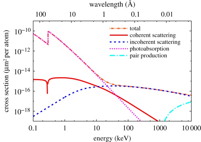
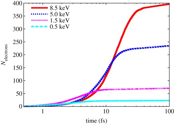
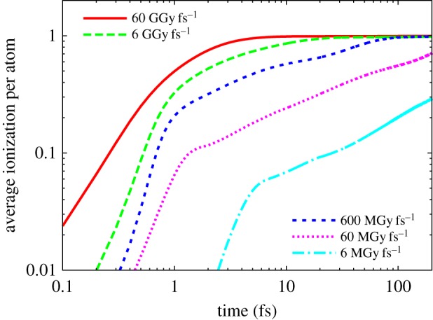
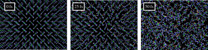
X-ray free-electron lasers have opened up the possibility of structure determination of protein crystals at room temperature, free of radiation damage. The femtosecond-duration pulses of these sources enable diffraction signals to be collected from samples at doses of 1000 MGy or higher. The sample is vaporized by the intense pulse, but not before the scattering that gives rise to the diffraction pattern takes place. Consequently, only a single flash diffraction pattern can be recorded from a crystal, giving rise to the method of serial crystallography where tens of thousands of patterns are collected from individual crystals that flow across the beam and the patterns are indexed and aggregated into a set of structure factors. The high-dose tolerance and the many-crystal averaging approach allow data to be collected from much smaller crystals than have been examined at synchrotron radiation facilities, even from radiation-sensitive samples. Here, we review the interaction of intense femtosecond X-ray pulses with materials and discuss the implications for structure determination. We identify various dose regimes and conclude that the strongest achievable signals for a given sample are attained at the highest possible dose rates, from highest possible pulse intensities.

Keywords: X-ray lasers; protein crystallography; radiation damage.


Figures

Figure 1.
Figure 1.
Atomic cross sections of neutral carbon for photoabsorption, elastic scattering, and inelastic (Compton) scattering. The carbon K-shell absorption edge is visible at 284 eV photon energy. The cross sections are plotted in the unusual units of µm2 per atom, because this shows the inverse of how many photons are required per square micrometre to photoionize or scatter from any atom in the beam. (1 barn = 10−24 cm2 = 10−16 μm2). (Online version in colour.)
Figure 2.
Figure 2.
The cascade of electrons generated by a single electron of energy 500, 1.5, 5 and 8 keV in a urea crystal (CON2H4) as calculated by molecular dynamics code [40,41]. The number of secondary electrons generated is plotted as a function of time after the photon absorption event. After 100 fs, there is an electron generated per 21 eV of absorbed energy. The majority of secondary electrons are created in less than 20 fs. (Online version in colour.)
Figure 3.
Figure 3.
The evolution of the average ionization of atoms in a sample illuminated by an X-ray pulse at various dose rates, as calculated by plasma dynamics code. The ionization per atom is normalized to 1, corresponding to full ionization in all atoms. (Online version in colour.)
Figure 4.
Figure 4.
Molecular dynamics simulations of the evolution of a urea crystal exposed to a 50 fs X-ray pulse of 5 × 10¹² 9 keV photons focused to 1 μm2. Views of the crystal are shown before illumination, after 25 fs, and after 50 fs. This intensity corresponds to a dose rate of 50 MGy fs1. Reproduced from [46]. (Online version in colour.)
Figure 5.
Figure 5.
Plots of the RMS atomic displacement as a function of time during pulses at various dose rates, as calculated by plasma dynamics code in a sample matching the composition of photosystem I and which is much larger than the photoelectron range [46]. The curves are averaged over photon energies from 2 to 9 keV. (Online version in colour.)
Figure 6.
Figure 6.
Magnitude of the effective form factor of Fe atoms in a protein sample as a function of pulse fluence (or equivalently, dose) for a photon energy of 8.1 keV (trial mode). Reproduced with permission from [27]. (Online version in colour.)
Figure 7.
Figure 7.
Simulated diffraction patterns of a 5 × 5 × 5 unit cell cubic crystal undergoing an X-ray induced explosion, following the dynamics shown in figure 5. (a) The undamaged pattern simulated with no atomic displacements; (b) the pattern simulated with 1 Å RMS atomic displacement and (c) the pulse-integrated pattern where the RMS displacement reached 1 Å at the end of the pulse. The unit cell length is 25 Å. (Online version in colour.)
Figure 8.
Figure 8.
The effective fluence (or total diffracted signal) that can be achieved in a single pulse is a product of the pulse intensity (or equivalently dose rate) and the time the sample diffracts (the turn-off time). The turn-off time decreases with increasing dose rate, but the result is an increase in total achievable signal. (Online version in colour.)

References

    1. Henderson R. 1995. The potential and limitations of neutrons, electrons and X-rays for atomic resolution microscopy of unstained biological molecules. Q. Rev. Biophys. 28, 171–193. (10.1017/S003358350000305X) - DOI - PubMed
    1. Owen RL, Rudino-Pinera E, Garman EF. 2006. Experimental determination of the radiation dose limit for cryocooled protein crystals. Proc. Natl Acad. Sci. USA 103, 4912–4917. (10.1073/pnas.0600973103) - DOI - PMC - PubMed
    1. Howells MR, et al. 2009. An assessment of the resolution limitation due to radiation-damage in X-ray diffraction microscopy. J. Electr. Spectrosc. Relat. Phenom. 170, 4–12. (10.1016/j.elspec.2008.10.008) - DOI - PMC - PubMed
    1. Holton JM. 2009. A beginner's guide to radiation damage. J. Synchronton Radiat. 16, 133–142. (10.1107/S0909049509004361) - DOI - PMC - PubMed
    1. Chapman HN, et al. 2011. Femtosecond X-ray protein nanocrystallography. Nature 470, 73–77. (10.1038/nature09750) - DOI - PMC - PubMed

Publication types

LinkOut - more resources