Review
. 2014 Jul 17;369(1647):20130314.
doi: 10.1098/rstb.2013.0314.

Femtosecond crystallography of membrane proteins in the lipidic cubic phase

Affiliations
Review

Femtosecond crystallography of membrane proteins in the lipidic cubic phase

Wei Liu et al. Philos Trans R Soc Lond B Biol Sci. .

Abstract

Despite recent technological advances in heterologous expression, stabilization and crystallization of membrane proteins (MPs), their structural studies remain difficult and require new transformative approaches. During the past two years, crystallization in lipidic cubic phase (LCP) has started gaining a widespread acceptance, owing to the spectacular success in high-resolution structure determination of G protein-coupled receptors (GPCRs) and to the introduction of commercial instrumentation, tools and protocols. The recent appearance of X-ray free-electron lasers (XFELs) has enabled structure determination from substantially smaller crystals than previously possible with minimal effects of radiation damage, offering new exciting opportunities in structural biology. The unique properties of LCP material have been exploited to develop special protocols and devices that have established a new method of serial femtosecond crystallography of MPs in LCP (LCP-SFX). In this method, microcrystals are generated in LCP and streamed continuously inside the same media across the intersection with a pulsed XFEL beam at a flow rate that can be adjusted to minimize sample consumption. Pioneering studies that yielded the first room temperature GPCR structures, using a few hundred micrograms of purified protein, validate the LCP-SFX approach and make it attractive for structure determination of difficult-to-crystallize MPs and their complexes with interacting partners. Together with the potential of femtosecond data acquisition to interrogate unstable intermediate functional states of MPs, LCP-SFX holds promise to advance our understanding of this biomedically important class of proteins.

Keywords: G protein-coupled receptor; X-ray free-electron laser; lipidic cubic phase; membrane proteins; serial femtosecond crystallography.


Figures

Figure 1.
Figure 1.
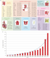
Progress in MP structure determination using LCP crystallization. (a) Gallery of MPs, structures of which were solved using LCP crystallization. For each unique MP, the highest resolution and corresponding PDB ID are shown. (b) Increase in the number of unique MPs structures obtained by LCP crystallization with time. Continuous curve represents an exponential fit.
Figure 2.
Figure 2.
A schematic workflow associated with the LCP-SFX method. (a) MP microcrystals are grown in LCP (red) inside a syringe mixer. (b) LCP with microcrystals is streamed from an LCP injector and intersects with an XFEL beam focused to about 1.5 μm diameter. Diffraction snapshots are recorded at 120 Hz by the Cornell-SLAC pixel array detector. (c) Millions of snapshots are processed to extract patterns with crystal diffraction and index these patterns, followed by integration and merging of individual reflections to derive structure factors. (d) These structure factors are then used to solve and refine the MP structure.
Figure 3.
Figure 3.
First GPCR structures obtained by LCP-SFX. (a) Comparison between XFEL (light red) and synchrotron (blue) structures of 5-HT2B/ergotamine. Ergotamine is shown as sticks along with 2mFo-DFc electron density map around it, contoured at 1σ. (b) Smoothened receptor in complex with cyclopamine. A long cavity inside the receptor is highlighted in blue. Zoomed insert shows the cyclopamine binding site with 2mFo-DFc electron density map contoured at 1σ. Horizontal lines indicate approximate membrane boundaries.

References

    1. Yildirim MA, Goh KI, Cusick ME, Barabasi AL, Vidal M. 2007. Drug–target network. Nat. Biotechnol. 25, 1119–1126. (10.1038/nbt1338) - DOI - PubMed
    1. Deisenhofer J, Epp O, Miki K, Huber R, Michel H. 1985. Structure of the protein subunits in the photosynthetic reaction centre of Rhodopseudomonas viridis at 3 Å resolution. Nature 318, 618–624. (10.1038/318618a0) - DOI - PubMed
    1. Kang HJ, Lee C, Drew D. 2013. Breaking the barriers in membrane protein crystallography. Int. J. Biochem. Cell. Biol. 45, 636–644. (10.1016/j.biocel.2012.12.018) - DOI - PubMed
    1. Katritch V, Cherezov V, Stevens RC. 2013. Structure-function of the G protein-coupled receptor superfamily. Annu. Rev. Pharmacol. Toxicol. 53, 531–556. (10.1146/annurev-pharmtox-032112-135923) - DOI - PMC - PubMed
    1. Kloppmann E, Punta M, Rost B. 2012. Structural genomics plucks high-hanging membrane proteins. Curr. Opin. Struct. Biol. 22, 326–332. (10.1016/j.sbi.2012.05.002) - DOI - PMC - PubMed

Publication types