. 2014 Jul 17;369(1647):20130500.
doi: 10.1098/rstb.2013.0500.

7 Å resolution in protein two-dimensional-crystal X-ray diffraction at Linac Coherent Light Source

Affiliations

7 Å resolution in protein two-dimensional-crystal X-ray diffraction at Linac Coherent Light Source

Bill Pedrini et al. Philos Trans R Soc Lond B Biol Sci. .

Abstract

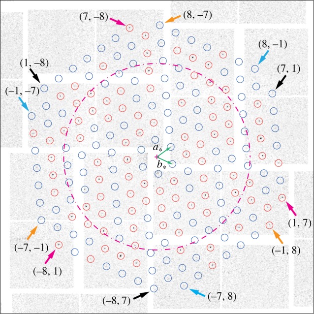
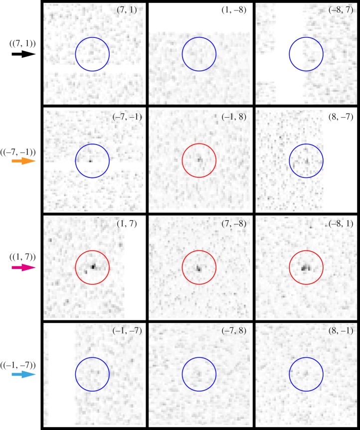
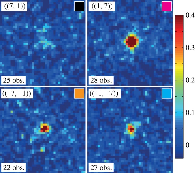
Membrane proteins arranged as two-dimensional crystals in the lipid environment provide close-to-physiological structural information, which is essential for understanding the molecular mechanisms of protein function. Previously, X-ray diffraction from individual two-dimensional crystals did not represent a suitable investigational tool because of radiation damage. The recent availability of ultrashort pulses from X-ray free-electron lasers (XFELs) has now provided a means to outrun the damage. Here, we report on measurements performed at the Linac Coherent Light Source XFEL on bacteriorhodopsin two-dimensional crystals mounted on a solid support and kept at room temperature. By merging data from about a dozen single crystal diffraction images, we unambiguously identified the diffraction peaks to a resolution of 7 Å, thus improving the observable resolution with respect to that achievable from a single pattern alone. This indicates that a larger dataset will allow for reliable quantification of peak intensities, and in turn a corresponding increase in the resolution. The presented results pave the way for further XFEL studies on two-dimensional crystals, which may include pump-probe experiments at subpicosecond time resolution.

Keywords: X-ray diffraction; X-ray free-electron laser; bacteriorhodopsin; crystallographic data analysis; two-dimensional protein crystal.


Figures

Figure 1.
Figure 1.
Example of a diffraction image from a single bR two-dimensional crystal. The dashed ring corresponds to 10 Å resolution. The circles mark the positions of expected diffraction peaks at lower than 7 Å resolution. The precise orientation of the peak lattice was derived from the positions of the prominent, easily identifiable peaks encircled in red. The basis vectors (a*, b*) of the two-dimensional reciprocal space lattice are shown as green arrows. The small arrows mark the position of peaks in the classes ((7, 1)) (black), ((1, 7)) (magenta), ((−7, −1)) (orange) and ((−1, −7)) (cyan), each class consisting of three equivalent peaks.
Figure 2.
Figure 2.
Magnifications of the diffraction image of figure 1 around peak positions in classes ((7, 1)), ((−7, −1)), ((1, 7)) and ((−1, −7)). The colour coding given by the arrows on the left is the same as in figure 1.
Figure 3.
Figure 3.
Examples of ‘image sector sums’ (see text) for the four peak classes ((7, 1)), ((−7, −1)), ((1, 7)) and ((−1, −7)), all at 7.2 Å resolution. For each peak, the number of observations is indicated, and the colour in the small box at the top right of each panel corresponds to that of the arrows in figure 1. The intensity colour scale is the same for all four panels. Maximum intensity is about 40 times the background noise, calculated as the average on all image sector sums of the local noise level measured away from the central peak region.

References

    1. Rosenbaum DM, Rasmussen SGF, Kobilka BK. 2009. The structure and function of G-protein-coupled receptors. Nature 459, 356–363. ( 10.1038/nature08144) - DOI - PMC - PubMed
    1. Deupi X, Standfuss J, Schertler G. 2012. Conserved activation paths in G-protein-coupled receptors. Biochem. Soc. Trans. 40, 383–388. ( 10.1042/BST20120001) - DOI - PubMed
    1. Fujiyoshi Y. 2011. Electron crystallography for structural and functional studies of membrane proteins. J. Electron Microsc. 60, S149–S159. ( 10.1093/jmicro/dfr008) - DOI - PubMed
    1. Srivastava SK, Gayathri S, Manjasetty BA, Gopal B. 2012. Analysis of conformational variation in macromolecular structural models. PLoS ONE 7, e39993 ( 10.1371/journal.pone.0039993) - DOI - PMC - PubMed
    1. Unwin PNT, Henderson R. 1975. Three-dimensional model of purple membrane obtained by electron microscopy. Nature 257, 28–32. ( 10.1038/257028a0) - DOI - PubMed

Publication types

Substances

LinkOut - more resources